bs-1389R [Primary Antibody]
Rabbit  Anti-Cyclin G  Polyclonal Antibody
www.bioss.com.cn
sales@bioss.com.cn
techsupport@bioss.com.cn
400-901-9800
DATASHEET

Host: Rabbit

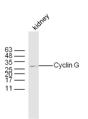
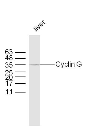
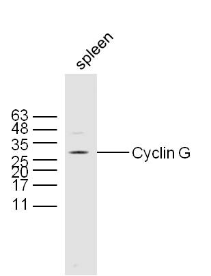
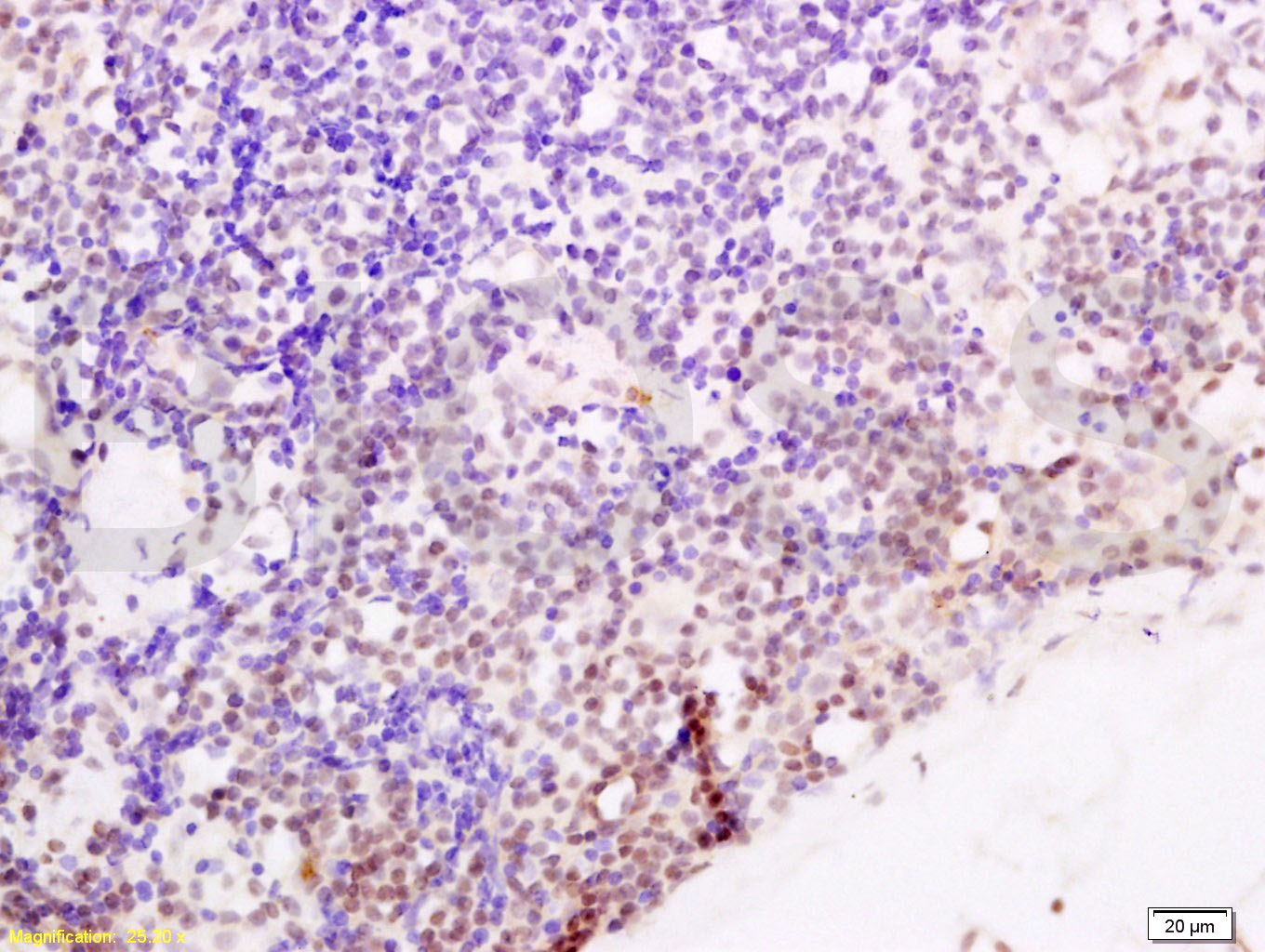
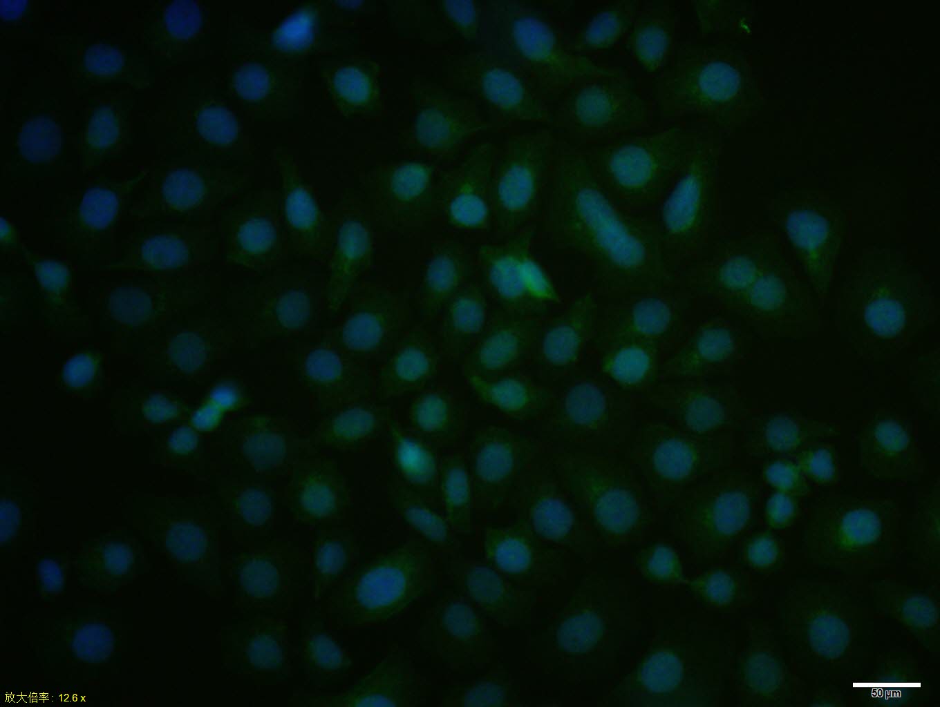
Target Protein: Cyclin G

IR: Immunogen Range:181-295/295

Clonality: Polyclonal

Isotype: IgG

Entrez Gene: 900

Swiss Prot: P51959

Source: KLH conjugated synthetic peptide derived from human Cyclin G:181-295/295 

Purification: affinity purified by Protein A

Storage: 0.01M TBS(pH7.4) with 1% BSA, 0.03% Proclin300 and 50% Glycerol. Shipped at 4℃. Store at -20 °C for one year. Avoid repeated freeze/thaw cycles.

Background: Cyclin G contains a typical N terminal cyclin box and a carboxy terminal domain sequence homologous to the tyrosine phosphorylation site of the epidermal growth factor receptor. Cyclin G2 shares 53% amino acid sequence identity with cyclin G1. Peak expression of cyclin G2 is seen in late S phase, as opposed to cyclin G1 expression, which is constitutive.

Size: 100ul

Concentration: 1mg/ml

Applications: WB=1:500-2000,IHC-P=1:100-500,IHC-F=1:100-500,ICC=1:100,IF=1:100-500,ELISA=1:5000-10000

Cross Reactive Species: Human,Mouse (predicted: Rat,Rabbit,Pig,Cow,Chicken,Dog,Horse)

For research use only. Not intended for diagnostic or therapeutic use.

VALIDATION IMAGES
Sample: Kidney (Mouse) Lysate at 40 ug Primary: Anti-Cyclin G (bs-1389R) at 1/300 dilution Secondary: IRDye800CW Goat Anti-Rabbit IgG at 1/20000 dilution Predicted band size: 34 kD Observed band size: 34/30 kD
Sample: Liver (Mouse) Lysate at 40 ug Primary: Anti-Cyclin G (bs-1389R) at 1/300 dilution Secondary: IRDye800CW Goat Anti-Rabbit IgG at 1/20000 dilution Predicted band size: 34 kD Observed band size: 34/30 kD
Sample: Spleen (Mouse) Lysate at 40 ug Primary: Anti-Cyclin G (bs-1389R) at 1/300 dilution Secondary: IRDye800CW Goat Anti-Rabbit IgG at 1/20000 dilution Predicted band size: 34 kD Observed band size: 34/30 kD
Tissue/cell: human endometrium carcinoma; 4% Paraformaldehyde-fixed and paraffin-embedded; Antigen retrieval: citrate buffer ( 0.01M, pH 6.0 ), Boiling bathing for 15min; Block endogenous peroxidase by 3% Hydrogen peroxide for 30min; Blocking buffer (normal goat serum,C-0005) at 37℃ for 20 min; Incubation: Anti-Cyclin G Polyclonal Antibody, Unconjugated(bs-1389R) 1:200, overnight at 4°C, followed by conjugation to the secondary antibody(SP-0023) and DAB(C-0010) staining
HepG2 cell; 4% Paraformaldehyde-fixed; Triton X-100 at room temperature for 20 min; Blocking buffer (normal goat serum, C-0005) at 37°C for 20 min; Antibody incubation with (Cyclin G) polyclonal Antibody, Unconjugated (bs-1389R) 1:100, 90 minutes at 37°C; followed by a conjugated Goat Anti-Rabbit IgG antibody at 37°C for 90 minutes, DAPI (blue, C02-04002) was used to stain the cell nuclei.
 
PRODUCT SPECIFIC PUBLICATIONS