bs-23550R [Primary Antibody]
Rabbit  Anti-NCR1  Polyclonal Antibody
www.bioss.com.cn
sales@bioss.com.cn
techsupport@bioss.com.cn
400-901-9800
DATASHEET

Host: Rabbit

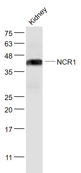
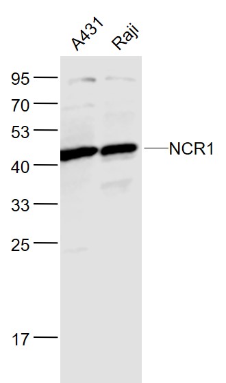
Target Protein: NCR1

IR: Immunogen Range:1-100/304

Clonality: Polyclonal

Isotype: IgG

Entrez Gene: 9437

Swiss Prot: O76036

Source: KLH conjugated synthetic peptide derived from human NCR1:1-100/304 

Purification: affinity purified by Protein A

Storage: 0.01M TBS(pH7.4) with 1% BSA, 0.03% Proclin300 and 50% Glycerol. Shipped at 4℃. Store at -20 °C for one year. Avoid repeated freeze/thaw cycles.

Background: The natural cytotoxicity receptors (NCRs) are a recently characterized family of Ig-like activation receptors that appear to be major triggering receptors in tumor cell recognition. NCR1 is a glycoprotein that has two extracellular Ig-like domains followed by a ~40 amino acid residue stalk region, a type I transmembrane domain, and a short cytoplasmic tail. NCR1 has been shown to represent a novel NK cell-specific molecule involved in human NK cell activation. NCR1 has been implicated in NK cell-mediated lysis of several autologous tumor cells and pathogen-infected cell lines.

Size: 200ul

Concentration: 1mg/ml

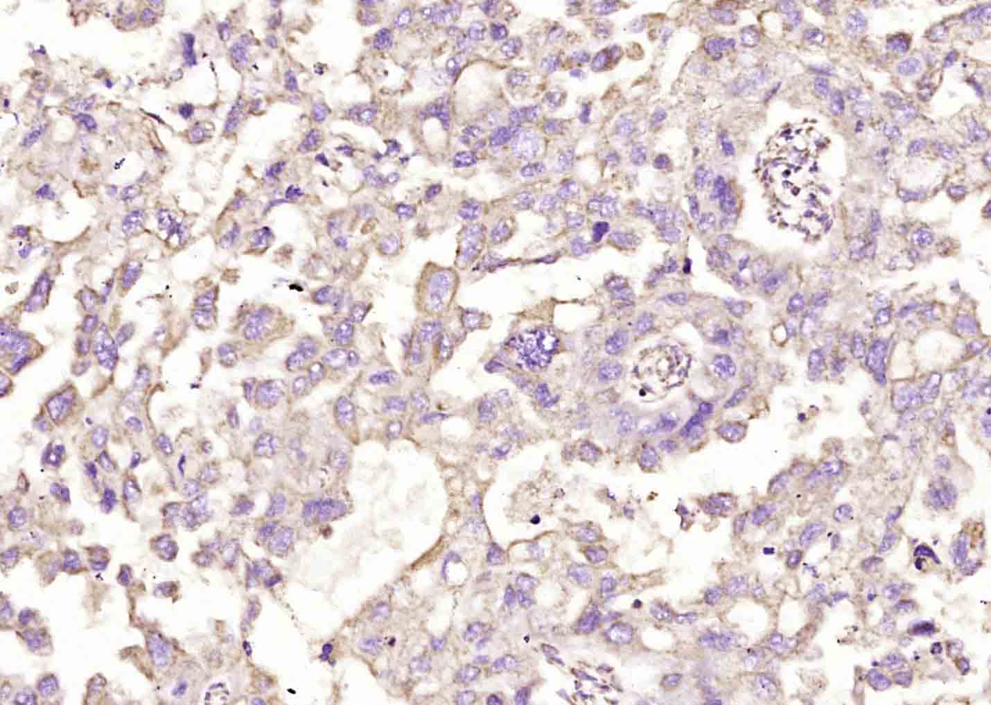
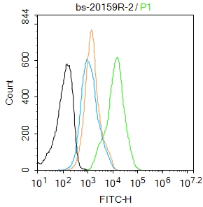
Applications: WB=1:500-2000,IHC-P=1:100-500,IHC-F=1:100-500,Flow-Cyt=2ug/Test,ICC=1:100-500,IF=1:100-500,ELISA=1:5000-10000

Cross Reactive Species: Human,Mouse

For research use only. Not intended for diagnostic or therapeutic use.

VALIDATION IMAGES
Sample: Kidney (Mouse) Lysate at 40 ug Primary: Anti- NCR1 (bs-23550R) at 1/1000 dilution Secondary: IRDye800CW Goat Anti-Rabbit IgG at 1/20000 dilution Predicted band size: 31 kD Observed band size: 46 kD
Sample: A431 (Human) Cell Lysate at 30 ug Raji (Human) Cell Lysate at 30 ug Primary: Anti- NCR1 (bs-23550R) at 1/500 dilution Secondary: IRDye800CW Goat Anti-Rabbit IgG at 1/20000 dilution Predicted band size: 31 kD Observed band size: 46 kD
Paraformaldehyde-fixed, paraffin embedded (human skin cancer); Antigen retrieval by boiling in sodium citrate buffer (pH6.0) for 15min; Block endogenous peroxidase by 3% hydrogen peroxide for 20 minutes; Blocking buffer (normal goat serum) at 37°C for 30min; Antibody incubation with (NCR1) Polyclonal Antibody, Unconjugated (bs-23550R) at 1:200 overnight at 4°C, followed by operating according to SP Kit(Rabbit) (sp-0023) instructionsand DAB staining.
Blank control:Mouse spleen. Primary Antibody (green line): Rabbit Anti-NCR1 antibody (bs-23550R) Dilution: 2μg /10^6 cells; Isotype Control Antibody (orange line): Rabbit IgG . Secondary Antibody : Goat anti-rabbit IgG-AF488 Dilution: 1μg /test. Protocol The cells were fixed with 4% PFA (10min at room temperature)and then permeabilized with 90% ice-cold methanol for 20 min at-20℃. The cells were then incubated in 5%BSA to block non-specific protein-protein interactions for 30 min at room temperature .Cells stained with Primary Antibody for 30 min at room temperature. The secondary antibody used for 40 min at room temperature. Acquisition of 20,000 events was performed.
Blank control:Mouse kidney. Primary Antibody (green line): Rabbit Anti-NCR1 antibody (bs-23550R) Dilution: 2μg /10^6 cells; Isotype Control Antibody (orange line): Rabbit IgG . Secondary Antibody : Goat anti-rabbit IgG-AF488 Dilution: 1μg /test. Protocol The cells were fixed with 4% PFA (10min at room temperature)and then permeabilized with 90% ice-cold methanol for 20 min at -20℃.The cells were then incubated in 5%BSA to block non-specific protein-protein interactions for 30 min at room temperature .Cells stained with Primary Antibody for 30 min at room temperature. The secondary antibody used for 40 min at room temperature. Acquisition of 20,000 events was performed.
 
PRODUCT SPECIFIC PUBLICATIONS