sales@bioss.com.cn
techsupport@bioss.com.cn
400-901-9800
Host: Rabbit
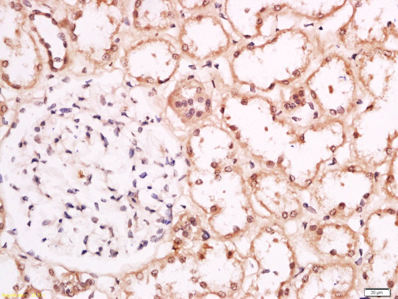
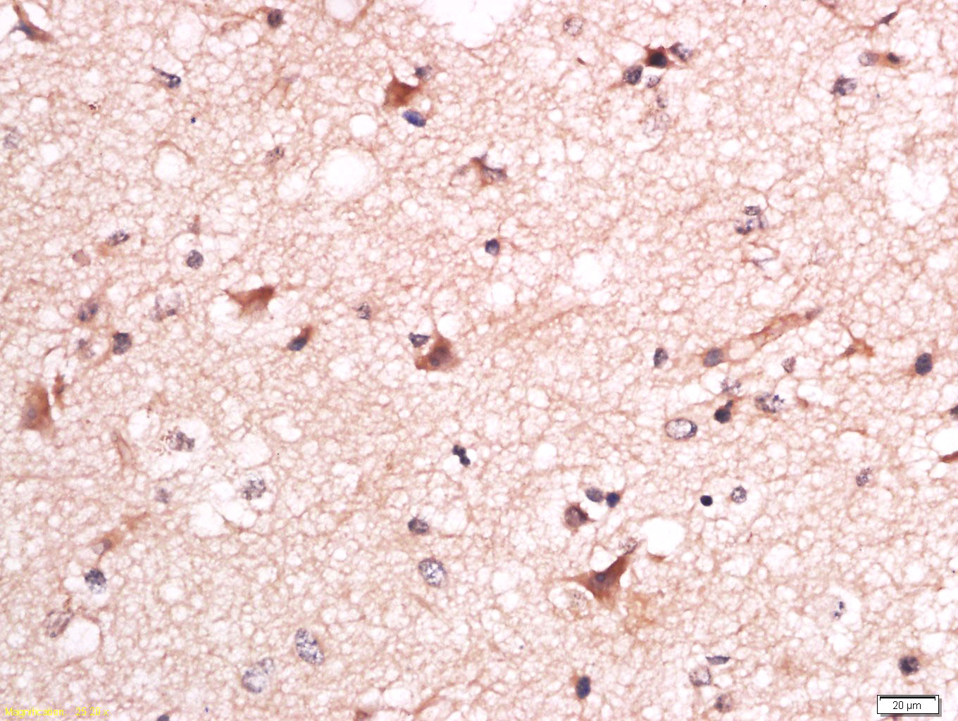
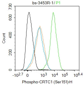
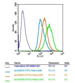
Target Protein: Phospho-CRTC1 (Ser151) Rabbit pAb
IR: Immunogen Range:TN(p-S)DS
Clonality: Polyclonal
Isotype: IgG
Entrez Gene: 23373
Swiss Prot: Q6UUV9
Source: KLH conjugated Synthesised phosphopeptide derived from human Torc1 around the phosphorylation site of Ser151:TN(p-S)DS
Purification: affinity purified by Protein A
Storage: 0.01M TBS (pH7.4) with 1% BSA, 0.02% Proclin300 and 50% Glycerol. Shipped at 4℃. Store at -20℃ for one year. Avoid repeated freeze/thaw cycles.
Background: which activates transcription through both consensus and variant cAMP response element (CRE) sites. MECT1 does not appear to modulate CREB1 DNA-binding activity but enhances the interaction of CREB1 with TAF4/TAFII-130. MECT1 translocates with MAML2 (MasterMind-Like Protein 2) to yield a fusion oncogene: t(11;19) (q21;p13). This translocation occurs in mucoepidermoid carcinomas, benign Warthin tumors and clear cell hidradenomas. The novel fusion product that results disrupts the Notch signaling pathway. The fusion protein consists of the N-terminus of MECT1 joined to the C-terminus of MAML2. The reciprocal fusion protein consisting of the N-terminus of MAML2 joined to the C-terminus of MECT1 has been detected in a small number of mucoepidermoid carcinomas. Multiple isoforms have been reported for the MECT1 protein.
Size: 200ul
Concentration: 1mg/ml
Applications: IHC-P=1:100-500,IHC-F=1:100-500,IF=1:100-500,Flow-Cyt=1μg /Test
Cross Reactive Species: Human (predicted: Mouse,Rat,Rabbit,Pig,Cow,Chicken,Dog,Horse)
For research use only. Not intended for diagnostic or therapeutic use.