sales@bioss.com.cn
techsupport@bioss.com.cn
400-901-9800
Host: Rabbit
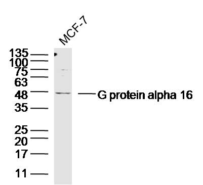
Target Protein: G protein alpha 16
IR: Immunogen Range:301-374/374
Clonality: Polyclonal
Isotype: IgG
Entrez Gene: 2769
Swiss Prot: P30679
Source: KLH conjugated synthetic peptide derived from human G protein alpha 16:301-374/374
Purification: affinity purified by Protein A
Storage: 0.01M TBS(pH7.4) with 1% BSA, 0.03% Proclin300 and 50% Glycerol. Shipped at 4℃. Store at -20 °C for one year. Avoid repeated freeze/thaw cycles.
Background: Heterotrimeric G proteins function to relay information from cell surface receptors to intracellular effectors (1). Each of a very broad range of receptors specifically detects an extracellular stimulus (a photon, pheromone, odorant, hormone or neurotransmitter) while the effectors (i.e., adenylyl cyclase), which act to generate one or more intracellular messengers, are less numerous. In mammals, G protein alpha, Beta and Gamma polypeptides are encoded by at least 16, 4 and 7 genes, respectively (2-5). Most interest in G proteins has been focused on their a subunits, since these proteins bind and hydrolyze GTP and most obviously regulate the activity of the best studied effectors. Four distinct classes of G alpha subunits have been identified; these include Gs, Gi, Gq and Ga 12/13 (3,4). The Gi class comprises all the known a subunits that are susceptible to pertussis toxin modifications, including Ga i-1, Ga i-2, Ga i-3, Ga o, Ga t1, Ga t2, Ga z and Ga gust (4). Of these, the three Ga i subtypes function to open atrial potassium channels (6). Ga 16 is a member of the Gq subfamily and is expressed specifically in hematopoietic cells (7).
Size: 50ul
Concentration: 1mg/ml
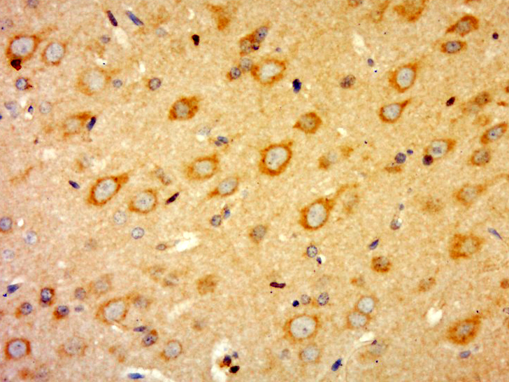
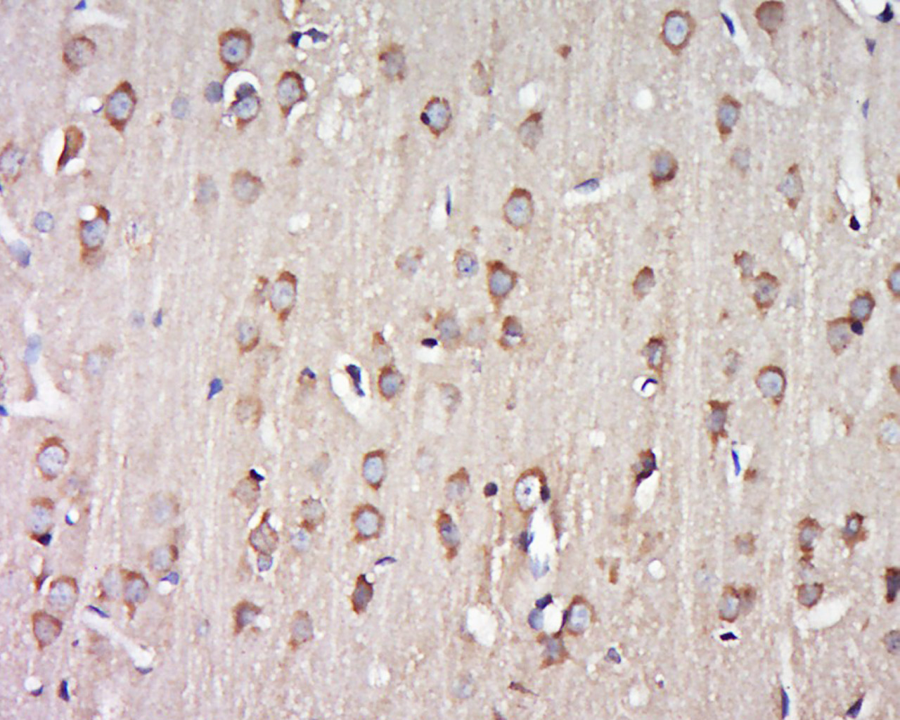
Applications: WB=1:500-2000,IHC-P=1:100-500,IHC-F=1:100-500,ICC=1:100-500,IF=1:100-500,ELISA=1:5000-10000
Cross Reactive Species: Human,Rat (predicted: Mouse,Rabbit,Pig,Sheep,Cow,Dog,Horse)
For research use only. Not intended for diagnostic or therapeutic use.