sales@bioss.com.cn
techsupport@bioss.com.cn
400-901-9800
Host: Rabbit
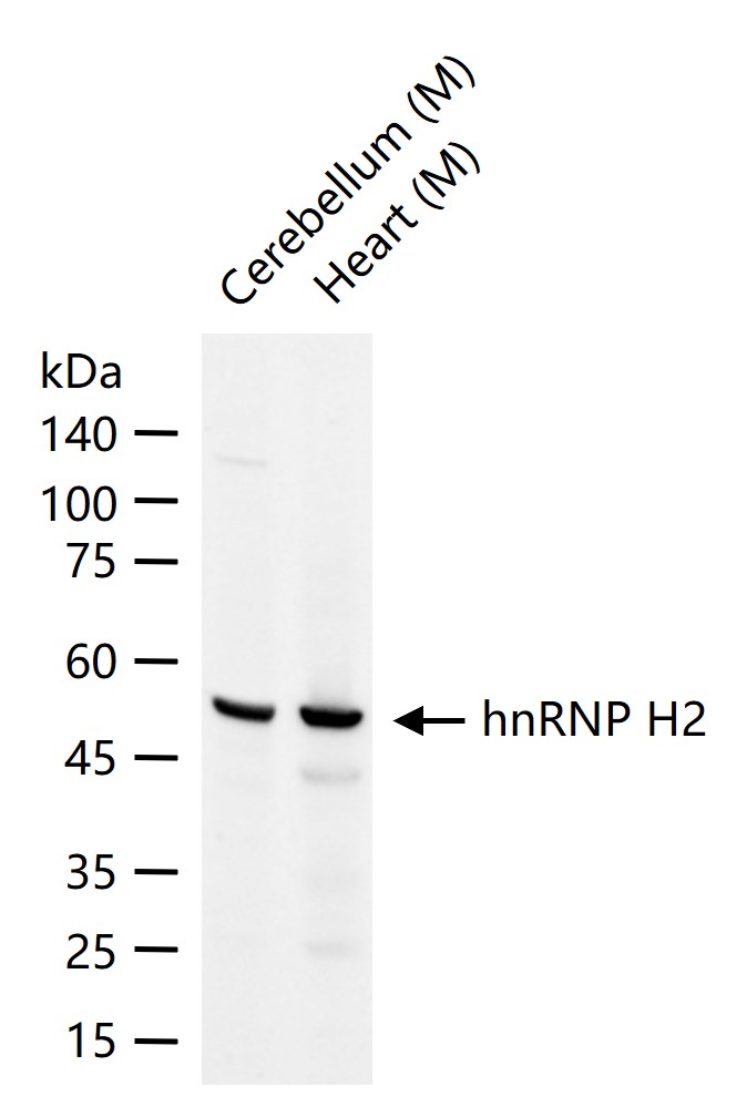
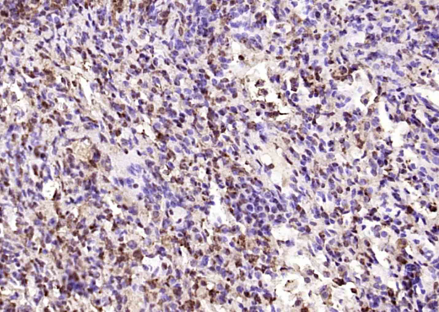
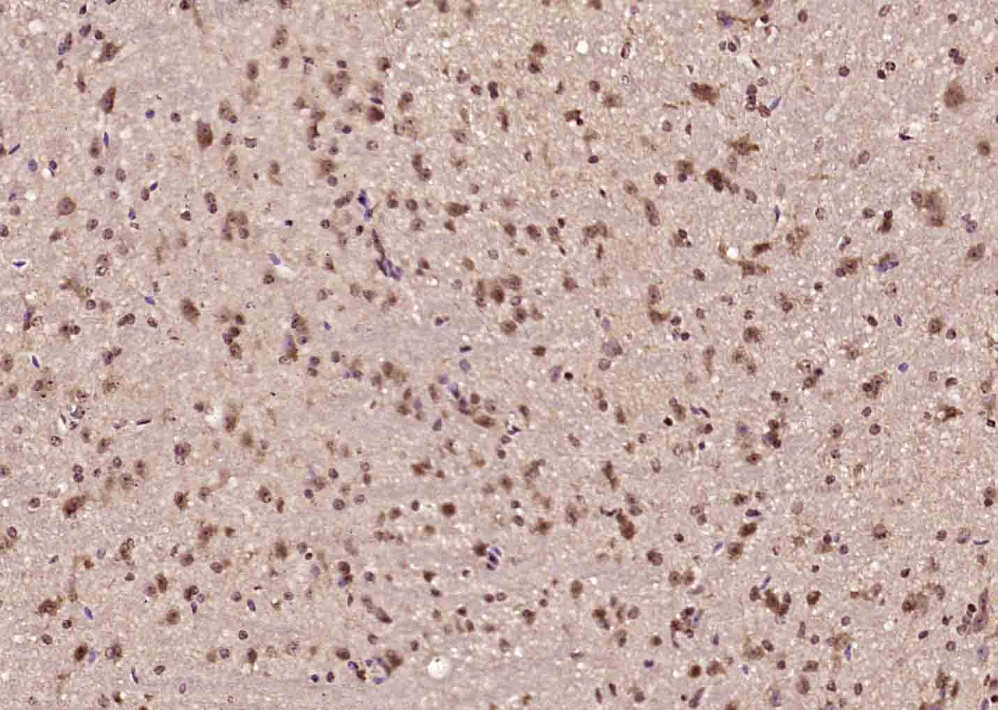
Target Protein: hnRNP H2 Rabbit pAb
IR: Immunogen Range:101-200/449
Clonality: Polyclonal
Isotype: IgG
Entrez Gene: 3188
Swiss Prot: P55795
Source: KLH conjugated synthetic peptide derived from human hnRNP H2:101-200/449
Purification: affinity purified by Protein A
Storage: 0.01M TBS (pH7.4) with 1% BSA, 0.02% Proclin300 and 50% Glycerol. Shipped at 4℃. Store at -20℃ for one year. Avoid repeated freeze/thaw cycles.
Background: This gene belongs to the subfamily of ubiquitously expressed heterogeneous nuclear ribonucleoproteins (hnRNPs). The hnRNPs are RNA binding proteins and they complex with heterogeneous nuclear RNA (hnRNA). These proteins are associated with pre-mRNAs in the nucleus and appear to influence pre-mRNA processing and other aspects of mRNA metabolism and transport. While all of the hnRNPs are present in the nucleus some seem to shuttle between the nucleus and the cytoplasm. The hnRNP proteins have distinct nucleic acid binding properties. The protein encoded by this gene has three repeats of quasi-RRM domains that binds to RNAs. It is very similar to the family member HNRPH1. This gene is thought to be involved in Fabray disease and X-linked agammaglobulinemia phenotype. Alternative splicing results in multiple transcript variants encoding the same protein. Read-through transcription between this locus and the ribosomal protein L36a gene has been observed. [provided by RefSeq, Jan 2011]
Size: 50ul
Concentration: 1mg/ml
Applications: WB=1:500-2000,IHC-P=1:100-500,IHC-F=1:100-500,IF=1:100-500
Cross Reactive Species: Mouse (predicted: Human,Rat,Pig)
For research use only. Not intended for diagnostic or therapeutic use.