bsm-52537R [Primary Antibody]
Rabbit  Anti-GART Recombinant Rabbit mAb   Antibody
www.bioss.com.cn
sales@bioss.com.cn
techsupport@bioss.com.cn
400-901-9800
DATASHEET

Host: Rabbit

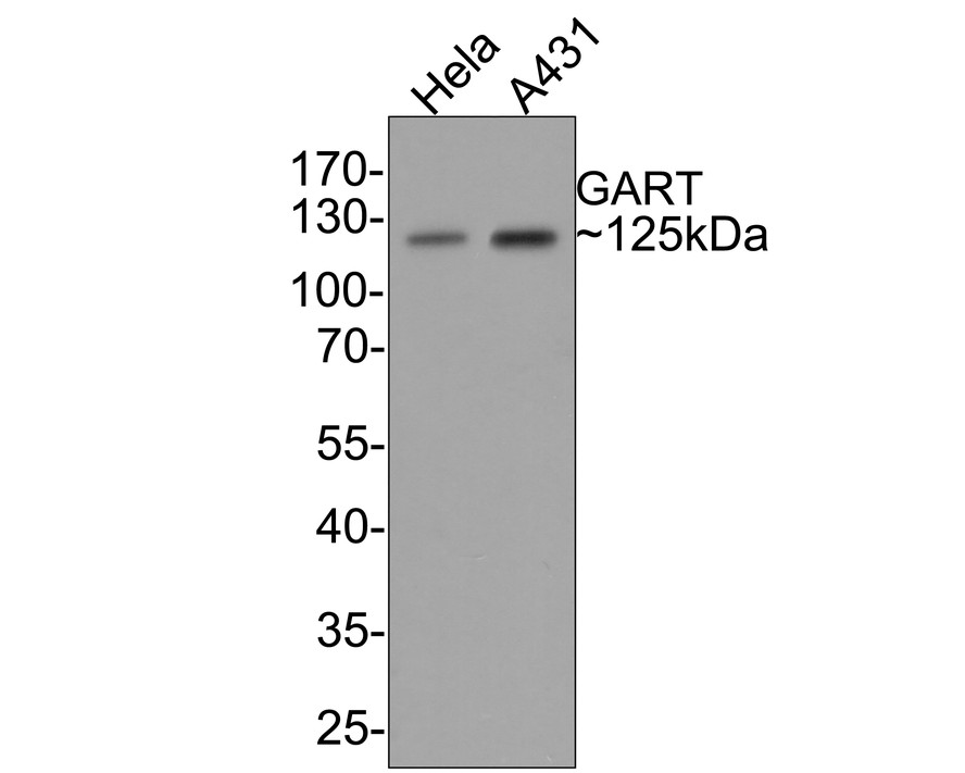
Target Protein: GART Recombinant Rabbit mAb

IR: Immunogen Range:570-750/1010

Clonality:

Isotype: IgG

Entrez Gene: 2618

Swiss Prot: P22102

Source: Recombinant Human GART protein :570-750/1010 

Purification: affinity purified by Protein A

Storage: 0.01M TBS (pH7.4) with 1% BSA, 0.02% Proclin300 and 50% Glycerol. Shipped at 4℃. Store at -20℃ for one year. Avoid repeated freeze/thaw cycles.

Background: Purines are critical for energy metabolism, cell signaling and cell reproduction and also function as precursors for coenzymes, energy transfer molecules, regulatory factors and proteins involved in RNA and DNA synthesis. GART (GAR transformylase), also referred to as AIRS, GARS, PAIS, PGFT, PRGS or GARTF, is 1,010 amino acids in length and is a key folate-dependent trifunctional enzyme with phosphoribosylglycinamide formyltransferase, phosphoribosylglycinamide synthetase and AICAR (phosphoribosylaminoimidazole synthetase) activity required for de novo purine biosynthesis. Cancer cells require considerable amounts of purines to sustain their accelerated growth and GART is, therefore, a target for cancer chemotherapy. GART is highly conserved in vertebrates. Two isoforms of GART are expressed due to alternative splicing events.

Size: 100ul

Concentration: 1mg/ml

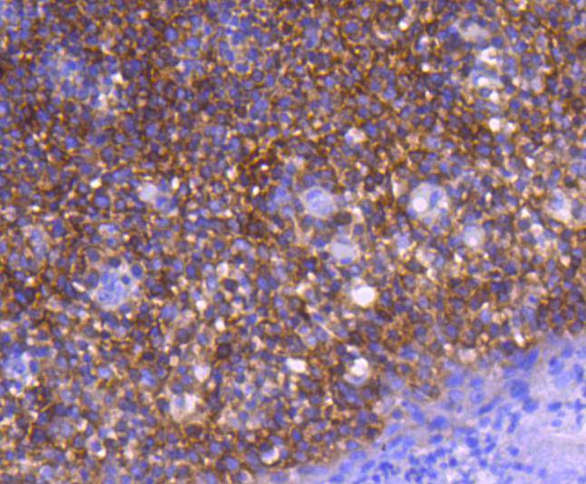
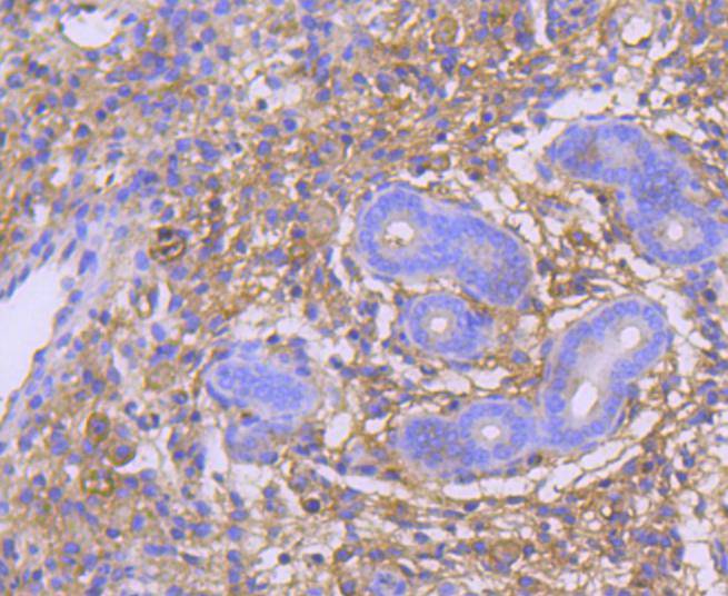
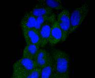
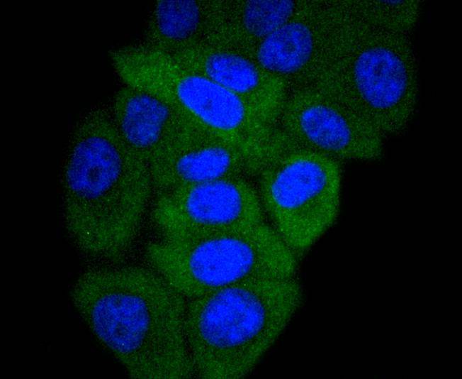
Applications: WB=1:500,IHC-P=1:100-500,IHC-F=1:400-800,IF=1:100-500,ICC/IF=1:50-200

Cross Reactive Species: Human,Mouse (predicted: Rat)

For research use only. Not intended for diagnostic or therapeutic use.

VALIDATION IMAGES
Western blot analysis of GART on different lysates with Rabbit anti-GART antibody at 1/500 dilution. Lane 1: Hela cell lysate Lane 2: A431 cell lysate Lysates/proteins at 10 碌g/Lane. Predicted band size: 108 kDa Observed band size: 125 kDa Exposure time: 30 seconds; 10% SDS-PAGE gel.
Immunohistochemical analysis of paraffin-embedded human tonsil tissue using anti-GART antibody. The section was pre-treated using heat mediated antigen retrieval with Tris-EDTA buffer (pH 9.0) for 20 minutes.The tissues were blocked in 1% BSA for 30 minutes at room temperature, washed with ddH2O and PBS, and then probed with the primary antibody (bsm-52537R, 1/50) for 30 minutes at room temperature. The detection was performed using an HRP conjugated compact polymer system. DAB was used as the chromogen. Tissues were counterstained with hematoxylin and mounted with DPX.
Immunohistochemical analysis of paraffin-embedded mouse uterus tissue using anti-GART antibody. The section was pre-treated using heat mediated antigen retrieval with Tris-EDTA buffer (pH 9.0) for 20 minutes.The tissues were blocked in 1% BSA for 30 minutes at room temperature, washed with ddH2O and PBS, and then probed with the primary antibody (ET1610-68, 1/50) for 30 minutes at room temperature. The detection was performed using an HRP conjugated compact polymer system. DAB was used as the chromogen. Tissues were counterstained with hematoxylin and mounted with DPX.
ICC staining of GART in SW480 cells (green). Formalin fixed cells were permeabilized with 0.1% Triton X-100 in TBS for 10 minutes at room temperature and blocked with 10% negative goat serum for 15 minutes at room temperature. Cells were probed with the primary antibody ( bsm-52537R, 1/50) for 1 hour at room temperature, washed with PBS. Alexa Fluor®488 conjugate-Goat anti-Rabbit IgG was used as the secondary antibody at 1/1,000 dilution. The nuclear counter stain is DAPI (blue).
ICC staining of GART in Hela cells (green). Formalin fixed cells were permeabilized with 0.1% Triton X-100 in TBS for 10 minutes at room temperature and blocked with 10% negative goat serum for 15 minutes at room temperature. Cells were probed with the primary antibody (ET1610-68, 1/50) for 1 hour at room temperature, washed with PBS. Alexa Fluor®488 conjugate-Goat anti-Rabbit IgG was used as the secondary antibody at 1/1,000 dilution. The nuclear counter stain is DAPI (blue).
ICC staining of GART in HepG2 cells (green). Formalin fixed cells were permeabilized with 0.1% Triton X-100 in TBS for 10 minutes at room temperature and blocked with 10% negative goat serum for 15 minutes at room temperature. Cells were probed with the primary antibody (ET1610-68, 1/50) for 1 hour at room temperature, washed with PBS. Alexa Fluor®488 conjugate-Goat anti-Rabbit IgG was used as the secondary antibody at 1/1,000 dilution. The nuclear counter stain is DAPI (blue).
 
PRODUCT SPECIFIC PUBLICATIONS