bs-0932R [Primary Antibody]
Rabbit  Anti-WDR26 Rabbit pAb  Polyclonal Antibody
www.bioss.com.cn
sales@bioss.com.cn
techsupport@bioss.com.cn
400-901-9800
DATASHEET

Host: Rabbit

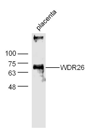
Target Protein: WDR26 Rabbit pAb

IR: Immunogen Range:101-200/514

Clonality: Polyclonal

Isotype: IgG

Entrez Gene: 80232

Swiss Prot: Q9H7D7

Source: KLH conjugated synthetic peptide derived from human WDR26:101-200/514 

Purification: affinity purified by Protein A

Storage: 0.01M TBS (pH7.4) with 1% BSA, 0.02% Proclin300 and 50% Glycerol. Shipped at 4℃. Store at -20℃ for one year. Avoid repeated freeze/thaw cycles.

Background: This gene encodes a member of the WD repeat protein family. WD repeats are minimally conserved regions of approximately 40 amino acids typically bracketed by gly-his and trp-asp (GH-WD), which may facilitate formation of heterotrimeric or multiprotein complexes. Members of this family are involved in a variety of cellular processes, including cell cycle progression, signal transduction, apoptosis, and gene regulation. Two transcript variants encoding two different isoforms have been found for this gene. [provided by RefSeq].

Size: 100ul

Concentration: 1mg/ml

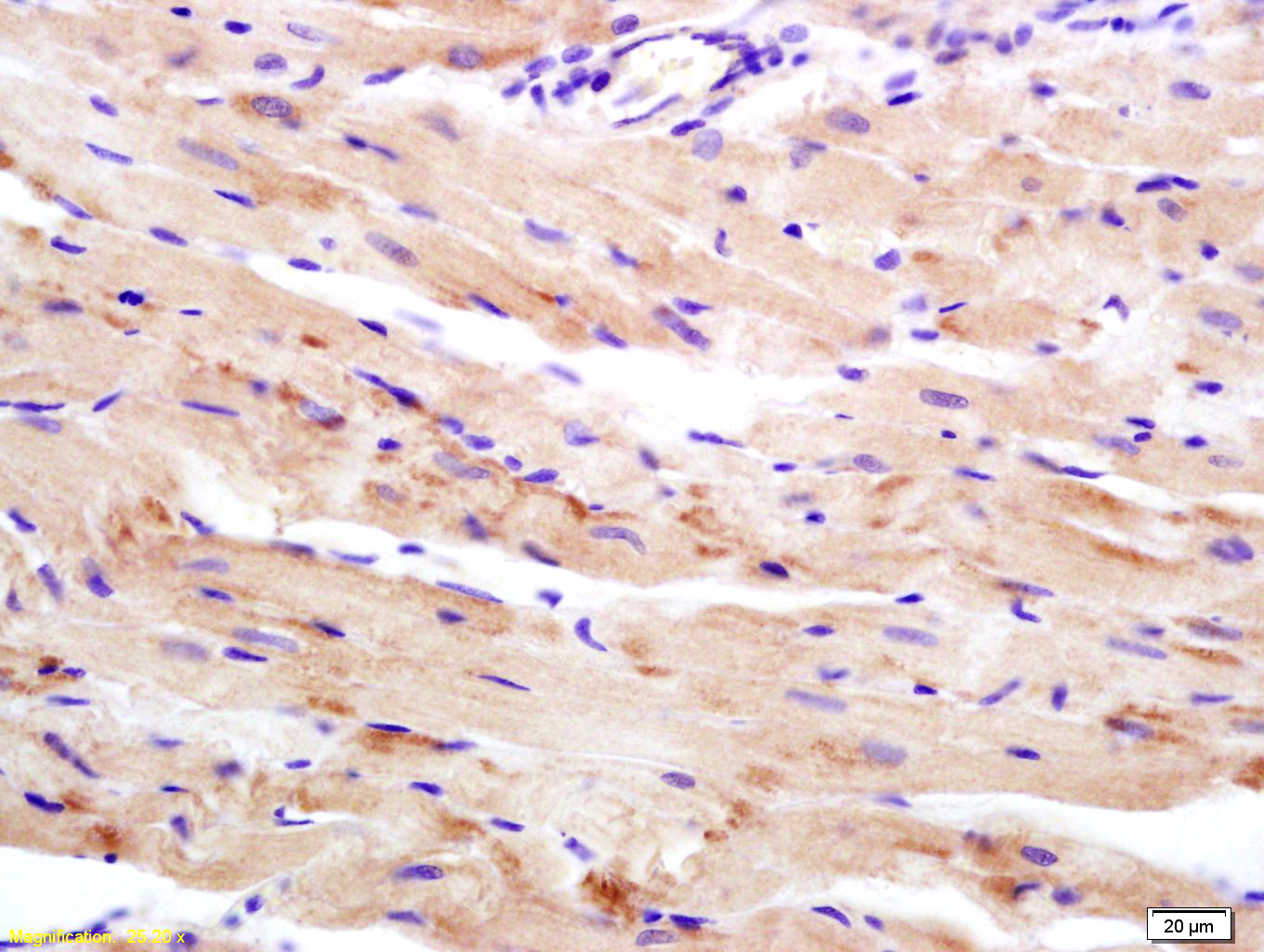
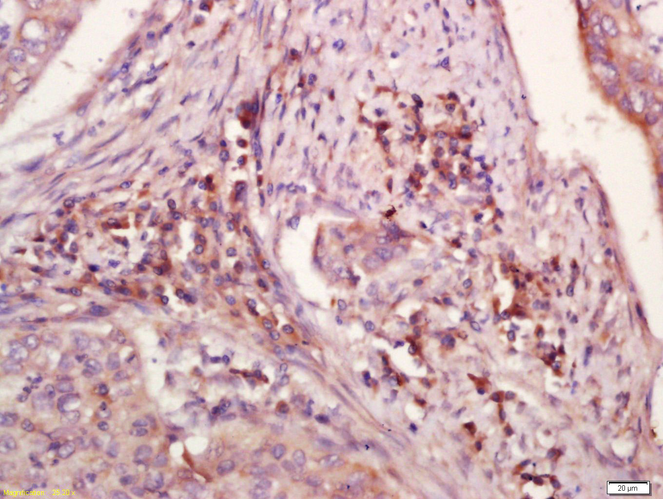
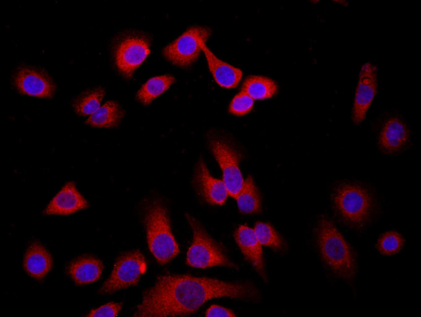
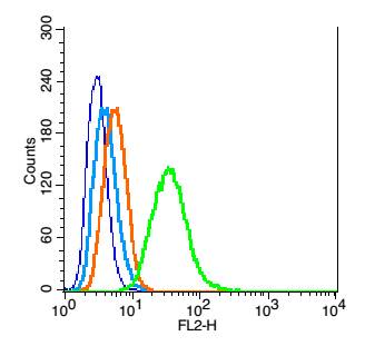
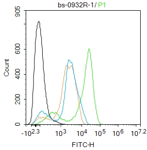
Applications: WB=1:500-2000,IHC-P=1:100-500,IHC-F=1:100-500,IF=1:100-500,Flow-Cyt=1µg/Test,ICC/IF=1:100

Cross Reactive Species: Human,Mouse,Rat (predicted: Cow,Chicken,Dog,Horse)

For research use only. Not intended for diagnostic or therapeutic use.

VALIDATION IMAGES
Sample: Placenta (Mouse) Lysate at 40 ug Primary: Anti-WDR26 (bs-0932R) at 1/300 dilution Secondary: IRDye800CW Goat Anti-Rabbit IgG at 1/20000 dilution Predicted band size: 72 kD Observed band size: 70 kD
Tissue/cell: rat heart tissue; 4% Paraformaldehyde-fixed and paraffin-embedded; Antigen retrieval: citrate buffer ( 0.01M, pH 6.0 ), Boiling bathing for 15min; Block endogenous peroxidase by 3% Hydrogen peroxide for 30min; Blocking buffer (normal goat serum,C-0005) at 37℃ for 20 min; Incubation: Anti-WDR26 Polyclonal Antibody, Unconjugated(bs-0932R) 1:200, overnight at 4°C, followed by conjugation to the secondary antibody(SP-0023) and DAB(C-0010) staining
Tissue/cell: human laryngocarcinoma; 4% Paraformaldehyde-fixed and paraffin-embedded; Antigen retrieval: citrate buffer ( 0.01M, pH 6.0 ), Boiling bathing for 15min; Block endogenous peroxidase by 3% Hydrogen peroxide for 30min; Blocking buffer (normal goat serum,C-0005) at 37℃ for 20 min; Incubation: Anti-WDR26 Polyclonal Antibody, Unconjugated(bs-0932R) 1:200, overnight at 4°C, followed by conjugation to the secondary antibody(SP-0023) and DAB(C-0010) staining
Tissue/cell: MCF7; 4% Paraformaldehyde-fixed; Triton X-100 at room temperature for 20 min; Blocking buffer (normal goat serum, C-0005) at 37°C for 20 min; Antibody incubation with (WDR26) Polyclonal Antibody, Unconjugated (bs-0932R) 1:200, 90 minutes at 37°C; followed by a conjugated Goat Anti-Rabbit IgG antibody (bs-0295G-FITC) at 37°C for 90 minutes, DAPI (blue, C02-04002) was used to stain the cell nuclei.
Blank control: RSC96(blue), the cells were fixed with 2% paraformaldehyde (10 min) and then permeabilized with ice-cold 90% methanol for 30 min on ice. Isotype Control Antibody: Rabbit IgG(orange) ; Secondary Antibody: Goat anti-rabbit IgG-PE(white blue), Dilution: 1:200 in 1 X PBS containing 0.5% BSA ; Primary Antibody Dilution: 1μg in 100 μL1X PBS containing 0.5% BSA(green).
Blank control:THP-1. Primary Antibody (green line): Rabbit Anti-WDR26 antibody (bs-0932R) Dilution: 1ug/Test; Secondary Antibody : Goat anti-rabbit IgG-FITC Dilution: 0.5ug/Test. Protocol The cells were fixed with 4% PFA (10min at room temperature)and then permeabilized with 90% ice-cold methanol for 20 min at -20℃.The cells were then incubated in 5%BSA to block non-specific protein-protein interactions for 30 min at room temperature .Cells stained with Primary Antibody for 30 min at room temperature. The secondary antibody used for 40 min at room temperature. Acquisition of 20,000 events was performed.
 
PRODUCT SPECIFIC PUBLICATIONS