bs-1468R [Primary Antibody]
Rabbit  Anti-MDR1/P Glycoprotein Rabbit pAb  Polyclonal Antibody
www.bioss.com.cn
sales@bioss.com.cn
techsupport@bioss.com.cn
400-901-9800
DATASHEET

Host: Rabbit

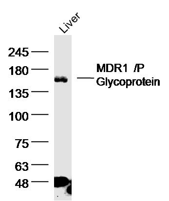
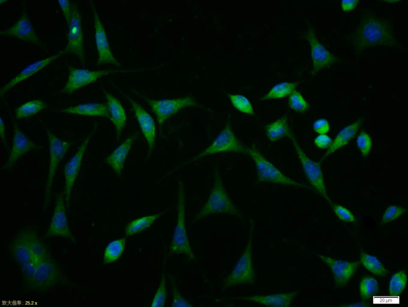
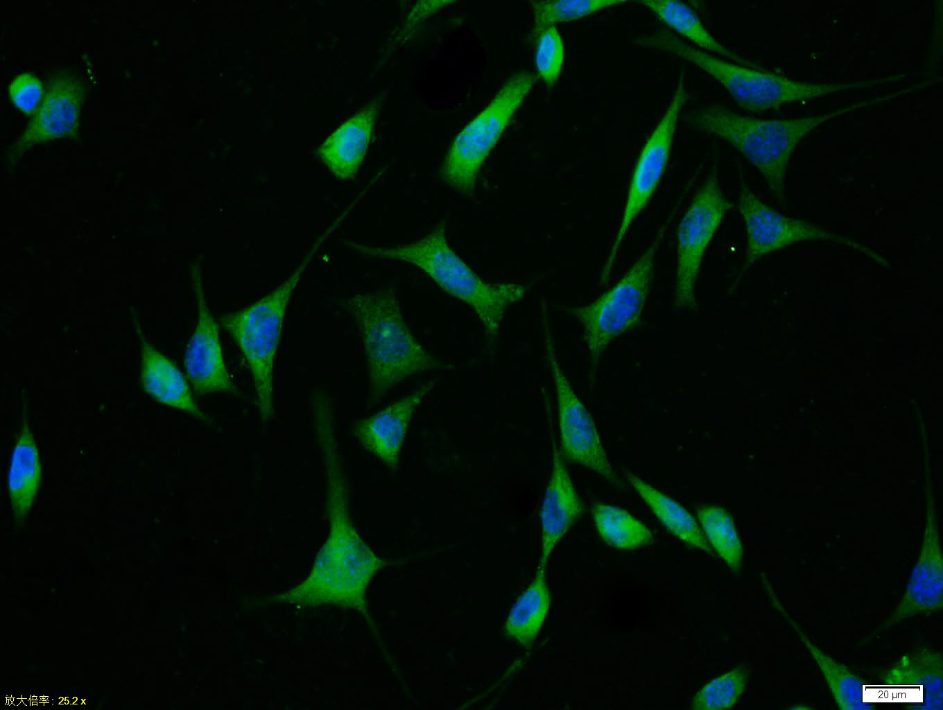
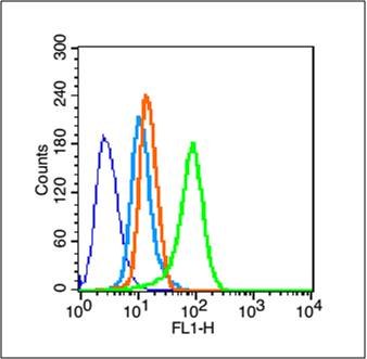
Target Protein: MDR1/P Glycoprotein Rabbit pAb

IR: Immunogen Range:1051-1280/1280

Clonality: Polyclonal

Isotype: IgG

Entrez Gene: 5243

Swiss Prot: P08183

Source: KLH conjugated synthetic peptide derived from human MDR1:1051-1280/1280 

Purification: affinity purified by Protein A

Storage: 0.01M TBS (pH7.4) with 1% BSA, 0.02% Proclin300 and 50% Glycerol. Shipped at 4℃. Store at -20℃ for one year. Avoid repeated freeze/thaw cycles.

Background: P Glycoprotein, the product of the MDR1 gene, is expressed in distinct non-malignant cells, typically cells with secretory and excretory functions. It is assumed to function as an ATP-dependent drug efflux pump with broad substrate specificity. The highest expression of P Glycoprotein has been observed in kidney (proximal tubules), liver (bile canaliculi), adrenal gland and intestine, suggesting that the primary role of P Glycoprotein is in the normal secretion of physiological metabolites and ingested chemicals into bile, urine and the lumen of the intestinal tract. Elevated levels of P Glycoprotein have also been reported in multidrug-resistant cell lines and in colon, endometrial, ovarian, and breast tumors, as well as in sarcomas and leukemias / lymphomas.

Size: 200ul

Concentration: 1mg/ml

Applications: WB=1:500-2000,Flow-Cyt=1μg/Test,ICC/IF=1:100

Cross Reactive Species: Human,Mouse,Rat

For research use only. Not intended for diagnostic or therapeutic use.

VALIDATION IMAGES
Sample: liver (Rat) Lysate at 40 ug Primary: Anti-MDR1'P Glycoprotein (bs-1468R)at 1/300 dilution Secondary: IRDye800CW Goat Anti-Rabbit IgG at 1/20000 dilution Predicted band size: 141kD Observed band size: 141 kD
Tissue/cell:SH-SY5Y cell; 4% Paraformaldehyde-fixed; Triton X-100 at room temperature for 20 min; Blocking buffer (normal goat serum,C-0005) at 37°C for 20 min; Antibody incubation with (MDR1/P Glycoprotein) polyclonal Antibody, Unconjugated (bs-1468R) 1:100, 90 minutes at 37°C; followed by a FITC conjugated Goat Anti-Rabbit IgG antibody at 37°C for 90 minutes, DAPI (blue, C02-04002) was used to stain the cell nuclei.
Blank control (blue line): Hela (blue). Primary Antibody (green line): Rabbit Anti-MDR1 antibody (bs-1468R) Dilution: 1μg /10^6 cells; Isotype Control Antibody (orange line): Rabbit IgG . Secondary Antibody (white blue line): Goat anti-rabbit IgG-FITC Dilution: 1μg /test. Protocol The cells were fixed with 70% methanol (Overnight at -20℃) and then permeabilized with ice-cold 90% methanol for 30 min on ice. Cells stained with Primary Antibody for 30 min at room temperature. The cells were then incubated in 1 X PBS/2%BSA/10% goat serum to block non-specific protein-protein interactions followed by the antibody for 15 min at room temperature. The secondary antibody used for 40 min at room temperature. Acquisition of 20,000 events was performed.
 
PRODUCT SPECIFIC PUBLICATIONS