bs-3389R [Primary Antibody]
Rabbit  Anti-Phospho-RPS6 (Ser240 + Ser244) Rabbit pAb  Polyclonal Antibody
www.bioss.com.cn
sales@bioss.com.cn
techsupport@bioss.com.cn
400-901-9800
DATASHEET

Host: Rabbit

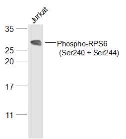
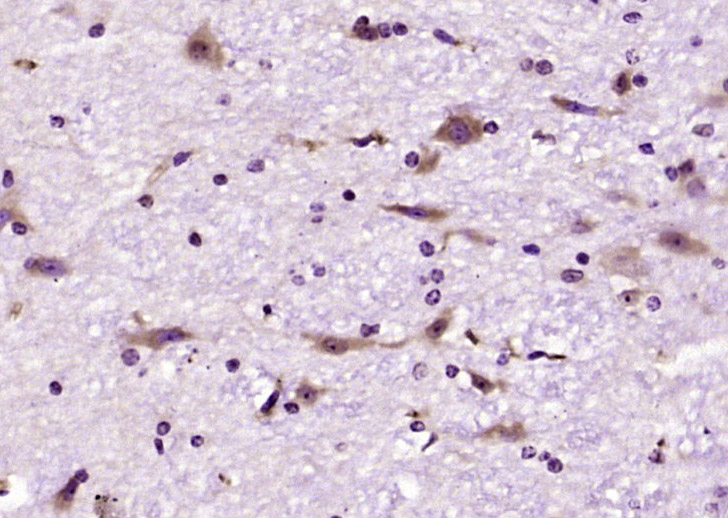
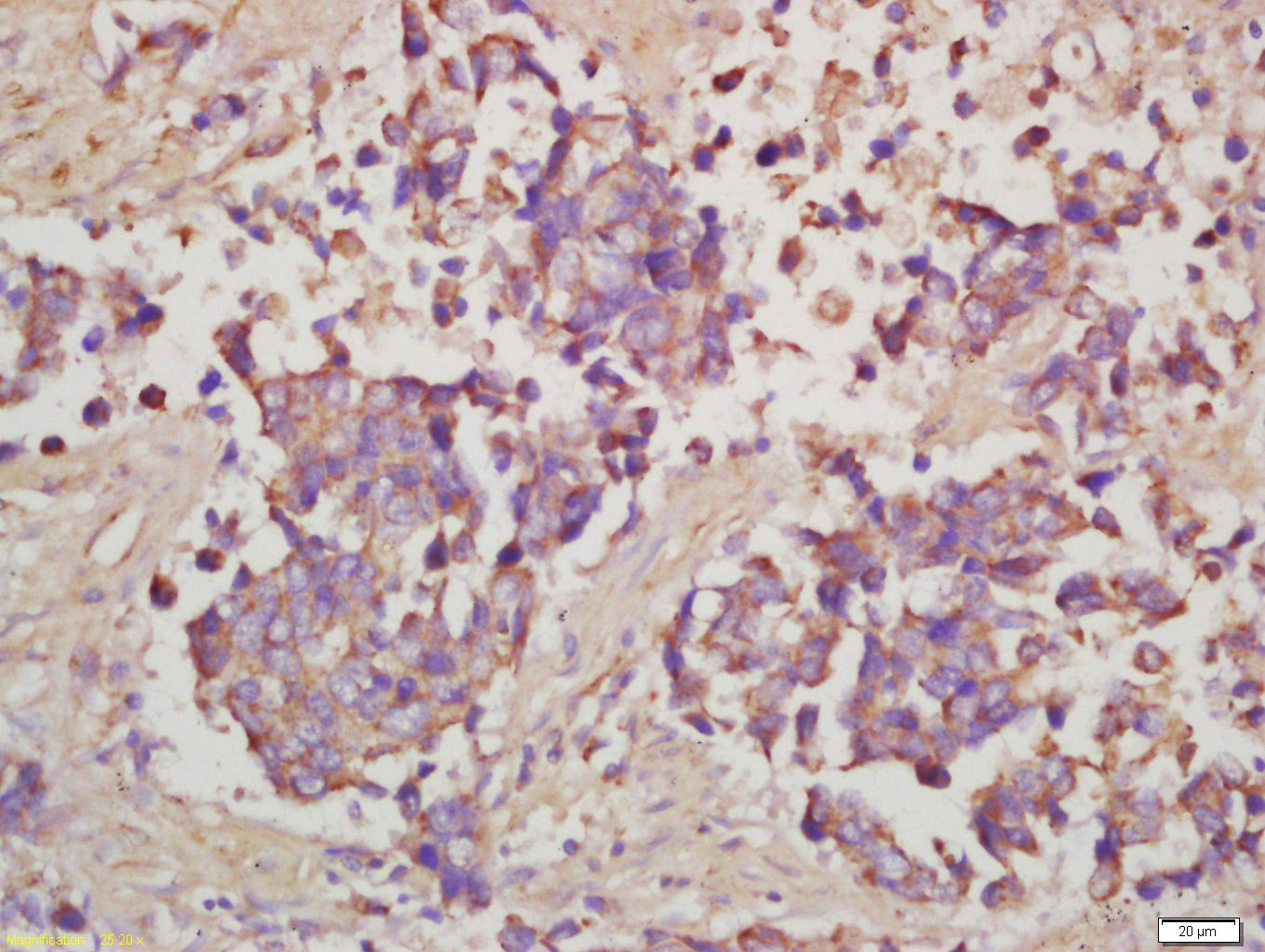
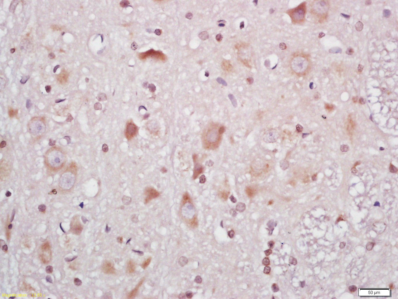
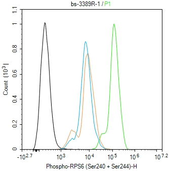
Target Protein: Phospho-RPS6 (Ser240 + Ser244) Rabbit pAb

IR: Immunogen Range:RA(p-S)TSK(p-S)ES

Clonality: Polyclonal

Isotype: IgG

Entrez Gene: 6194

Swiss Prot: P62753

Source: KLH conjugated Synthesised phosphopeptide derived from human RPS6 around the phosphorylation site of Ser240/244:RA(p-S)TSK(p-S)ES 

Purification: affinity purified by Protein A

Storage: 0.01M TBS (pH7.4) with 1% BSA, 0.02% Proclin300 and 50% Glycerol. Shipped at 4℃. Store at -20℃ for one year. Avoid repeated freeze/thaw cycles.

Background: Ribosomes, the organelles that catalyze protein synthesis, consist of a small 40S subunit and a large 60S subunit. Together these subunits are composed of 4 RNA species and approximately 80 structurally distinct proteins. This gene encodes a cytoplasmic ribosomal protein that is a component of the 40S subunit. The protein belongs to the S6E family of ribosomal proteins. It is the major substrate of protein kinases in the ribosome, with subsets of five C-terminal serine residues phosphorylated by different protein kinases. Phosphorylation is induced by a wide range of stimuli, including growth factors, tumor-promoting agents, and mitogens. Dephosphorylation occurs at growth arrest. The protein may contribute to the control of cell growth and proliferation through the selective translation of particular classes of mRNA. As is typical for genes encoding ribosomal proteins, there are multiple processed pseudogenes of this gene dispersed through the genome. [provided by RefSeq, Jul 2008]

Size: 50ul

Concentration: 1mg/ml

Applications: WB=1:100-1000,IHC-P=1:100-500,IHC-F=1:100-500,IF=1:100-500,Flow-Cyt=1ug/Test

Cross Reactive Species: Human,Mouse,Rat

For research use only. Not intended for diagnostic or therapeutic use.

VALIDATION IMAGES
Sample: A431(Human) Cell Lysate at 30 ug Primary: Anti-Phospho-RPS6 (Ser240 + Ser244) (bs-3389R) at 1/500 dilution Secondary: IRDye800CW Goat Anti-Rabbit IgG at 1/20000 dilution Predicted band size: 29 kD Observed band size: 29 kD
Sample: Jurkat(Human) Cell Lysate at 30 ug Primary: Anti-Phospho-RPS6 (Ser240 + Ser244) (bs-3389R) at 1/500 dilution Secondary: IRDye800CW Goat Anti-Rabbit IgG at 1/20000 dilution Predicted band size: 29 kD Observed band size: 29 kD
Paraformaldehyde-fixed, paraffin embedded (mouse cerebellum tissue); Antigen retrieval by boiling in sodium citrate buffer (pH6.0) for 15min; Block endogenous peroxidase by 3% hydrogen peroxide for 20 minutes; Blocking buffer (normal goat serum) at 37°C for 30min; Antibody incubation with(Phospho-RPS6(Ser240+Ser244)) Polyclonal Antibody, Unconjugated (bs-3389R) at 1:200 overnight at 4°C, followed by operating according to SP Kit(Rabbit) (sp-0023) instructionsand DAB staining.
Tissue/cell: human lung carcinoma; 4% Paraformaldehyde-fixed and paraffin-embedded; Antigen retrieval: citrate buffer ( 0.01M, pH 6.0 ), Boiling bathing for 15min; Block endogenous peroxidase by 3% Hydrogen peroxide for 30min; Blocking buffer (normal goat serum,C-0005) at 37℃ for 20 min; Incubation: Anti-Phospho-RPS6 (Ser240+Ser244) Polyclonal Antibody, Unconjugated(bs-3389R) 1:200, overnight at 4°C, followed by conjugation to the secondary antibody(SP-0023) and DAB(C-0010) staining
Tissue/cell: rat brain tissue; 4% Paraformaldehyde-fixed and paraffin-embedded; Antigen retrieval: citrate buffer ( 0.01M, pH 6.0 ), Boiling bathing for 15min; Block endogenous peroxidase by 3% Hydrogen peroxide for 30min; Blocking buffer (normal goat serum,C-0005) at 37℃ for 20 min; Incubation: Anti-Phospho-RPS6 (Ser240+Ser244) Polyclonal Antibody, Unconjugated(bs-3389R) 1:200, overnight at 4°C, followed by conjugation to the secondary antibody(SP-0023) and DAB(C-0010) staining
Blank control(black line):HepG2. Primary Antibody (green line): Rabbit Anti-Phospho-RPS6 (Ser240 + Ser244) antibody (bs-3389R) Dilution:1ug/Test; Secondary Antibody(white blue line): Goat anti-rabbit IgG-AF488 Dilution: 0.5ug/Test. Isotype control(orange line): Normal Rabbit IgG Protocol The cells were fixed with 4% PFA (10min at room temperature)and then permeabilized with 90% ice-cold methanol for 20 min at -20℃, The cells were then incubated in 5%BSA to block non-specific protein-protein interactions for 30 min at room temperature .Cells stained with Primary Antibody for 30 min at room temperature. The secondary antibody used for 40 min at room temperature. Acquisition of 20,000 events was performed.
 
PRODUCT SPECIFIC PUBLICATIONS