bs-1867R [Primary Antibody]
Rabbit  Anti-PD-1  Polyclonal Antibody
www.bioss.com.cn
sales@bioss.com.cn
techsupport@bioss.com.cn
400-901-9800
DATASHEET

Host: Rabbit

Target Protein: PD-1

IR: Immunogen Range:201-288/288

Clonality: Polyclonal

Isotype: IgG

Entrez Gene: 5133

Swiss Prot: Q15116

Source: KLH conjugated synthetic peptide derived from human PD-1:201-288/288 

Purification: affinity purified by Protein A

Storage: 0.01M TBS(pH7.4) with 1% BSA, 0.03% Proclin300 and 50% Glycerol. Shipped at 4℃. Store at -20 °C for one year. Avoid repeated freeze/thaw cycles.

Background: Programmed cell death protein 1 (PDCD1) is an immune-inhibitory receptor expressed in activated T cells; it is involved in the regulation of T-cell functions, including those of effector CD8+ T cells. In addition, this protein can also promote the differentiation of CD4+ T cells into T regulatory cells. PDCD1 is expressed in many types of tumors including melanomas, and has demonstrated to play a role in anti-tumor immunity. Moreover, this protein has been shown to be involved in safeguarding against autoimmunity, however, it can also contribute to the inhibition of effective anti-tumor and anti-microbial immunity. [provided by RefSeq, Aug 2020]

Size: 50ul

Concentration: 1mg/ml
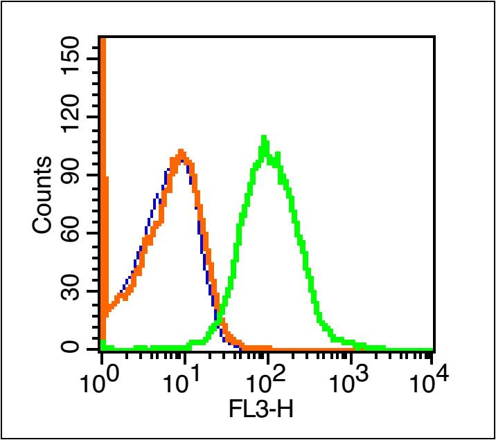
Applications: WB=1:500-2000,IHC-P=1:100-500,IHC-F=1:100-500,Flow-Cyt=1μg /test,IF=1:100-500,ELISA=1:5000-10000

Cross Reactive Species: Human,Mouse,Rat (predicted: Rabbit,Cow,Horse)

For research use only. Not intended for diagnostic or therapeutic use.

VALIDATION IMAGES
Sample: Raji(Human) Cell Lysate at 30 ug Thymus(Mouse) Lysate at 40 ug Lymph node(Mouse) Lysate at 40 ug Spleen(Mouse) Lysate at 40 ug Raw264.7(Mouse) Cell Lysate at 40 ug Thymus (Rat) Lysate at 40 ug Primary: Anti-PD-1 (bs-1867R) at 1/1000 dilution Secondary: IRDye800CW Goat Anti-Rabbit IgG at 1/20000 dilution Predicted band size: 55 kD Observed band size: 58 kD
Sample: Hela(Human) Cell Lysate at 30 ug MOLT-4(Human) Cell Lysate at 30 ug Jurkat(Human) Cell Lysate at 30 ug Primary: Anti-PD-1 (bs-1867R) at 1/500 dilution Secondary: IRDye800CW Goat Anti-Rabbit IgG at 1/20000 dilution Predicted band size: 32 kD Observed band size: 55 kD
Paraformaldehyde-fixed, paraffin embedded (human tonsil); Antigen retrieval by boiling in sodium citrate buffer (pH6.0) for 15min; Block endogenous peroxidase by 3% hydrogen peroxide for 20 minutes; Blocking buffer (normal goat serum) at 37°C for 30min; Antibody incubation with (PD-1 ) Polyclonal Antibody, Unconjugated (bs-1867R) at 1:200 overnight at 4°C, followed by operating according to SP Kit(Rabbit) (sp-0023) instructionsand DAB staining.
Paraformaldehyde-fixed, paraffin embedded (rat spleen); Antigen retrieval by boiling in sodium citrate buffer (pH6.0) for 15min; Block endogenous peroxidase by 3% hydrogen peroxide for 20 minutes; Blocking buffer (normal goat serum) at 37°C for 30min; Antibody incubation with (PD-1 ) Polyclonal Antibody, Unconjugated (bs-1867R) at 1:200 overnight at 4°C, followed by operating according to SP Kit(Rabbit) (sp-0023) instructionsand DAB staining.
Blank control (blue line): Mouse spleen cells(blue). Primary Antibody (green line): Rabbit Anti-PD-1/PE-CY7 Conjugated antibody (bs-1867R-PE-CY7) Dilution: 1μg /10^6 cells; Isotype Control Antibody (orange line): Rabbit IgG-PE-CY7 . Protocol The cells were fixed with 70% ice-cold methanol overnight at 4℃ . The cells were then incubated in 1 X PBS/2%BSA/10% goat serum to block non-specific protein-protein interactions followed by the antibody for 15 min at room temperature. Cells stained with Primary Antibody for 30 min at room temperature.Acquisition of 20,000 events was performed.
 
PRODUCT SPECIFIC PUBLICATIONS