sales@bioss.com.cn
techsupport@bioss.com.cn
400-901-9800
Host: Rabbit
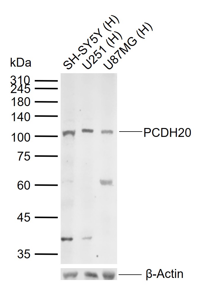
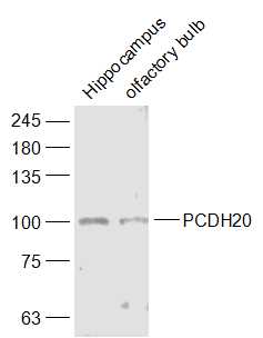
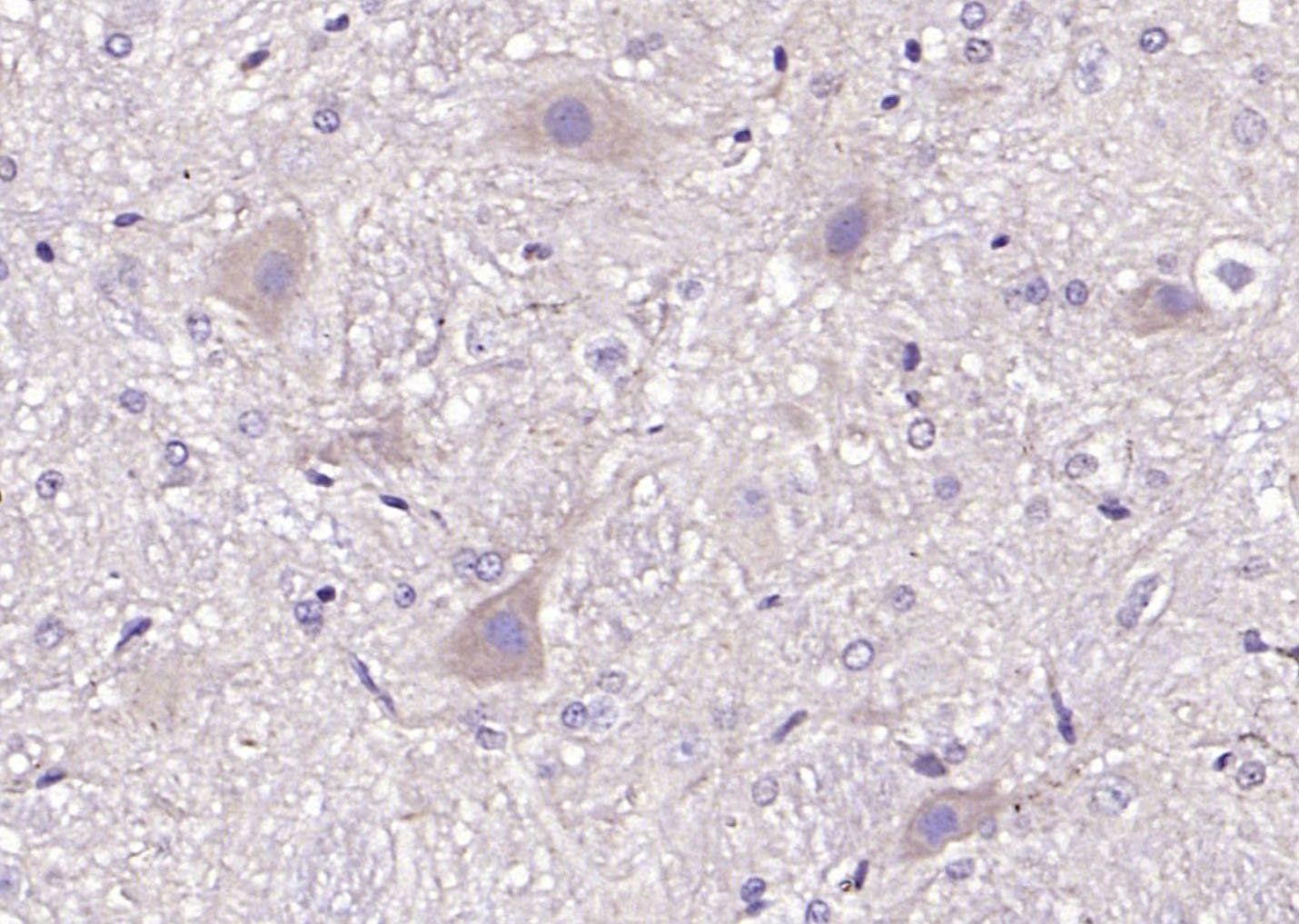
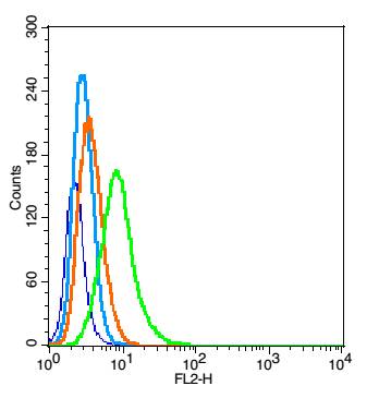
Target Protein: PCDH20 Rabbit pAb
IR: Immunogen Range:611-710/924
Clonality: Polyclonal
Isotype: IgG
Entrez Gene: 64881
Swiss Prot: Q8N6Y1
Source:
KLH conjugated synthetic peptide derived from human PCDH20:611-710/924
Purification: affinity purified by Protein A
Storage: 0.01M TBS (pH7.4) with 1% BSA, 0.02% Proclin300 and 50% Glycerol. Shipped at 4℃. Store at -20℃ for one year. Avoid repeated freeze/thaw cycles.
Background: As a subfamily of the cadherin superfamily, protocadherins are cadherin-like cell adhesion proteins that contain up to seven extracellular domains and are predominantly expressed in the nervous system. Importantly, the adhesion mechanism of protocadherins is distinct from classic cadherins. Through inactivation or overexpression, several protocadherins have been implicated in a variety of cancers. Protocadherin-20 (PCDH20), also known as protocadherin-13, is a 924 amino acid protein containing 6 cadherin domains and potentially functioning as a calcium-dependent cell-adhesion protein. In non-small cell lung cancer cell lines, a homozygous loss of PCDH20 was identified through either deletion of one allele and methylation of the other or methylation of both alleles. Hypermethylation of PCDH20 is associated with worse prognosis and clinical outcome, suggesting that PCDH20 may function as a tumor suppressor.
Size: 100ul
Concentration: 1mg/ml
Applications: WB=1:500-2000,IHC-P=1:100-500,IHC-F=1:100-500,IF=1:100-500,Flow-Cyt=1μg/Test
Cross Reactive Species: Human,Mouse,Rat (predicted: Rabbit,Horse)
For research use only. Not intended for diagnostic or therapeutic use.