bs-11320R [Primary Antibody]
Rabbit  Anti-MNX1/HLXB9 Rabbit pAb  Polyclonal Antibody
www.bioss.com.cn
sales@bioss.com.cn
techsupport@bioss.com.cn
400-901-9800
DATASHEET

Host: Rabbit

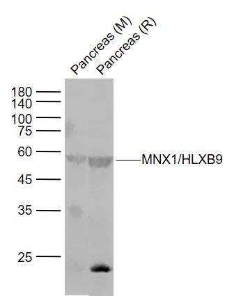
Target Protein: MNX1/HLXB9 Rabbit pAb

IR: Immunogen Range:231-330/401

Clonality: Polyclonal

Isotype: IgG

Entrez Gene: 3110

Swiss Prot: P50219

Source: KLH conjugated synthetic peptide derived from human HLXB9:231-330/401 

Purification: affinity purified by Protein A

Storage: 0.01M TBS (pH7.4) with 1% BSA, 0.02% Proclin300 and 50% Glycerol. Shipped at 4℃. Store at -20℃ for one year. Avoid repeated freeze/thaw cycles.

Background: The HB9 homeobox transcription factor regulates gene expression during embryonic development and also in specific adult tissues. HB9 gene mutations are implicated in Curriano syndrome, which is characterized by a triad consisting of a presacral tumor, sacral agenesis and anorectal malformation. In human bone marrow cells, HB9 expression directly correlates with CD34 expression. Furthermore, HB9 expression increases in CD34+ cells that are treated with IL-3 and granulocyte macrophage-colony-stimulating factor. Early in murine development, HB9 is expressed in pancreatic buds (dorsal and ventral) with subsequent expression in differentiating beta cells in the islets of Langerhans. The dorsal lobe of the pancreas fails to form in HB9(-) mice; the resultant pancreas has smaller islets of Langerhans and less beta cells than normal pancreas. The HB9 gene is expressed in the human adult pancreas. In the developing vertebrate embryo, the HB9 gene plays an essential role in motor neuron differentiation. The motor columns of HB9(-) mice are disorganized, lacking phrenic and abducens nerves and exhibiting intercostal nerve defects.

Size: 100ul

Concentration: 1mg/ml

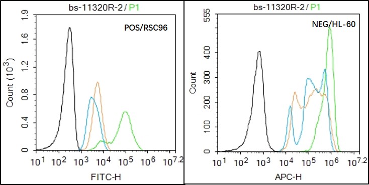
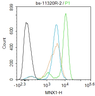
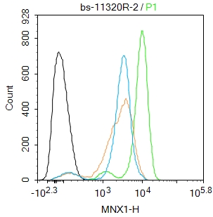
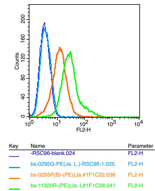
Applications: WB=1:500-2000,Flow-Cyt=1ug/test,ICC/IF=1:100

Cross Reactive Species: Human,Mouse,Rat

For research use only. Not intended for diagnostic or therapeutic use.

VALIDATION IMAGES
Sample: Lane 1: Pancreas (Mouse) Lysate at 40 ug Lane 2: Pancreas (Rat) Lysate at 40 ug Primary: Anti-MNX1/HLXB9 (bs-11320R) at 1/1000 dilution Secondary: IRDye800CW Goat Anti-Rabbit IgG at 1/20000 dilution Predicted band size: 55 kD Observed band size: 55 kD
K562 cell; 4% Paraformaldehyde-fixed; Triton X-100 at room temperature for 20 min; Blocking buffer (normal goat serum, C-0005) at 37°C for 20 min; Antibody incubation with (MNX1) polyclonal Antibody, Unconjugated (bs-11320R) 1:100, 90 minutes at 37°C; followed by a conjugated Goat Anti-Rabbit IgG antibody at 37°C for 90 minutes, DAPI (blue, C02-04002) was used to stain the cell nuclei.
Black line : Positive blank control RSC96); Negative blank control (HL60) Green line : Primary Antibody (Rabbit Anti- HLXB9 antibody (bs-11320R) ) Orange line:Isotype Control Antibody (Rabbit IgG) . Blue line : Secondary Antibody (Goat anti-rabbit IgG-AF488) RSC96(Positive)and HL60(Negative control)cells (black) were fixed with 4% PFA for 10min at room temperature, permeabilized with 90% ice-cold methanol for 20 min at -20℃, and incubated in 5% BSA blocking buffer for 30 min at room temperature. Cells were then stained with HLXB9 Antibody(bs-11320R)at 1:50 dilution in blocking buffer and incubated for 30 min at room temperature, washed twice with 2% BSA in PBS, followed by secondary antibody(blue) incubation for 40 min at room temperature. Acquisitions of 20,000 events were performed. Cells stained with primary antibody (green), and isotype control (orange).
Blank control: K562. Primary Antibody (green line): Rabbit Anti-MNX1 antibody (bs-11320R) Dilution: 2μg /10^6 cells; Isotype Control Antibody (orange line): Rabbit IgG . Secondary Antibody : Goat anti-rabbit IgG-FITC Dilution: 0.5μg /test. Protocol The cells were fixed with 4% PFA (10min at room temperature)and then permeabilized with 90% ice-cold methanol for 20 min at -20℃. The cells were then incubated in 5%BSA to block non-specific protein-protein interactions for 30 min at room temperature .Cells stained with Primary Antibody for 30 min at room temperature. The secondary antibody used for 40 min at room temperature. Acquisition of 20,000 events was performed.
Positive control: RSC96 Isotype Control Antibody: Rabbit IgG ; Secondary Antibody: Goat anti-rabbit IgG-FITC, Dilution: 1:100 in 1 X PBS containing 0.5% BSA ; Primary Antibody Dilution: 6μg in 100 μL1X PBS containing 0.5% BSA.
 
PRODUCT SPECIFIC PUBLICATIONS