bs-11510R [Primary Antibody]
Rabbit  Anti-TTC8 Rabbit pAb  Polyclonal Antibody
www.bioss.com.cn
sales@bioss.com.cn
techsupport@bioss.com.cn
400-901-9800
DATASHEET

Host: Rabbit

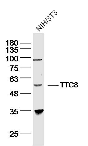
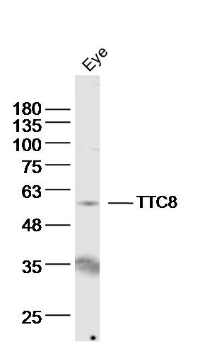
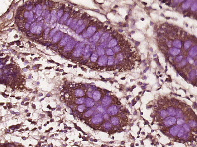
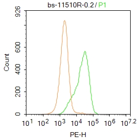
Target Protein: TTC8 Rabbit pAb

IR: Immunogen Range:251-330/541

Clonality: Polyclonal

Isotype: IgG

Entrez Gene: 123016

Swiss Prot: Q8TAM2

Source: KLH conjugated synthetic peptide derived from human BBS8:251-330/541 

Purification: affinity purified by Protein A

Storage: 0.01M TBS (pH7.4) with 1% BSA, 0.02% Proclin300 and 50% Glycerol. Shipped at 4℃. Store at -20℃ for one year. Avoid repeated freeze/thaw cycles.

Background: Bardet-Biedl syndrome (BBS) is a pleiotropic genetic disorder characterized by obesity, photoreceptor degeneration, polydactyly, hypogenitalism, renal abnormalities, and developmental delay. BBS patients also have an increased risk of developing diabetes, hypertension, and congenital heart defects. BBS is a heterogeneous disorder mapping to eight genetic loci and encoding eight proteins, BBS1-BBS8. Five BBS proteins encode basal body or cilia proteins, suggesting that BBS is a ciliary dysfunction disorder. BBS2 contains two overlapping genes: BBS2L1 and BBS2L2. BBSL1 was re-named BBS7, whereas BBS2L2 independently funcitons as BBS1. BBS7 contains 672 amino acids and is expressed at low to moderate levels in most human tissues.

Size: 200ul

Concentration: 1mg/ml

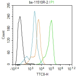
Applications: WB=1:500-2000,IHC-P=1:100-500,IHC-F=1:100-500,IF=1:100-500,Flow-Cyt=0.2ug/test

Cross Reactive Species: Human,Mouse (predicted: Rat,Rabbit,Pig,Sheep,Cow,Chicken,Dog,Horse)

For research use only. Not intended for diagnostic or therapeutic use.

VALIDATION IMAGES
Sample: NIH/3T3 (human)cell Lysate at 40 ug Primary: Anti- TTC8 (bs-11510R)at 1/300 dilution Secondary: IRDye800CW Goat Anti-Rabbit IgG at 1/20000 dilution Predicted band size: 61kD Observed band size: 58 kD
Sample: eye (mouse) Lysate at 40 ug Primary: Anti- TTC8 (bs-11510R)at 1/300 dilution Secondary: IRDye800CW Goat Anti-Rabbit IgG at 1/20000 dilution Predicted band size: 61kD Observed band size: 58 kD
Paraformaldehyde-fixed, paraffin embedded (Mouse stomach); Antigen retrieval by boiling in sodium citrate buffer (pH6.0) for 15min; Block endogenous peroxidase by 3% hydrogen peroxide for 20 minutes; Blocking buffer (normal goat serum) at 37°C for 30min; Antibody incubation with (TTC8) Polyclonal Antibody, Unconjugated (bs-11510R) at 1:400 overnight at 4°C, followed by operating according to SP Kit(Rabbit) (sp-0023) instructionsand DAB staining.
Paraformaldehyde-fixed, paraffin embedded (Human colon cancer); Antigen retrieval by boiling in sodium citrate buffer (pH6.0) for 15min; Block endogenous peroxidase by 3% hydrogen peroxide for 20 minutes; Blocking buffer (normal goat serum) at 37°C for 30min; Antibody incubation with (TTC8) Polyclonal Antibody, Unconjugated (bs-11510R) at 1:400 overnight at 4°C, followed by operating according to SP Kit(Rabbit) (sp-0023) instructionsand DAB staining.
Blank control:A549. Primary Antibody (green line): Rabbit Anti-TTC8 antibody (bs-11510R) Dilution: 1μg /10^6 cells; Isotype Control Antibody (orange line): Rabbit IgG . Secondary Antibody : Goat anti-rabbit IgG-PE Dilution:0.2μg /test. Protocol The cells were fixed with 4% PFA (10min at room temperature)and then permeabilized with 20% PBST for 20 min at room temperature. The cells were then incubated in 5% BSA to block non-specific protein-protein interactions for 30 min at at room temperature .Cells stained with Primary Antibody for 30 min at room temperature. The secondary antibody used for 40 min at room temperature. Acquisition of 20,000 events was performed.
Blank control:SH-SY5Y. Primary Antibody (green line): Rabbit Anti-Iba1 antibody (bs-11510R) Dilution: 2ug/Test; Secondary Antibody : Goat anti-rabbit IgG-FITC Dilution: 0.5ug/Test. Protocol The cells were fixed with 4% PFA (10min at room temperature)and then permeabilized with 90% ice-cold methanol for 20 min at -20℃.The cells were then incubated in 5%BSA to block non-specific protein-protein interactions for 30 min at room temperature .Cells stained with Primary Antibody for 30 min at room temperature. The secondary antibody used for 40 min at room temperature. Acquisition of 20,000 events was performed.
 
PRODUCT SPECIFIC PUBLICATIONS