bs-12146R [Primary Antibody]
Rabbit  Anti-Neurabin 2 Rabbit pAb  Polyclonal Antibody
www.bioss.com.cn
sales@bioss.com.cn
techsupport@bioss.com.cn
400-901-9800
DATASHEET

Host: Rabbit

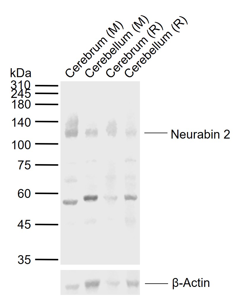
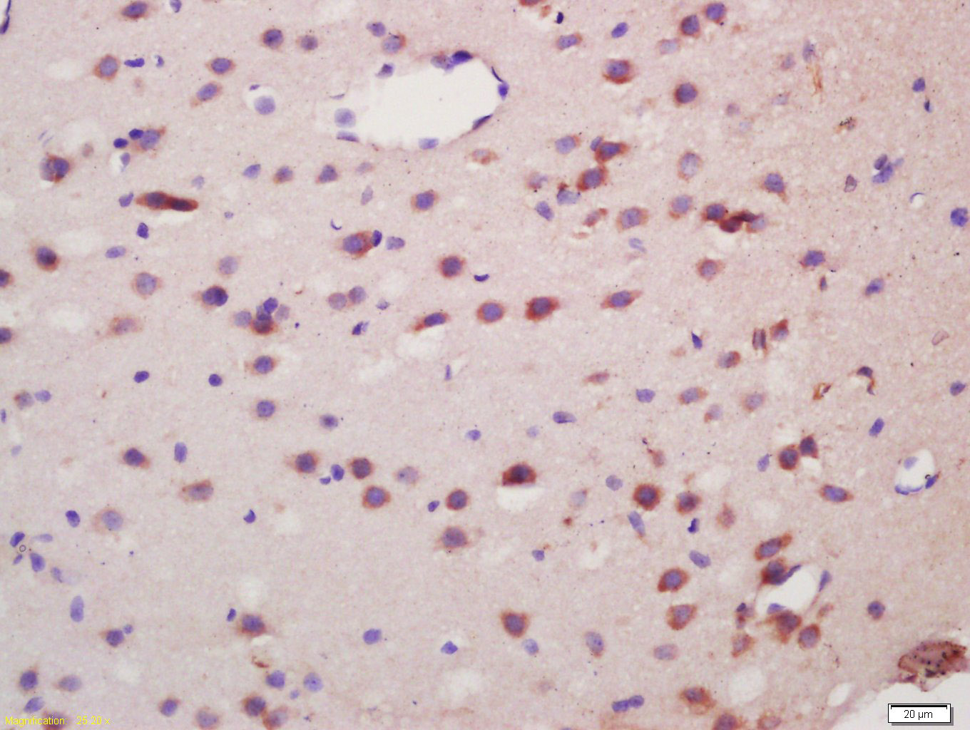
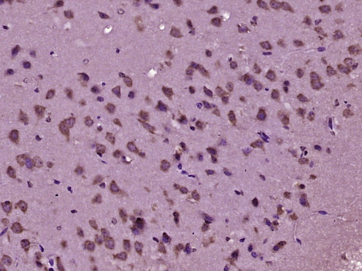
Target Protein: Neurabin 2 Rabbit pAb

IR: Immunogen Range:358-460/815

Clonality: Polyclonal

Isotype: IgG

Entrez Gene: 84687

Swiss Prot: Q96SB3

Source: KLH conjugated synthetic peptide derived from human Spinophilin/Neurabin 2:358-460/815 

Purification: affinity purified by Protein A

Storage: 0.01M TBS (pH7.4) with 1% BSA, 0.02% Proclin300 and 50% Glycerol. Shipped at 4℃. Store at -20℃ for one year. Avoid repeated freeze/thaw cycles.

Background: Neurabin-II, also called spinophilin, interacts with actin and PP-1 in dendritic spines of the central nervous system (1,2). The gene encoding human neurabin-II maps to chromosome 17q21-q22 (2). The structural characteristics of neurabin-II include one F-actin binding domain at the N-terminal region, a predicted coiled-coil struture at the C-terminal, one PDZ domain at the middle region, and a domain known to interact with transmembrane proteins (1). Neurabin-II bundles actin fliaments in vitro (1). In vivo, spinophilin localizes to the cortical sites of actin filaments and to the sites of active membrane remodelling (4). Neurabin-II also forms a complex with the catalytic subunit of PP1 and modulates PP1 enzymatic activity in vitro (2). Neurabin-II localizes to the head of dendritic spines (2) and aids in the ability of PP-1 to regulate the activity of a-amino-3-hydroxy-5-methyl-4-isoxazolepropionic acid (AMPA) and N-methyl-D-asparate (NMDA) receptors (3). In this manner, neurabin-II modulates both glutamatergic synaptic transmission and dendritic morphology (3). Synergistic interactions between spinophilin and human tumor supressor ARF suggest a role for neurabin-II in cell growth (5).

Size: 50ul

Concentration: 1mg/ml

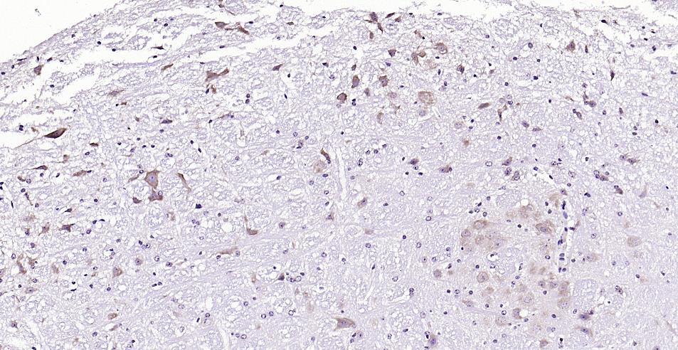
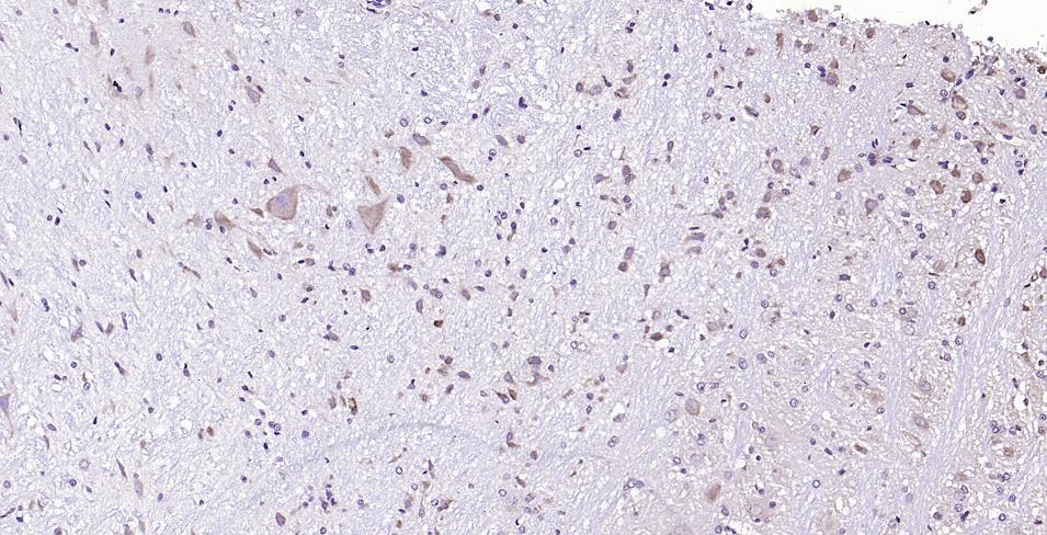
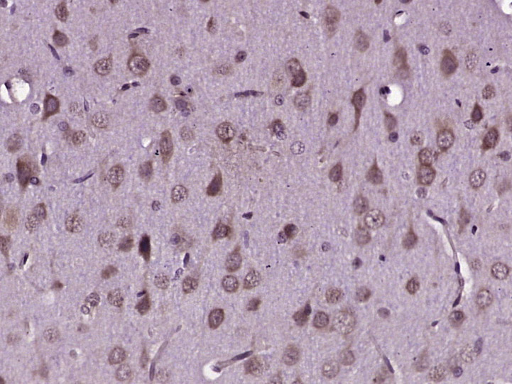
Applications: WB=1:500-2000,IHC-P=1:100-500,IHC-F=1:100-500,IF=1:100-500

Cross Reactive Species: Mouse,Rat (predicted: Human,Pig,Sheep,Dog)

For research use only. Not intended for diagnostic or therapeutic use.

VALIDATION IMAGES
Sample: Lane 1: Mouse Cerebrum tissue lysates Lane 2: Mouse Cerebellum tissue lysates Lane 3: Rat Cerebrum tissue lysates Lane 4: Rat Cerebellum tissue lysates Primary: Anti-Neurabin 2 (bs-12146R) at 1/1000 dilution Secondary: IRDye800CW Goat Anti-Rabbit IgG at 1/20000 dilution Predicted band size: 89 kDa Observed band size: 115 kDa
Paraformaldehyde-fixed, paraffin embedded (mouse cerebellum); Antigen retrieval by boiling in sodium citrate buffer (pH6.0) for 15min; Block endogenous peroxidase by 3% hydrogen peroxide for 20 minutes; Blocking buffer (normal goat serum) at 37°C for 30min; Antibody incubation with (Neurabin 2) Polyclonal Antibody, Unconjugated (bs-12146R) at 1:200 overnight at 4°C, followed by operating according to SP Kit(Rabbit) (sp-0023) instructionsand DAB staining.
Paraformaldehyde-fixed, paraffin embedded (Rat cerebellum); Antigen retrieval by boiling in sodium citrate buffer (pH6.0) for 15min; Block endogenous peroxidase by 3% hydrogen peroxide for 20 minutes; Blocking buffer (normal goat serum) at 37°C for 30min; Antibody incubation with (Neurabin 2) Polyclonal Antibody, Unconjugated (bs-12146R) at 1:200 overnight at 4°C, followed by operating according to SP Kit(Rabbit) (sp-0023) instructionsand DAB staining.
Paraformaldehyde-fixed, paraffin embedded (rat brain tissue); Antigen retrieval by boiling in sodium citrate buffer (pH6.0) for 15min; Block endogenous peroxidase by 3% hydrogen peroxide for 20 minutes; Blocking buffer (normal goat serum) at 37°C for 30min; Antibody incubation with (Neurabin 2) Polyclonal Antibody, Unconjugated (bs-12146R) at 1:400 overnight at 4°C, followed by operating according to SP Kit(Rabbit) (sp-0023) instructionsand DAB staining.
Tissue/cell: rat brain tissue; 4% Paraformaldehyde-fixed and paraffin-embedded; Antigen retrieval: citrate buffer ( 0.01M, pH 6.0 ), Boiling bathing for 15min; Block endogenous peroxidase by 3% Hydrogen peroxide for 30min; Blocking buffer (normal goat serum,C-0005) at 37℃ for 20 min; Incubation: Anti-Neurabin 2 Polyclonal Antibody, Unconjugated(bs-12146R) 1:200, overnight at 4°C, followed by conjugation to the secondary antibody(SP-0023) and DAB(C-0010) staining
Paraformaldehyde-fixed, paraffin embedded (mouse brain tissue); Antigen retrieval by boiling in sodium citrate buffer (pH6.0) for 15min; Block endogenous peroxidase by 3% hydrogen peroxide for 20 minutes; Blocking buffer (normal goat serum) at 37°C for 30min; Antibody incubation with (Neurabin 2) Polyclonal Antibody, Unconjugated (bs-12146R) at 1:400 overnight at 4°C, followed by operating according to SP Kit(Rabbit) (sp-0023) instructionsand DAB staining.
 
PRODUCT SPECIFIC PUBLICATIONS