DATASHEET
Host:
Mouse
Target Protein:
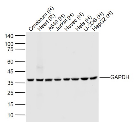
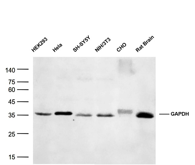
GAPDH Mouse mAb, Loading Control
IR:
Immunogen Range:
Clonality:
Monoclonal
Isotype:
IgG
Entrez Gene:
2597
Swiss Prot:
P04406
Source:
Recombinded Human GAPDH:
Purification:
affinity purified by Protein G
Storage:
0.01M TBS (pH7.4) with 1% BSA, 0.02% Proclin300 and 50% Glycerol. Shipped at 4℃. Store at -20℃ for one year. Avoid repeated freeze/thaw cycles.
Background:
Loading Control
Glyceraldehyde 3 phosphate dehydrogenase (GAPDH) is well known as one of the key enzymes involved in glycolysis. As well as functioning as a glycolytic enzyme in cytoplasm, recent evidence suggests that mammalian GAPDH is also involved in a great number of intracellular proceses such as membrane fusion, microtubule bundling, phosphotransferase activity, nuclear RNA export, DNA replication, and DNA repair. During the last decade a lot of data appeared concerning the role of GAPDH in different pathologies including prostate cancer progression, programmed neuronal cell death, age related neuronal diseases, such as Alzheimer's and Huntington's disease. GAPDH is expressed in all cells. It is constitutively expressed in almost all tissues at high levels. There are however some physiological factors such as hypoxia and diabetes that increase GAPDH expression in certain cell types. GAPDH molecule is composed of four 36kDa subunits.
Size:
50ul
Concentration:
1mg/ml
Applications:
WB=1:5000-500000
Cross Reactive Species:
Human,Mouse,Rat,Hamster (predicted: Rabbit,Pig,Sheep,Chicken,Dog,Monkey)
For research use only. Not intended for diagnostic or therapeutic use.