sales@bioss.com.cn
techsupport@bioss.com.cn
400-901-9800
Host: Rabbit
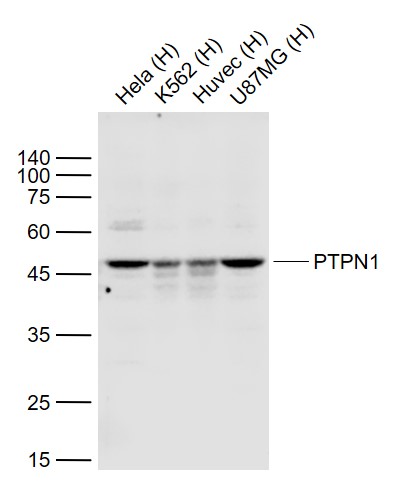
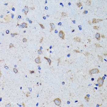
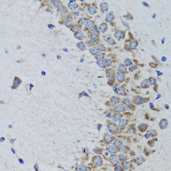
Target Protein: PTPN1 Rabbit pAb
IR: Immunogen Range:281-435/435
Clonality: Polyclonal
Isotype: IgG
Entrez Gene: 5770
Swiss Prot: P18031
Source: Recombinant human PTPN1:281-435/435
Purification: affinity purified by Protein A
Storage: 0.01M TBS (pH7.4) with 1% BSA, 0.02% Proclin300 and 50% Glycerol. Store at -20℃ for one year. Avoid repeated freeze/thaw cycles. The lyophilized antibody is stable at room temperature for at least one month and for greater than a year when kept at -20°C. When reconstituted in sterile pH 7.4 0.01M PBS or diluent of antib
Background:
PTP1B is the prototypic member of the protein tyrosine phosphatase (PTP) family, which comprises at least 37 proteins. The family is characterized by a catalytic phosphatase domain of approximately 240 amino acids, and includes both transmembrane and cytosolic enzymes. The PTPs have high substrate specificity for phosphotyrosyl proteins. At the primary sequence level, PTPs share little similarity with the protein serine phosphatases, protein threonine phosphatases, or the acid and alkaline phosphatases. PTP1B is a negative regulator of insulin and leptin signal transduction and is a drug discovery target in the fields of diabetes and obesity. Insulin also influences the expression of splice variants of PTP1B.
PTP1B also interacts with Insulin and EGF receptors, and undergoes phosphorylation after receptor stimulation. Tyrosine phosphorylation at Tyr-66, Tyr-152, and Tyr-153 occurs after insulin receptor activation, and tyrosine phosphorylation of Tyr-152 may be required for interactions with N-Cadherin. In addition, Akt can phosphorylate Ser-50 and this phosphorylation can reduce PTP1B activity.
Size: 50ul
Concentration: 1mg/ml
Applications: WB=1:500-2000,IHC-P=1:100-500,IHC-F=1:100-500,IF=1:100-500,Flow-Cyt=1ug/Test,ICC/IF=1:100
Cross Reactive Species: Human,Rat (predicted: Mouse,Rabbit,Pig,Cow,Horse)
For research use only. Not intended for diagnostic or therapeutic use.