sales@bioss.com.cn
techsupport@bioss.com.cn
400-901-9800
Host: Rabbit
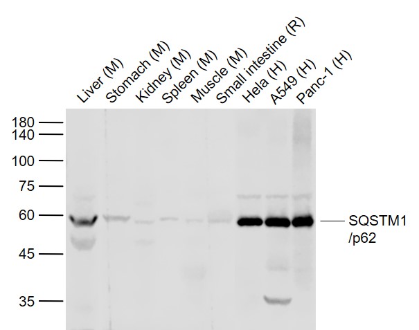
Target Protein: SQSTM1/p62 Rabbit pAb
IR: Immunogen Range:1-440
Clonality: Polyclonal
Isotype: IgG
Entrez Gene: 8878
Swiss Prot: Q13501
Source: Recombinant human SQSTM1/p62:1-440
Purification: affinity purified by Protein A
Storage: 0.01M TBS (pH7.4) with 1% BSA, 0.02% Proclin300 and 50% Glycerol. Store at -20℃ for one year. Avoid repeated freeze/thaw cycles. The lyophilized antibody is stable at room temperature for at least one month and for greater than a year when kept at -20°C. When reconstituted in sterile pH 7.4 0.01M PBS or diluent of antib
Background: This gene encodes a multifunctional protein that binds ubiquitin and regulates activation of the nuclear factor kappa-B (NF-kB) signaling pathway. The protein functions as a scaffolding/adaptor protein in concert with TNF receptor-associated factor 6 to mediate activation of NF-kB in response to upstream signals. Alternatively spliced transcript variants encoding either the same or different isoforms have been identified for this gene. Mutations in this gene result in sporadic and familial Paget disease of bone. [provided by RefSeq, Mar 2009]
Size: 50ul
Concentration: 1mg/ml
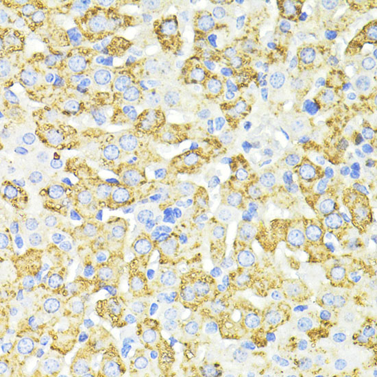
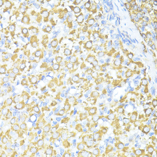
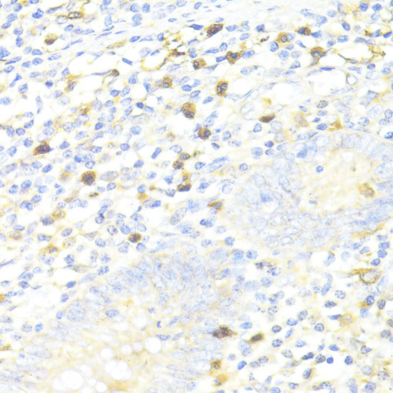
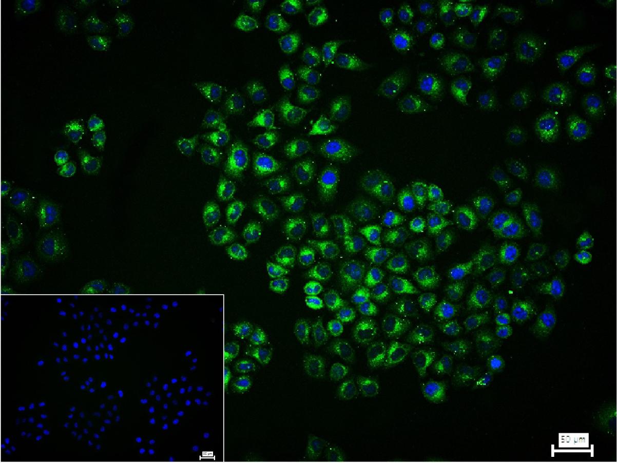
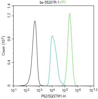
Applications: WB=1:500-2000,IHC-P=1:50-100,IHC-F=1:50-100,IF=1:50-100,Flow-Cyt=1:50-100,ICC/IF=1:50-200
Cross Reactive Species: Human,Mouse,Rat
For research use only. Not intended for diagnostic or therapeutic use.