sales@bioss.com.cn
techsupport@bioss.com.cn
400-901-9800
Host: Rabbit
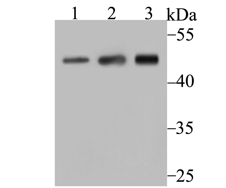
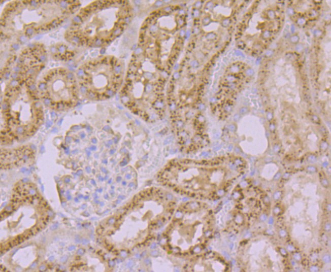
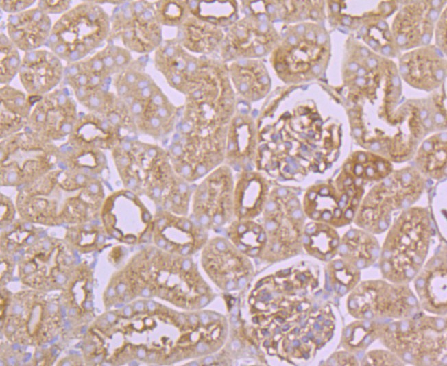
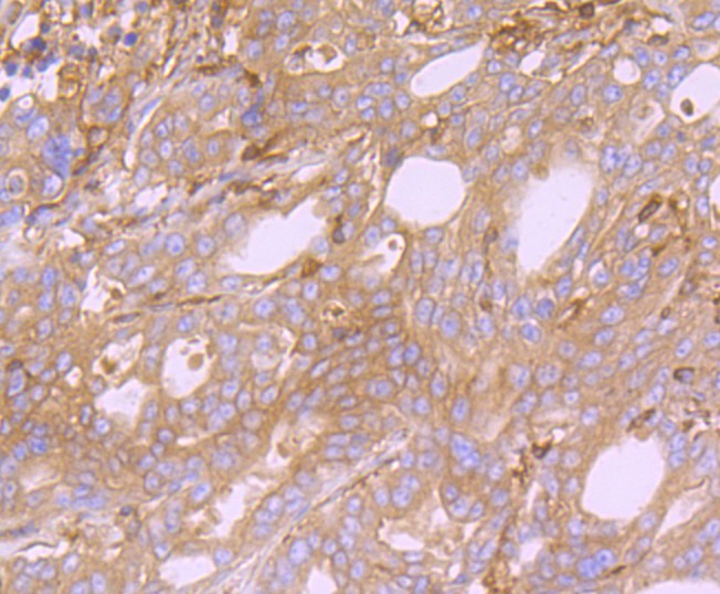
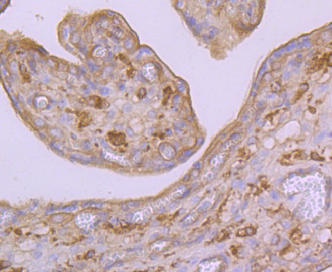
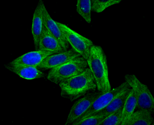
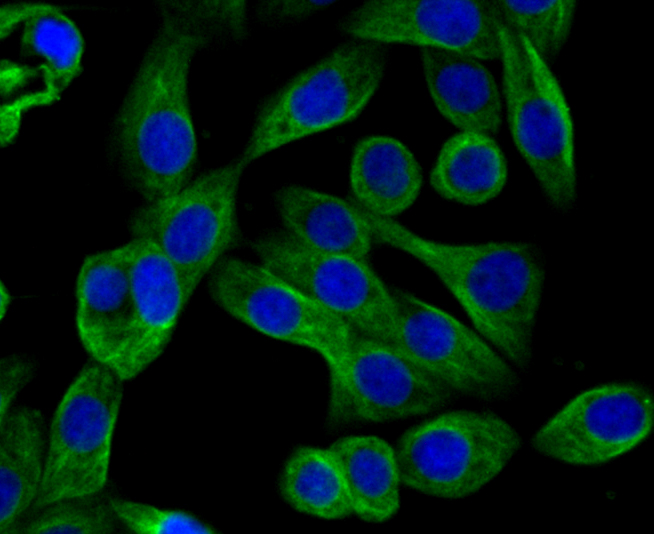
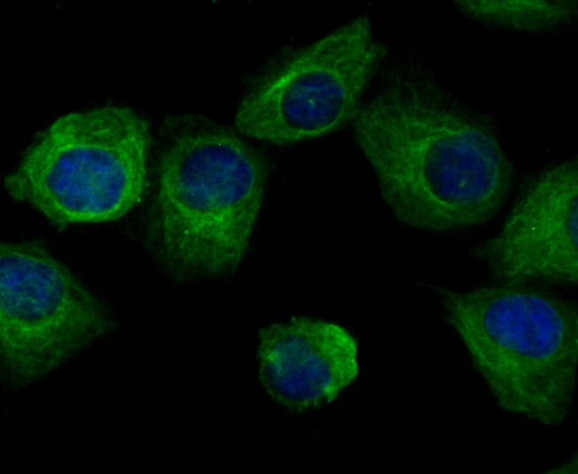
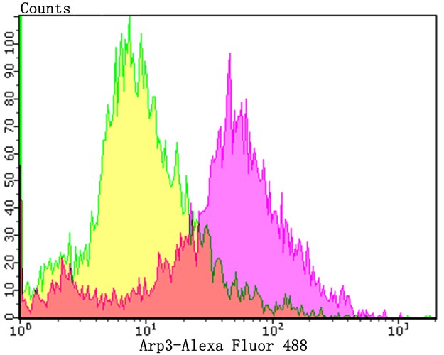
Target Protein: ARP3 Recombinant Rabbit mAb
IR: Immunogen Range:
Clonality:
Isotype: IgG
Entrez Gene: 10096
Swiss Prot: P61158
Source: Recombinant protein of C-terminal human Arp3.:
Purification: affinity purified by Protein A
Storage: 0.01M TBS (pH7.4) with 1% BSA, 0.02% Proclin300 and 50% Glycerol. Shipped at 4℃. Store at -20℃ for one year. Avoid repeated freeze/thaw cycles.
Background: Actin polymerization is required for a variety of cell functions, including chemotaxis, cell migration, cell adhesion, and platelet activation. Cells trigger actin polymerization through either the de novo nucleation of filaments from monomeric actin, the severing of existing filaments to create uncapped barbed ends, or the uncapping existing barbed ends. The nucleation of actin is a rate-limiting and unfavorable reaction in actin polymerization and therefore requires the involvement of the Arp2/3 complex, which helps create new filaments and promotes the end-to-side cross-linking of actin filaments into the branching meshwork. The Arp2/3 complex consists of the actin-related proteins Arp2 and Arp3, and various other accessory proteins. The Arp2/3 complex promotes actin nucleation by binding the pointed end of actin filaments, or by associating with the side of an existing filament, and nucleates growth in the barbed direction. In addition, the Arp2/3 complex also mediates actin cytoskeletal outgrowths that are regulated by the Rho family of small GTPases. In response to GTP-binding Cdc42, the Arp2/3 complex binds the Cdc42 substrates, namely the WASP proteins, and initiates the formation of lamellipodia and filopodia.
Size: 100ul
Concentration: 1mg/ml
Applications: WB=1:500-1000,IHC-P=1:50-200,IHC-F=1:20-200,IF=1:50-100,Flow-Cyt=1:50-100,ICC/IF=1:50-100
Cross Reactive Species: Human,Mouse,Rat
For research use only. Not intended for diagnostic or therapeutic use.