bsm-51466M [Primary Antibody]
Mouse  Anti-ALDH2 Mouse mAb  Monoclonal Antibody
www.bioss.com.cn
sales@bioss.com.cn
techsupport@bioss.com.cn
400-901-9800
DATASHEET

Host: Mouse

Target Protein: ALDH2 Mouse mAb

IR: Immunogen Range:

Clonality: Monoclonal

Isotype:

Entrez Gene: 217

Swiss Prot: P05091

Source: Recombinant human ALDH2.: 

Purification: affinity purified by Protein G

Storage: 0.01M TBS (pH7.4) with 1% BSA, 0.02% Proclin300 and 50% Glycerol. Shipped at 4℃. Store at -20℃ for one year. Avoid repeated freeze/thaw cycles.

Background: This protein belongs to the aldehyde dehydrogenase family of proteins. Aldehyde dehydrogenase is the second enzyme of the major oxidative pathway of alcohol metabolism. Two major liver isoforms of aldehyde dehydrogenase, cytosolic and mitochondrial, can be distinguished by their electrophoretic mobilities, kinetic properties, and subcellular localizations. Most Caucasians have two major isozymes, while approximately 50% of Orientals have the cytosolic isozyme but not the mitochondrial isozyme. A remarkably higher frequency of acute alcohol intoxication among Orientals than among Caucasians could be related to the absence of a catalytically active form of the mitochondrial isozyme. The increased exposure to acetaldehyde in individuals with the catalytically inactive form may also confer greater susceptibility to many types of cancer. This gene encodes a mitochondrial isoform, which has a low Km for acetaldehydes, and is localized in mitochondrial matrix. Alternative splicing results in multiple transcript variants encoding distinct isoforms.[provided by RefSeq, Mar 2011]

Size: 100ul

Concentration: 1mg/ml

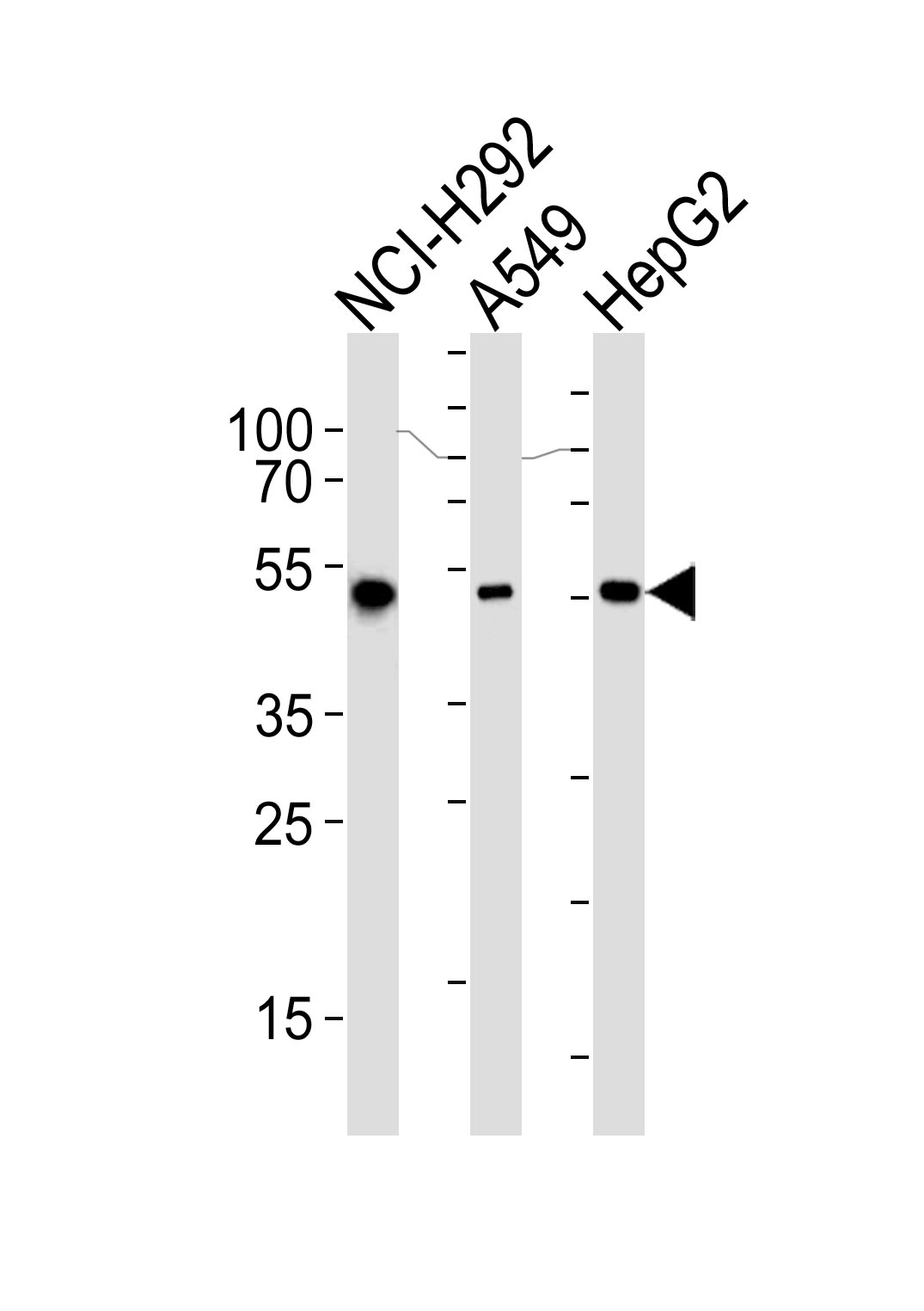
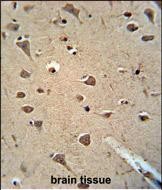
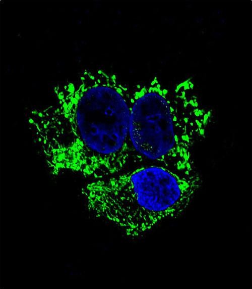
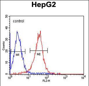
Applications: WB=1:500,IHC-P=1:400-800,IHC-F=1:400-800,IF=1:100-500,Flow-Cyt=1:50,ICC/IF=1:50

Cross Reactive Species: Human

For research use only. Not intended for diagnostic or therapeutic use.

VALIDATION IMAGES
Sample: Lane 1: NCI-H292 cell lysate Lane 2: A549 cell lysate Lane 3: HepG2 cell lysate Primary: Anti-ALDH2 (bsm-51466M) at 1/1000 dilution Secondary: IRDye800CW Goat Anti-Mouse IgG at 1/20000 dilution Predicted band size: 54 kD Observed band size: 54 kD
Paraformaldehyde-fixed, paraffin embedded (human brain ); Antigen retrieval by boiling in sodium citrate buffer (pH6.0) for 15min; Block endogenous peroxidase by 3% hydrogen peroxide for 20 minutes; Blocking buffer (normal goat serum) at 37°C for 30min; Antibody incubation with (ALDH2 ) Monoclonal Antibody, Unconjugated (bsm-51466M) at 1:200 overnight at 4°C, followed by operating according to SP Kit(Mouse)(sp-0024) instructionsand DAB staining.
HepG2 cell; 4% Paraformaldehyde-fixed; Triton X-100 at room temperature for 20 min; Blocking buffer (normal goat serum, C-0005) at 37°C for 20 min; Antibody incubation with (ALDH2) monoclonal Antibody, Unconjugated (bsm-51466M) 1:50, 90 minutes at 37°C; followed by a conjugated Goat Anti-Mouse IgG antibody at 37°C for 90 minutes, DAPI (blue, C02-04002) was used to stain the cell nuclei.
Blank control:HepG2. Primary Antibody (green line): Mouse Anti-ALDH2 antibody (bsm-51466M) Dilution: 1:50; Secondary Antibody : Goat anti-mouse IgG-PE Protocol The cells were fixed with 4% PFA (10min at room temperature)and then permeabilized with 90% ice-cold methanol for 20 min at-20℃.The cells were then incubated in 5%BSA to block non-specific protein-protein interactions for 30 min at room temperature .Cells stained with Primary Antibody for 30 min at room temperature. The secondary antibody used for 40 min at room temperature. Acquisition of 20,000 events was performed.
 
PRODUCT SPECIFIC PUBLICATIONS