sales@bioss.com.cn
techsupport@bioss.com.cn
400-901-9800
Host: Rabbit
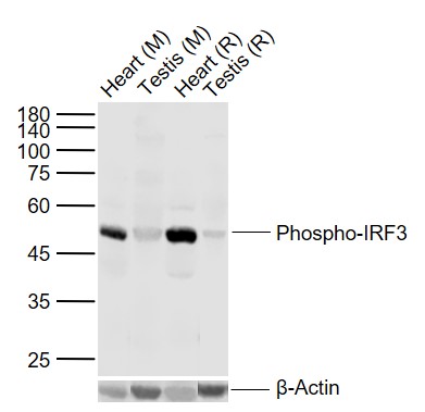
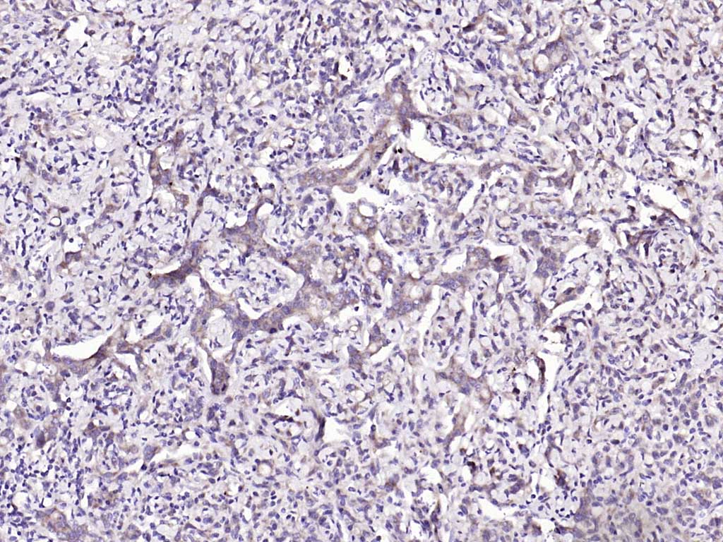
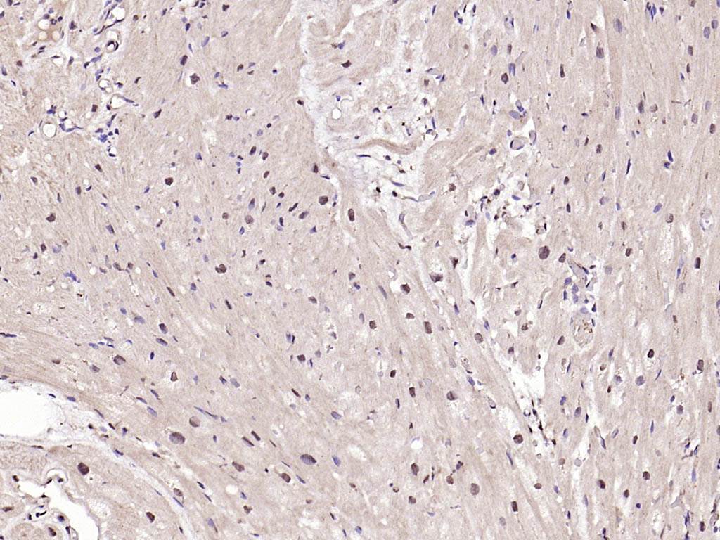
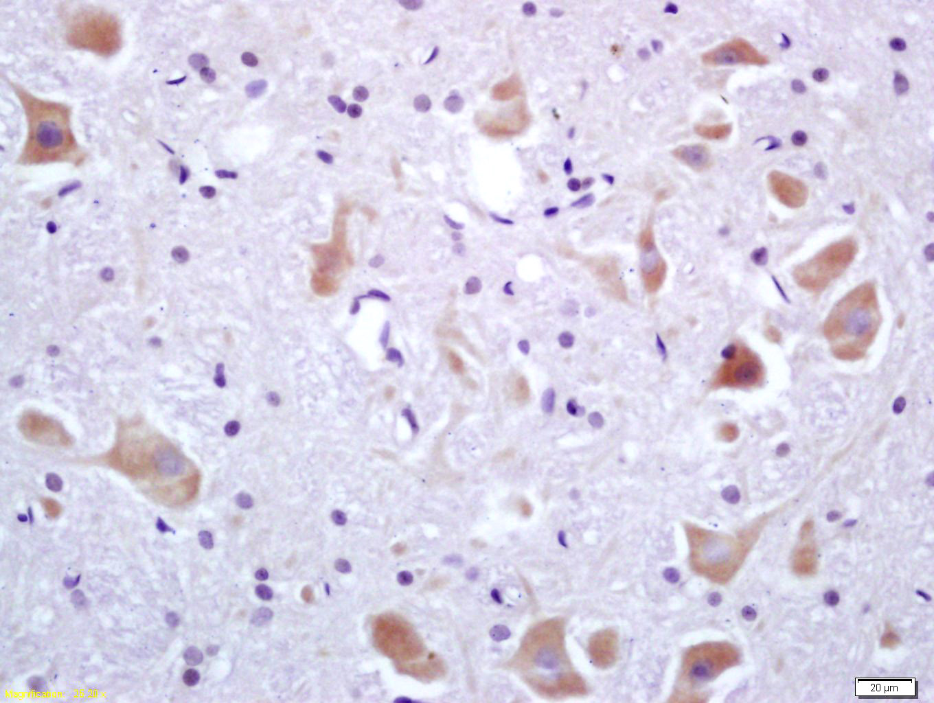
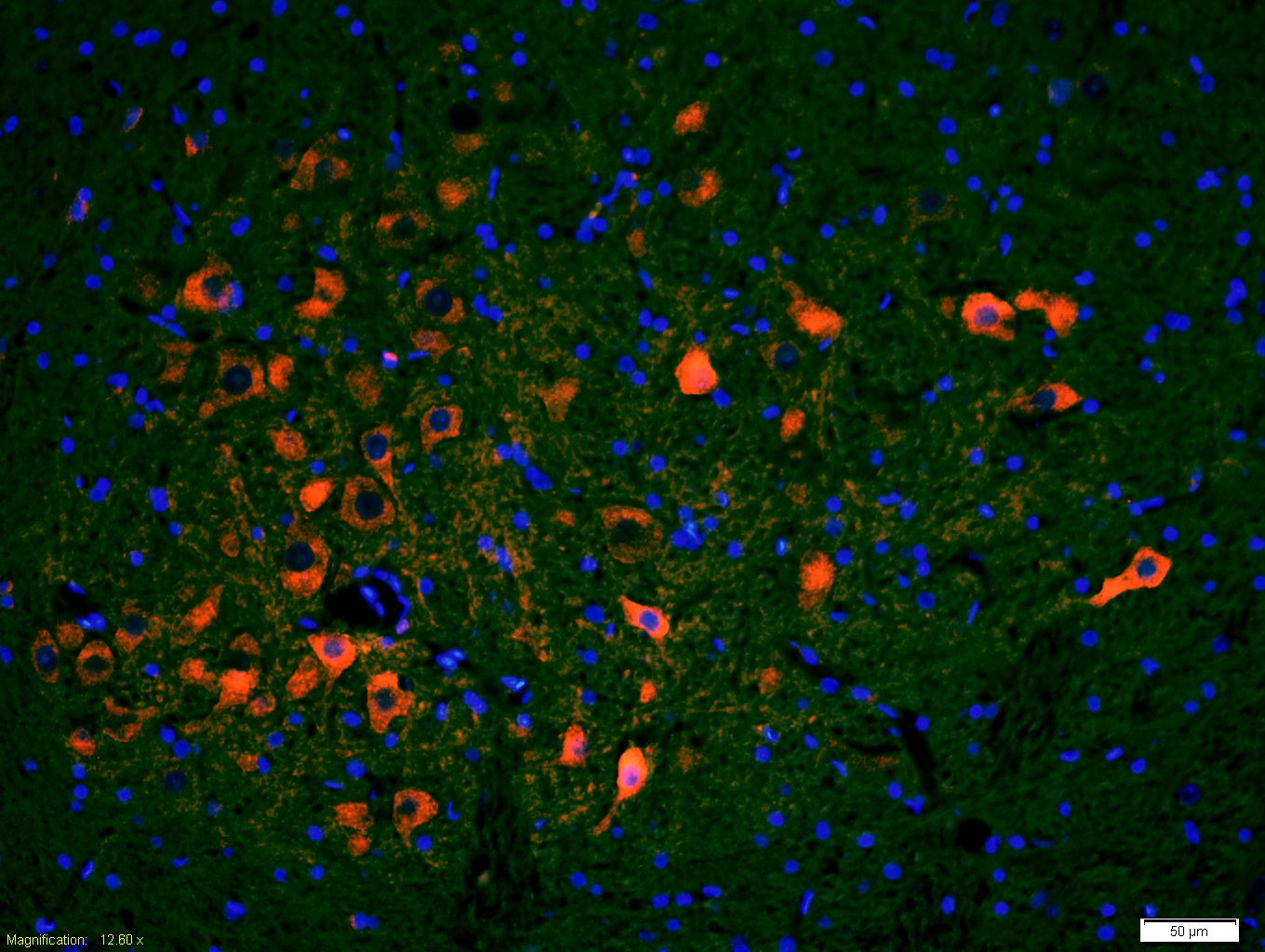
Target Protein: Phospho-IRF3 (Ser396) Rabbit pAb
IR: Immunogen Range:LHI(p-S)NS
Clonality: Polyclonal
Isotype: IgG
Entrez Gene: 3661
Swiss Prot: Q14653
Source: KLH conjugated synthesised phosphopeptide derived from human IRF3 around the phosphorylation site of Ser396:LHI(p-S)NS
Purification: affinity purified by Protein A
Storage: 0.01M TBS (pH7.4) with 1% BSA, 0.02% Proclin300 and 50% Glycerol. Shipped at 4℃. Store at -20℃ for one year. Avoid repeated freeze/thaw cycles.
Background: This gene encodes a member of the interferon regulatory transcription factor (IRF) family. The encoded protein is found in an inactive cytoplasmic form that upon serine/threonine phosphorylation forms a complex with CREBBP. This complex translocates to the nucleus and activates the transcription of interferons alpha and beta, as well as other interferon-induced genes. Alternatively spliced transcript variants encoding multiple isoforms have been observed for this gene. [provided by RefSeq, Nov 2011].
Size: 50ul
Concentration: 1mg/ml
Applications: WB=1:500-2000,IHC-P=1:100-500,IHC-F=1:100-500,IF=1:100-500
Cross Reactive Species: Human,Mouse,Rat (predicted: Pig,Sheep,Cow,Dog)
For research use only. Not intended for diagnostic or therapeutic use.