bs-3265R [Primary Antibody]
Rabbit  Anti-Phospho-Mcl1 (Ser159 + Thr163) Rabbit pAb  Polyclonal Antibody
www.bioss.com.cn
sales@bioss.com.cn
techsupport@bioss.com.cn
400-901-9800
DATASHEET

Host: Rabbit

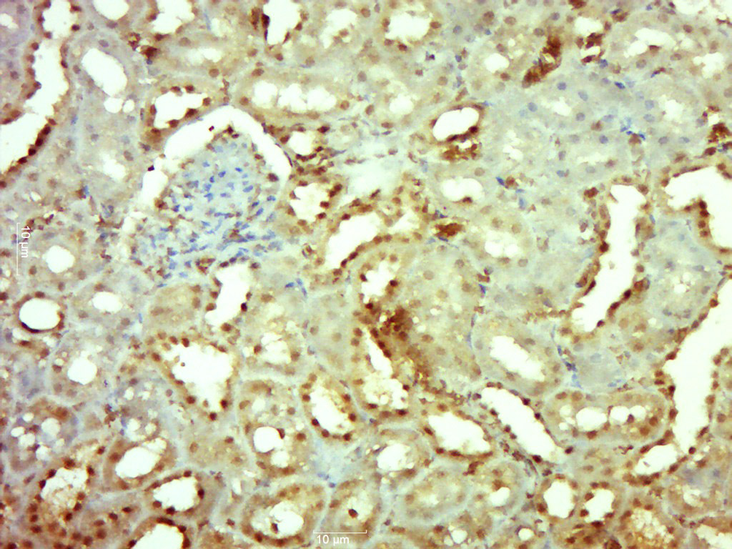
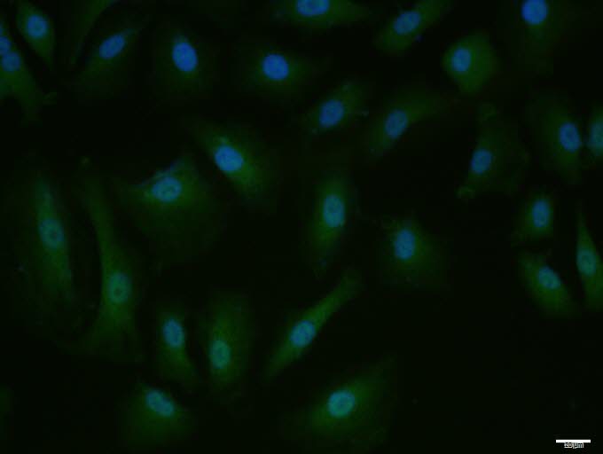
Target Protein: Phospho-Mcl1 (Ser159 + Thr163) Rabbit pAb

IR: Immunogen Range:DG(p-S)LPS(p-T)PP

Clonality: Polyclonal

Isotype: IgG

Entrez Gene: 4170

Swiss Prot: Q07820

Source: KLH conjugated synthesised phosphopeptide derived from human Mcl 1 around the phosphorylation site of Ser159/Thr163:DG(p-S)LPS(p-T)PP 

Purification: affinity purified by Protein A

Storage: 0.01M TBS (pH7.4) with 1% BSA, 0.02% Proclin300 and 50% Glycerol. Shipped at 4℃. Store at -20℃ for one year. Avoid repeated freeze/thaw cycles.

Background: Mcl1 is an anti-apoptotic member of Bcl2 family originally isolated from the ML1 human myeloid leukemia cell line during phorbol ester-induced differentiation along the monocyte/macrophage pathway. Mcl1 localizes to the mitochondria, interacts with and antagonizes pro-apoptotic Bcl2 family members, and inhibits apoptosis by a number of cytotoxic stimuli. It is involved in programing of differentiation and concomitant maintenance of viability but not of proliferation. Isoform 1 inhibits apoptosis while isoform 2 promotes it. Expression increases early during phorbol-ester induced differentiation along the monocyte/macrophage pathway in myeloid leukemia cell lines ML1.

Size: 100ul

Concentration: 1mg/ml

Applications: IHC-P=1:100-500,IHC-F=1:100-500,IF=1:100-500,Flow-Cyt=1ug/Test,ICC/IF=1:100

Cross Reactive Species: Human,Rat (predicted: Mouse,Cow,Dog,Horse)

For research use only. Not intended for diagnostic or therapeutic use.

VALIDATION IMAGES
Paraformaldehyde-fixed, paraffin embedded (Rat kidney); Antigen retrieval by boiling in sodium citrate buffer (pH6.0) for 15min; Block endogenous peroxidase by 3% hydrogen peroxide for 20 minutes; Blocking buffer (normal goat serum) at 37°C for 30min; Antibody incubation with (Phospho-Mcl1 (Ser159 + Thr163)) Polyclonal Antibody, Unconjugated (bs-3265R) at 1:500 overnight at 4°C, followed by a conjugated secondary (sp-0023) for 20 minutes and DAB staining.
A549 cell; 4% Paraformaldehyde-fixed; Triton X-100 at room temperature for 20 min; Blocking buffer (normal goat serum, C-0005) at 37°C for 20 min; Antibody incubation with (Phospho-Mcl1 (Ser159 + Thr163)) polyclonal Antibody, Unconjugated (bs-3265R) 1:100, 90 minutes at 37°C; followed by a conjugated Goat Anti-Rabbit IgG antibody at 37°C for 90 minutes, DAPI (blue, C02-04002) was used to stain the cell nuclei.
Blank control: K562. Primary Antibody (green line): Rabbit Anti-Phospho-Mcl1 (Ser159 + Thr163) antibody (bs-3265R) Dilution: 1μg /10^6 cells; Isotype Control Antibody (orange line): Rabbit IgG . Secondary Antibody : Goat anti-rabbit IgG-FITC Dilution: 1μg /test. Protocol The cells were fixed with 4% PFA (10min at room temperature)and then permeabilized with 90% ice-cold methanol for 20 min at-20℃. The cells were then incubated in 5%BSA to block non-specific protein-protein interactions for 30 min at room temperature .Cells stained with Primary Antibody for 30 min at room temperature. The secondary antibody used for 40 min at room temperature. Acquisition of 20,000 events was performed.
 
PRODUCT SPECIFIC PUBLICATIONS