bs-1188R [Primary Antibody]
Rabbit  Anti-FRA2 Rabbit pAb  Polyclonal Antibody
www.bioss.com.cn
sales@bioss.com.cn
techsupport@bioss.com.cn
400-901-9800
DATASHEET

Host: Rabbit

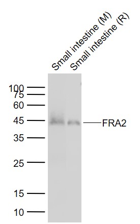
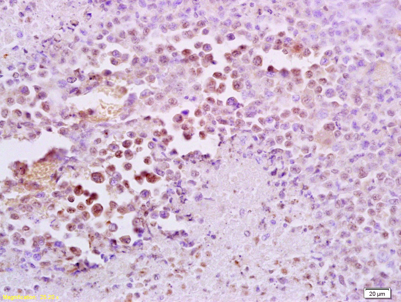
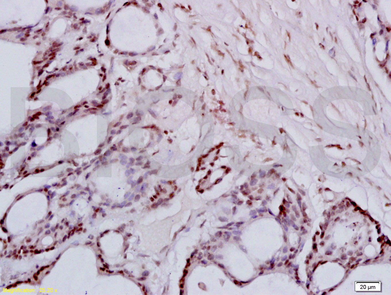
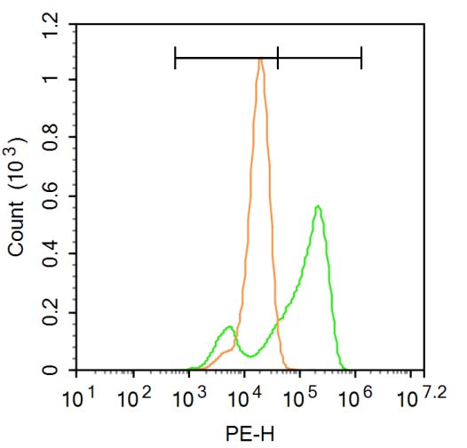
Target Protein: FRA2 Rabbit pAb

IR: Immunogen Range:231-326/326

Clonality: Polyclonal

Isotype: IgG

Entrez Gene: 2355

Swiss Prot: P15408

Source: KLH conjugated synthetic peptide derived from human FRA2:231-326/326 

Purification: affinity purified by Protein A

Storage: 0.01M TBS (pH7.4) with 1% BSA, 0.02% Proclin300 and 50% Glycerol. Shipped at 4℃. Store at -20℃ for one year. Avoid repeated freeze/thaw cycles.

Background: Fos and Jun dimerize to form Activator Protein 1 (AP1), a transcriptional factor that binds to the 12-O-tetradecanoylphorbol 13 acetate (TPA) response element (TRE) of several cellular and viral genes including human collagenase, metallothionein IIa, stromelysin, interleukin 2, SV40 and polyoma. Fos and Jun contain the 'leucine-zipper' motif that allows for dimerization and an adjacent basic domain required for biological activity. The functionally active form of Fos is in a heterodimer with a member of the Jun family. While Jun family members can form functional homodimers, studies indicate that Fos family members do not self-associate and therefore do not bind DNA on their own. The various dimers differ in their ability to transactivate AP1 dependent genes.

Size: 100ul

Concentration: 1mg/ml

Applications: WB=1:500-2000,IHC-P=1:100-500,IHC-F=1:100-500,IF=1:100-500,Flow-Cyt=3ug/test

Cross Reactive Species: Human,Mouse,Rat (predicted: Rabbit,Pig,Sheep,Cow,Chicken,Dog,GuineaPig)

For research use only. Not intended for diagnostic or therapeutic use.

VALIDATION IMAGES
Sample: Lane 1: Mouse Small intestine tissue lysates Lane 2: Rat Small intestine tissue lysates Primary: Anti-FRA2 (bs-1188R) at 1/1000 dilution Secondary: IRDye800CW Goat Anti-Rabbit IgG at 1/20000 dilution Predicted band size: 36 kD Observed band size: 44 kD
Tissue/cell: mouse lymphoma tissue; 4% Paraformaldehyde-fixed and paraffin-embedded; Antigen retrieval: citrate buffer ( 0.01M, pH 6.0 ), Boiling bathing for 15min; Block endogenous peroxidase by 3% Hydrogen peroxide for 30min; Blocking buffer (normal goat serum,C-0005) at 37℃ for 20 min; Incubation: Anti-FRA2/FOSL2 Polyclonal Antibody, Unconjugated(bs-1188R) 1:200, overnight at 4°C, followed by conjugation to the secondary antibody(SP-0023) and DAB(C-0010) staining
Tissue/cell: human colon carcinoma; 4% Paraformaldehyde-fixed and paraffin-embedded; Antigen retrieval: citrate buffer ( 0.01M, pH 6.0 ), Boiling bathing for 15min; Block endogenous peroxidase by 3% Hydrogen peroxide for 30min; Blocking buffer (normal goat serum,C-0005) at 37℃ for 20 min; Incubation: Anti-FRA2/FOSL2 Polyclonal Antibody, Unconjugated(bs-1188R) 1:200, overnight at 4°C, followed by conjugation to the secondary antibody(SP-0023) and DAB(C-0010) staining
Blank control: A549. Primary Antibody (green line): Rabbit Anti-FRA2 antibody (bs-1188R) Dilution: 1μg /10^6 cells; Isotype Control Antibody (orange line): Rabbit IgG . Secondary Antibody : Goat anti-rabbit IgG-PE Dilution: 3μg /test. Protocol The cells were fixed with 4% PFA (10min at room temperature)and then permeabilized with 90% ice-cold methanol for 20 min at-20℃. The cells were then incubated in 5% BSA to block non-specific protein-protein interactions for 30 min at at room temperature .Cells stained with Primary Antibody for 30 min at room temperature. The secondary antibody used for 40 min at room temperature. Acquisition of 20,000 events was performed.
 
PRODUCT SPECIFIC PUBLICATIONS