bs-0267R [Primary Antibody]
Rabbit  Anti-PRKCB Rabbit pAb  Polyclonal Antibody
www.bioss.com.cn
sales@bioss.com.cn
techsupport@bioss.com.cn
400-901-9800
DATASHEET

Host: Rabbit

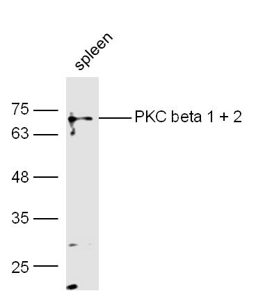
Target Protein: PRKCB Rabbit pAb

IR: Immunogen Range:601-673/673

Clonality: Polyclonal

Isotype: IgG

Entrez Gene: 5579

Swiss Prot: P05771

Source: KLH conjugated synthetic peptide derived from human PKC beta 1:601-673/673 

Purification: affinity purified by Protein A

Storage: 0.01M TBS (pH7.4) with 1% BSA, 0.02% Proclin300 and 50% Glycerol. Shipped at 4℃. Store at -20℃ for one year. Avoid repeated freeze/thaw cycles.

Background: Protein kinase C (PKC) is a family of serine- and threonine-specific protein kinases that can be activated by calcium and the second messenger diacylglycerol. PKC family members phosphorylate a wide variety of protein targets and are known to be involved in diverse cellular signaling pathways. PKC family members also serve as major receptors for phorbol esters, a class of tumor promoters. Each member of the PKC family has a specific expression profile and is believed to play a distinct role. The protein encoded by this gene is one of the PKC family members. It is a calcium-independent and phospholipid-dependent protein kinase. This kinase is important for T-cell activation. It is required for the activation of the transcription factors NF-kappaB and AP-1, and may link the T cell receptor (TCR) signaling complex to the activation of the transcription factors.

Size: 200ul

Concentration: 1mg/ml

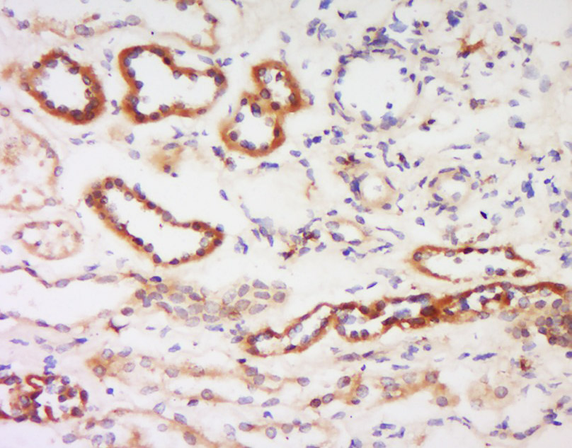
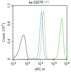
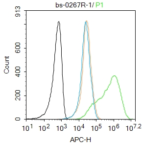
Applications: WB=1:500-2000,IHC-P=1:100-500,IHC-F=1:100-500,IF=1:100-500,Flow-Cyt=1:ug/Test

Cross Reactive Species: Human,Mouse,Rat (predicted: Rabbit,Pig,Cow)

For research use only. Not intended for diagnostic or therapeutic use.

VALIDATION IMAGES
Sample: spleen (Mouse) Lysate at 40 ug Primary: Anti-PKC beta 1 + 2 (bs-0267R) at 1/300 dilution Secondary: IRDye800CW Goat Anti-Rabbit IgG at 1/20000 dilution Predicted band size: 74 kD Observed band size: 74 kD
Tissue/cell: human kidney tissue; 4% Paraformaldehyde-fixed and paraffin-embedded; Antigen retrieval: citrate buffer ( 0.01M, pH 6.0 ), Boiling bathing for 15min; Block endogenous peroxidase by 3% Hydrogen peroxide for 30min; Blocking buffer (normal goat serum,C-0005) at 37℃ for 20 min; Incubation: Anti- PKC beta-1+2 Polyclonal Antibody, Unconjugated(bs-0267R) 1:200, overnight at 4°C, followed by conjugation to the secondary antibody(SP-0023) and DAB(C-0010) staining
Blank control (Black line): Molt4 (Black). Primary Antibody (green line):Rabbit Anti-PKC beta 1 + 2 antibody (bs-0267R) Dilution:1μg /10^6 cells; Isotype Control Antibody (orange line): Rabbit IgG . Secondary Antibody (white blue line): Goat anti-rabbit IgG-AF647 Dilution: 1μg /test. Protocol The cells were fixed with 4% PFA (10min at room temperature)and then permeabilized with 90% ice-cold methanol for 20 min at room temperature. The cells were then incubated in 5%BSA to block non-specific protein-protein interactions for 30 min at room temperature .Cells stained with Primary Antibody for 30 min at room temperature. The secondary antibody used for 40 min at room temperature. Acquisition of 20,000 events was performed.
Blank control:Molt4. Primary Antibody (green line): Rabbit Anti-PKC beta 1 + 2 antibody (bs-0267R) Dilution: 1μg /10^6 cells; Isotype Control Antibody (orange line): Rabbit IgG . Secondary Antibody : Goat anti-rabbit IgG-AF647 Dilution: 1μg /test. Protocol The cells were fixed with 4% PFA (10min at room temperature)and then permeabilized with 0.1% PBST for 20 min at room temperature. The cells were then incubated in 5%BSA to block non-specific protein-protein interactions for 30 min at room temperature .Cells stained with Primary Antibody for 30 min at room temperature. The secondary antibody used for 40 min at room temperature. Acquisition of 20,000 events was performed.
 
PRODUCT SPECIFIC PUBLICATIONS