VALIDATION IMAGES
Sample:
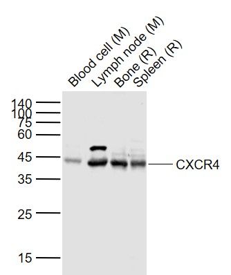
Lane 1: Blood cell (Mouse) Lysate at 40 ug
Lane 2: Lymph node (Mouse) Lysate at 40 ug
Lane 3: Bone (Rat) Lysate at 40 ug
Lane 4: Spleen (Rat) Lysate at 40 ug
Primary: Anti-CXCR4 (bs-20317R) at 1/1000 dilution
Secondary: IRDye800CW Goat Anti-Rabbit IgG at 1/20000 dilution
Predicted band size: 45 kD
Observed band size: 44 kD
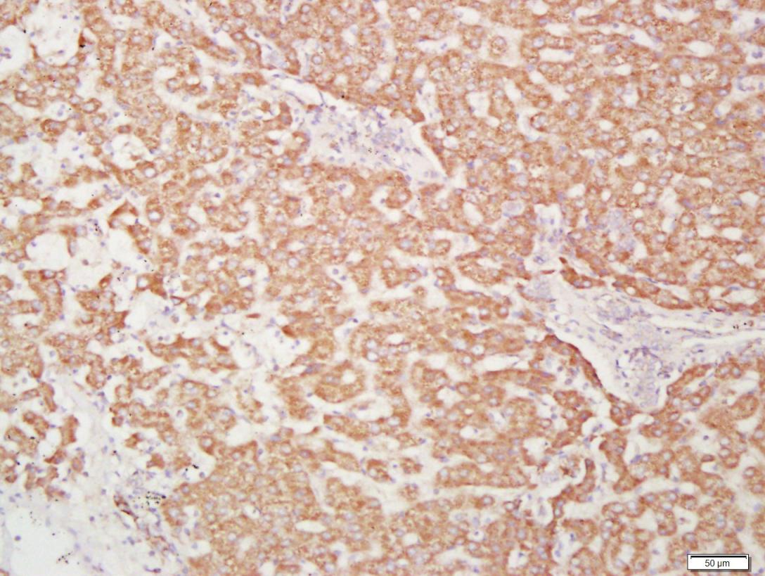
Paraformaldehyde-fixed, paraffin embedded Human Liver Cancer; Antigen retrieval by boiling in sodium citrate buffer (pH6.0) for 15 min; Antibody incubation with CXCR4 Polyclonal Antibody, Unconjugated (bs-1011R) at 1:400 overnight at 4°C, followed by conjugation to the bs-0295G-HRP and DAB (C-0010) staining.
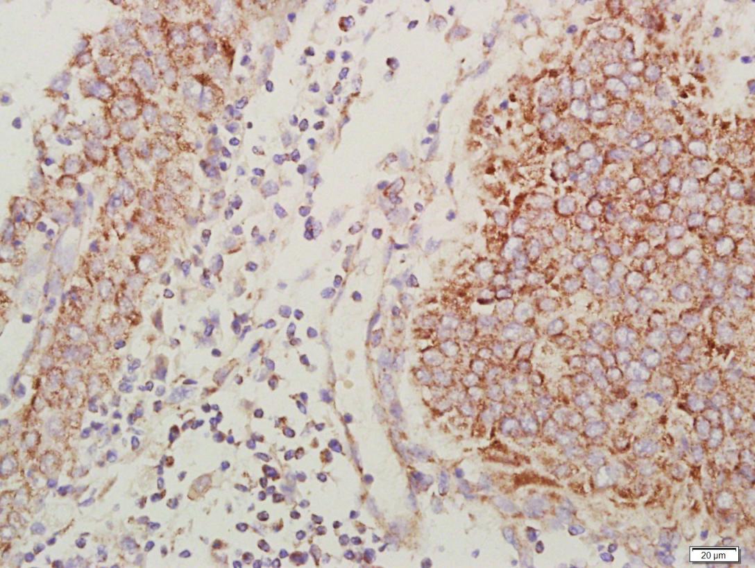
Paraformaldehyde-fixed, paraffin embedded Human Esophageal Cancer; Antigen retrieval by boiling in sodium citrate buffer (pH6.0) for 15 min; Antibody incubation with CXCR4 Polyclonal Antibody, Unconjugated (bs-1011R) at 1:400 overnight at 4°C, followed by conjugation to the bs-0295G-HRP and DAB (C-0010) staining.
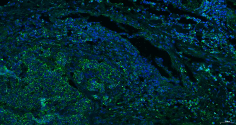
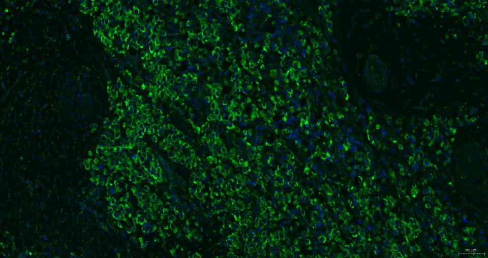
Paraformaldehyde-fixed, paraffin embedded Human esophageal cancer; Antigen retrieval by boiling in sodium citrate buffer (pH6.0) for 15 min; Antibody incubation with CXCR4 Polyclonal Antibody, Unconjugated (bs-1011R) at 1:200 overnight at 4°C. Followed by conjugated Goat Anti-Rabbit IgG antibody (green, bs-0295G-BF488), DAPI (blue, C02-04002) was used to stain the cell nuclei.
Paraformaldehyde-fixed, paraffin embedded Human Breast Cancer; Antigen retrieval by boiling in sodium citrate buffer (pH6.0) for 15 min; Antibody incubation with CXCR4 Polyclonal Antibody, Unconjugated (bs-1011R) at 1:200 overnight at 4°C. Followed by conjugated Goat Anti-Rabbit IgG antibody (green, bs-0295G-BF488), DAPI (blue, C02-04002) was used to stain the cell nuclei.
Paraformaldehyde-fixed, paraffin embedded Human prostate tumor; Antigen retrieval by boiling in sodium citrate buffer (pH6.0) for 15 min; Antibody incubation with CXCR4 Polyclonal Antibody, Unconjugated (bs-1011R) at 1:200 overnight at 4°C. Followed by conjugated Goat Anti-Rabbit IgG antibody (green, bs-0295G-BF488), DAPI (blue, C02-04002) was used to stain the cell nuclei.
Paraformaldehyde-fixed, paraffin embedded Human lung cancer; Antigen retrieval by boiling in sodium citrate buffer (pH6.0) for 15 min; Antibody incubation with CXCR4 Polyclonal Antibody, Unconjugated (bs-1011R) at 1:200 overnight at 4°C. Followed by conjugated Goat Anti-Rabbit IgG antibody (green, bs-0295G-BF488), DAPI (blue, C02-04002) was used to stain the cell nuclei.
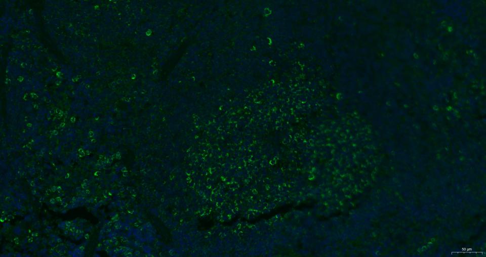
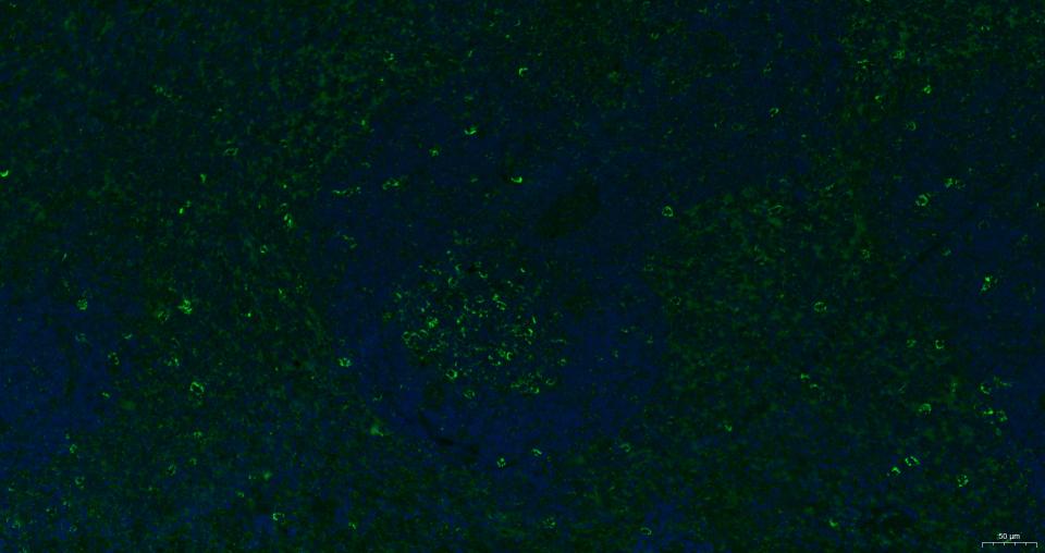
Paraformaldehyde-fixed, paraffin embedded Mouse spleen; Antigen retrieval by boiling in sodium citrate buffer (pH6.0) for 15 min; Antibody incubation with CXCR4 Polyclonal Antibody, Unconjugated (bs-1011R) at 1:200 overnight at 4°C. Followed by conjugated Goat Anti-Rabbit IgG antibody (green, bs-0295G-BF488), DAPI (blue, C02-04002) was used to stain the cell nuclei.
Paraformaldehyde-fixed, paraffin embedded Rat spleen; Antigen retrieval by boiling in sodium citrate buffer (pH6.0) for 15 min; Antibody incubation with CXCR4 Polyclonal Antibody, Unconjugated (bs-1011R) at 1:200 overnight at 4°C. Followed by conjugated Goat Anti-Rabbit IgG antibody (green, bs-0295G-BF488), DAPI (blue, C02-04002) was used to stain the cell nuclei.