sales@bioss.com.cn
techsupport@bioss.com.cn
400-901-9800
Host: Rabbit
Target Protein: MYLPF Rabbit pAb
IR: Immunogen Range:101-170/170
Clonality: Polyclonal
Isotype: IgG
Entrez Gene: 29895
Swiss Prot: Q96A32
Source: KLH conjugated synthetic peptide derived from human Fast skeletal myosin light chain 2:101-170/170
Purification: affinity purified by Protein A
Storage: 0.01M TBS (pH7.4) with 1% BSA, 0.02% Proclin300 and 50% Glycerol. Shipped at 4℃. Store at -20℃ for one year. Avoid repeated freeze/thaw cycles.
Background: Myosin is a highly conserved, ubiquitously expressed protein that interacts with Actin to generate the force for cellular movements. Conventional Myosins are hexameric proteins consisting of two heavy chain subunits, a pair of non-phosphorylatable light chain subunits and a pair of phosphorylatable light chain subunits. Three general classes of Myosin have been cloned: smooth muscle Myosins, striated muscle Myosins and non-muscle Myosins . Contractile activity in smooth muscle is regulated by the calcium/calmodulin-dependent phosphorylation of Myosin light chain (MLC) by Myosin light chain kinase. Myosin heavy chains, which are encoded by the MYH gene family, contain Actin-activated ATPase activity which generates the motor function of Myosin. Myosin heavy chains were initially isolated from a human fetal skeletal muscle and are the major determinant in the speed of contraction of skeletal muscle. Various isoforms of myosin heavy chains are differentially expressed depending on the functional activity of the muscle.
Size: 200ul
Concentration: 1mg/ml
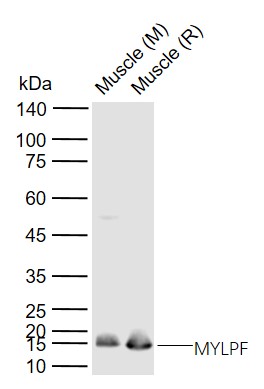
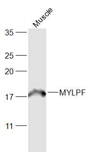
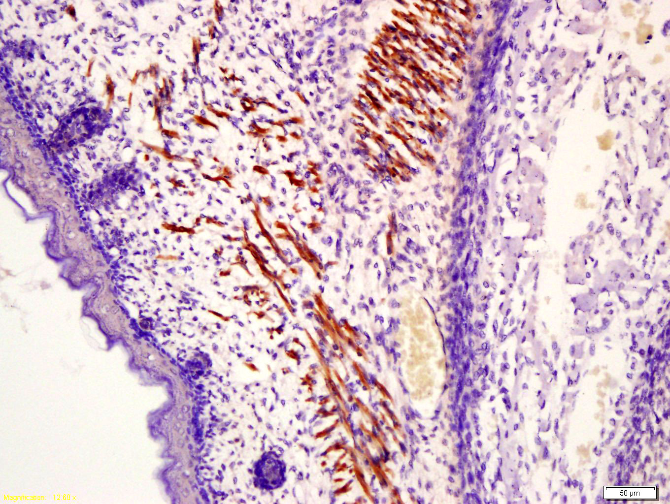
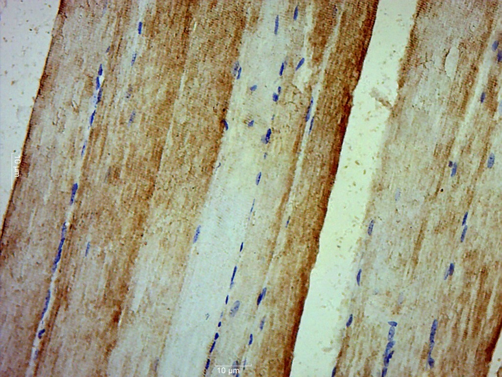
Applications: WB=1:500-2000,IHC-P=1:100-500,IHC-F=1:100-500,IF=1:100-500
Cross Reactive Species: Human,Mouse,Rat (predicted: Rabbit,Pig,Sheep,Cow,Dog,Horse)
For research use only. Not intended for diagnostic or therapeutic use.