bs-5292R [Primary Antibody]
Rabbit  Anti-phospho-DAXX (Ser517) Rabbit pAb  Polyclonal Antibody
www.bioss.com.cn
sales@bioss.com.cn
techsupport@bioss.com.cn
400-901-9800
DATASHEET

Host: Rabbit

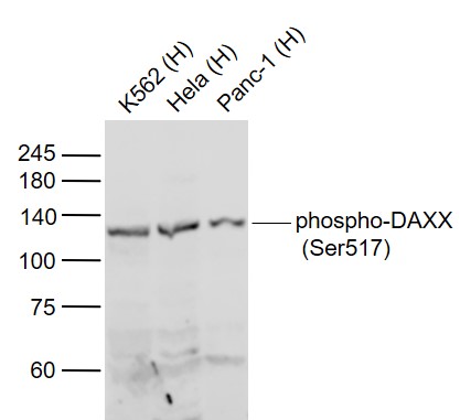
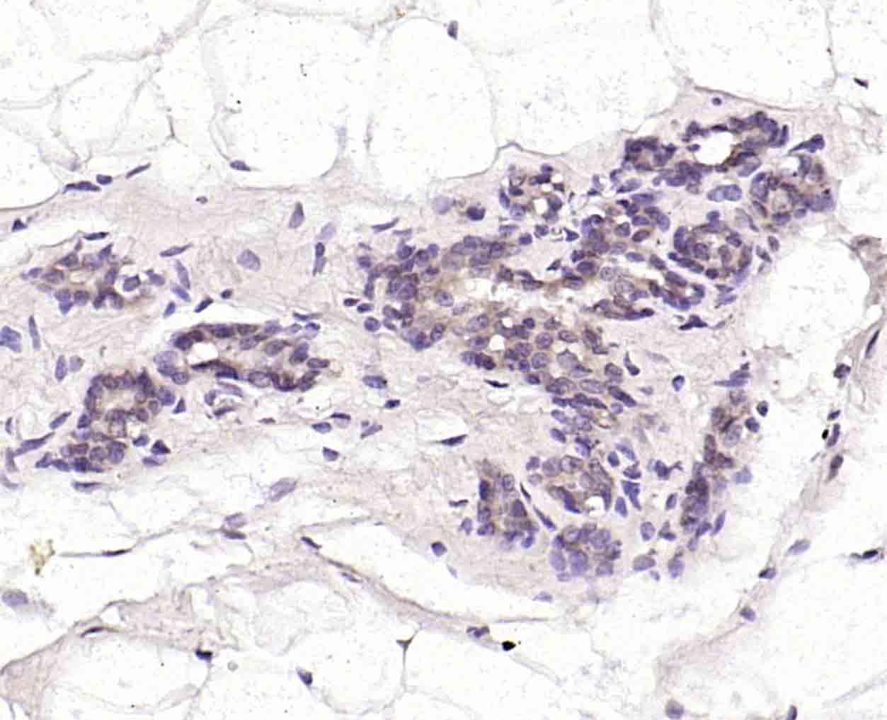
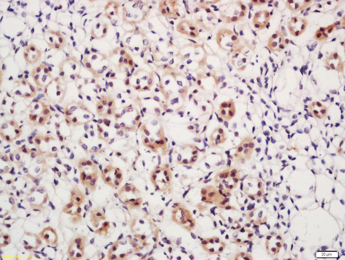
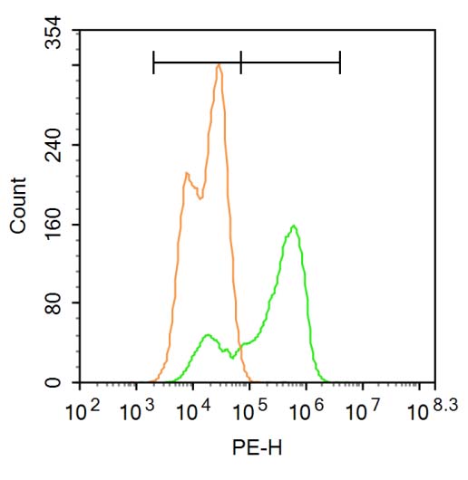
Target Protein: phospho-DAXX (Ser517) Rabbit pAb

IR: Immunogen Range:SR(p-S)SG

Clonality: Polyclonal

Isotype: IgG

Entrez Gene: 1616

Swiss Prot: Q9UER7

Source: KLH conjugated Synthesised phosphopeptide derived from human DAXX around the phosphorylation site of Ser517:SR(p-S)SG 

Purification: affinity purified by Protein A

Storage: 0.01M TBS (pH7.4) with 1% BSA, 0.02% Proclin300 and 50% Glycerol. Shipped at 4℃. Store at -20℃ for one year. Avoid repeated freeze/thaw cycles.

Background: Apoptosis, or programmed cell death, occurs during normal cellular differentiation and development of multicellular organisms. Apoptosis is induced by certain cytokines including TNF and Fas ligand of the TNF family through their death domain containing receptors, TNFR1 and Fas. Cell death signals are transduced by death domain (DD) containing adapter molecules and members of the ICE/CED3 protease family. A novel DD containing molecule was recently cloned from mouse, human and monkey and designated Daxx. Daxx is a death domain containing important intermediate in the Fas mediated apoptosis. Daxx binds specifically to the Fas death domain and enhances Fas induced apoptosis and activates the Jun N terminal kinase (JNK) pathway. It is widely expressed in fetal and adult human and mouse tissue, indicating its important function in Fas signaling pathways.

Size: 100ul

Concentration: 1mg/ml

Applications: WB=1:500-2000,IHC-P=1:100-500,IHC-F=1:100-500,IF=1:100-500,Flow-Cyt=3ug/Test

Cross Reactive Species: Human,Mouse,Rat (predicted: Pig)

For research use only. Not intended for diagnostic or therapeutic use.

VALIDATION IMAGES
Sample: Lane 1: K562 (Human) Cell Lysate at 30 ug Lane 2: Hela (Human) Cell Lysate at 30 ug Lane 3: Panc-1 (Human) Cell Lysate at 30 ug Primary: Anti-phospho-DAXX (Ser517) (bs-5292R) at 1/1000 dilution Secondary: IRDye800CW Goat Anti-Rabbit IgG at 1/20000 dilution Predicted band size: 110 kD Observed band size: 110 kD
Paraformaldehyde-fixed, paraffin embedded (rat breast ); Antigen retrieval by boiling in sodium citrate buffer (pH6.0) for 15min; Block endogenous peroxidase by 3% hydrogen peroxide for 20 minutes; Blocking buffer (normal goat serum) at 37°C for 30min; Incubation with (phospho-DAXX (Ser517) ) Polyclonal Antibody, Unconjugated (bs-5292R ) at 1:200 overnight at 4°C, followed by operating according to SP Kit(Rabbit) (sp-0023) instructionsand DAB staining.
Tissue/cell: mouse kidney tissue; 4% Paraformaldehyde-fixed and paraffin-embedded; Antigen retrieval: citrate buffer ( 0.01M, pH 6.0 ), Boiling bathing for 15min; Block endogenous peroxidase by 3% Hydrogen peroxide for 30min; Blocking buffer (normal goat serum,C-0005) at 37℃ for 20 min; Incubation: Anti-phospho-DAXX (Ser517) Polyclonal Antibody, Unconjugated(bs-5292R) 1:200, overnight at 4°C, followed by conjugation to the secondary antibody(SP-0023) and DAB(C-0010) staining
Blank control: Hela. Primary Antibody (green line): Rabbit Anti-phospho-DAXX (Ser517) antibody (bs-5292R) Dilution: 3μg /10^6 cells; Isotype Control Antibody (orange line): Rabbit IgG . Secondary Antibody : Goat anti-rabbit IgG-PE Dilution: 1μg /test. Protocol The cells were fixed with 4% PFA (10min at room temperature)and then permeabilized with ice-cold methanol for 20 min at-20℃.The cells were then incubated in 5%BSA to block non-specific protein-protein interactions for 30 min at at room temperature .Cells stained with Primary Antibody for 30 min at room temperature. The secondary antibody used for 40 min at room temperature. Acquisition of 20,000 events was performed.
 
PRODUCT SPECIFIC PUBLICATIONS