

| 产品编号 | bs-0189R |
| 英文名称 | alpha smooth muscle Actin Rabbit pAb |
| 中文名称 | 肌动蛋白α/α-SMA/α Actin抗体 |
| 别 名 | ACTSA; SMDYS; 0610041G09Rik; Actvs; SMAalpha; SMalphaA; a-SMA; alphaSMA; ACT-4; actin; ACTA_BOVIN; ACTA2; Alpha-actin-2; 3.6.4.-; ACTA_HUMAN; Cell growth-inhibiting gene 46 protein; ACTA_MOUSE; ACTA_RABIT; ACTA_RAT; α-Smooth Muscle Actin; α Smooth Muscle Actin; |
 |
Specific References (74) | bs-0189R has been referenced in 74 publications.
|
| 研究领域 | 肿瘤 细胞生物 免疫学 细胞骨架 |
| 抗体来源 | Rabbit |
| 克隆类型 | Polyclonal |
| 克 隆 号 | |
| 交叉反应 | Human,Mouse,Rat (predicted: Rabbit) |
| 产品应用 | WB=1:1000-5000,IHC-P=1:100-500,IHC-F=1:100-500,IF=1:100-500,Flow-Cyt=1μg/Test
not yet tested in other applications. optimal dilutions/concentrations should be determined by the end user. |
| 理论分子量 | 42 kDa |
| 检测分子量 | 42 |
| 细胞定位 | 细胞浆 |
| 性 状 | Liquid |
| 浓 度 | 1mg/ml |
| 免 疫 原 | KLH conjugated synthetic peptide derived from human Actin alpha: 301-375/375 |
| 亚 型 | IgG |
| 纯化方法 | affinity purified by Protein A |
| 缓 冲 液 | 0.01M TBS (pH7.4) with 1% BSA, 0.02% Proclin300 and 50% Glycerol. |
| 保存条件 | Shipped at 4℃. Store at -20℃ for one year. Avoid repeated freeze/thaw cycles. |
| 注意事项 | This product as supplied is intended for research use only, not for use in human, therapeutic or diagnostic applications. |
| PubMed | PubMed |
| 产品介绍 |
The product encoded by this gene belongs to the actin family of proteins, which are highly conserved proteins that play a role in cell motility, structure and integrity. Alpha, beta and gamma actin isoforms have been identified, with alpha actins being a major constituent of the contractile apparatus, while beta and gamma actins are involved in the regulation of cell motility. This actin is an alpha actin that is found in skeletal muscle. Mutations in this gene cause nemaline myopathy type 3, congenital myopathy with excess of thin myofilaments, congenital myopathy with cores, and congenital myopathy with fiber-type disproportion, diseases that lead to muscle fiber defects. [provided by RefSeq, Jul 2008] Function: Actins are highly conserved proteins that are involved in various types of cell motility and are ubiquitously expressed in all eukaryotic cells. Subunit: Polymerization of globular actin (G-actin) leads to a structural filament (F-actin) in the form of a two-stranded helix. Each actin can bind to 4 others. Subcellular Location: Cytoplasm, cytoskeleton. Post-translational modifications: Oxidation of Met-46 by MICALs (MICAL1, MICAL2 or MICAL3) to form methionine sulfoxide promotes actin filament depolymerization. Methionine sulfoxide is produced stereospecifically, but it is not known whether the (S)-S-oxide or the (R)-S-oxide is produced (By similarity). DISEASE: Note=ACTA2 mutations predispose patients to a variety of diffuse and diverse vascular diseases, premature onset coronary artery disease (CAD), premature ischemic strokes and Moyamoya disease. Defects in ACTA2 are the cause of familial aortic aneurysm thoracic type 6 (AAT6) [MIM:611788]. AATs are characterized by permanent dilation of the thoracic aorta usually due to degenerative changes in the aortic wall. They are primarily associated with a characteristic histologic appearance known as 'medial necrosis' or 'Erdheim cystic medial necrosis' in which there is degeneration and fragmentation of elastic fibers, loss of smooth muscle cells, and an accumulation of basophilic ground substance. Defects in ACTA2 are the cause of Moyamoya disease type 5 (MYMY5) [MIM:614042]. Moyamoya disease is a progressive cerebral angiopathy characterized by bilateral intracranial carotid artery stenosis and telangiectatic vessels in the region of the basal ganglia. The abnormal vessels resemble a 'puff of smoke' (moyamoya) on cerebral angiogram. Affected individuals can develop transient ischemic attacks and/or cerebral infarction, and rupture of the collateral vessels can cause intracranial hemorrhage. Hemiplegia of sudden onset and epileptic seizures constitute the prevailing presentation in childhood, while subarachnoid bleeding occurs more frequently in adults. Defects in ACTA2 are the cause of multisystemic smooth muscle dysfunction syndrome (MSMDYS) [MIM:613834]. MSMDYS is a syndrome characterized by dysfunction of smooth muscle cells throughout the body, leading to aortic and cerebrovascular disease, fixed dilated pupils, hypotonic bladder, malrotation, and hypoperistalsis of the gut and pulmonary hypertension. Similarity: Belongs to the actin family. SWISS: P62736 Gene ID: 59 Database links: Entrez Gene: 101021287 Baboon Entrez Gene: 59 Human Entrez Gene: 11475 Mouse Entrez Gene: 100009271 Rabbit Omim: 102620 Human SwissProt: P62736 Human SwissProt: P62737 Mouse SwissProt: P62740 Rabbit Unigene: 500483 Human Unigene: 213025 Mouse Unigene: 195319 Rat Unigene: 3114 Rat 结构蛋白(Structural Proteins) Actin α/α-Actin 是一种具有收缩能力的微丝蛋白,a-SMA广泛分布于几乎所有的肌型细胞中。Actin-α蛋白主要用于检测骨骼肌、平滑肌、血管平滑肌、心肌和肌原性肿瘤 包括:平滑肌瘤、平滑肌肉瘤、横纹肌肉瘤以及肌上细胞和肌上皮瘤。Actin(肌动蛋白)是在所有真核细胞中都表达的高度保守的蛋白质。它们沿微管组成了细胞骨架的主要成分。肌动蛋白至少表达为6种异构形式。它在心脏、骨骼横纹肌组织和某些平滑肌组织中表达,调节其收缩功能。有报导说肌动蛋白在乳房瘤中是高度磷酸化的。肌动蛋白的功能失调也会导致某种类型的心脏病。平滑肌α肌动蛋白使人更感兴趣,因为编码它的基因是相对局限于在血管平滑肌细胞中表达的少数几个基因之一。肌动蛋白是标记平滑肌和肌上皮细胞肿瘤的有效工具。 |
| 产品图片 |
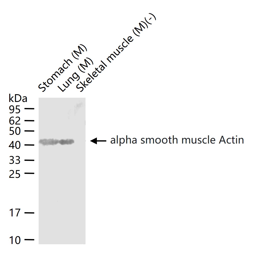
Sample:
Stomach (Mouse) Lysate at 40 ug
Lung (Mouse) Lysate at 40 ug
Skeletal muscle(-) (Mouse) Lysate at 40 ug
Primary: Anti-alpha smooth muscle Actin (bs-0189R) at 1/1000 dilution
Secondary: IRDye800CW Goat Anti-Rabbit IgG at 1/20000 dilution
Predicted band size: 42 kD
Observed band size: 42 kD
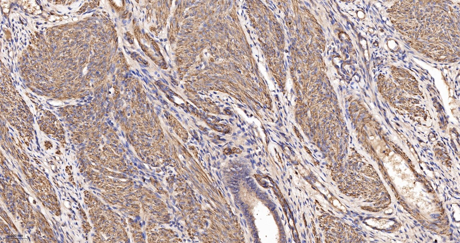
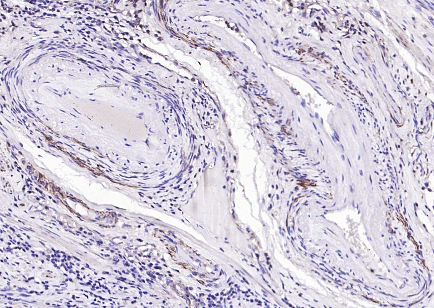
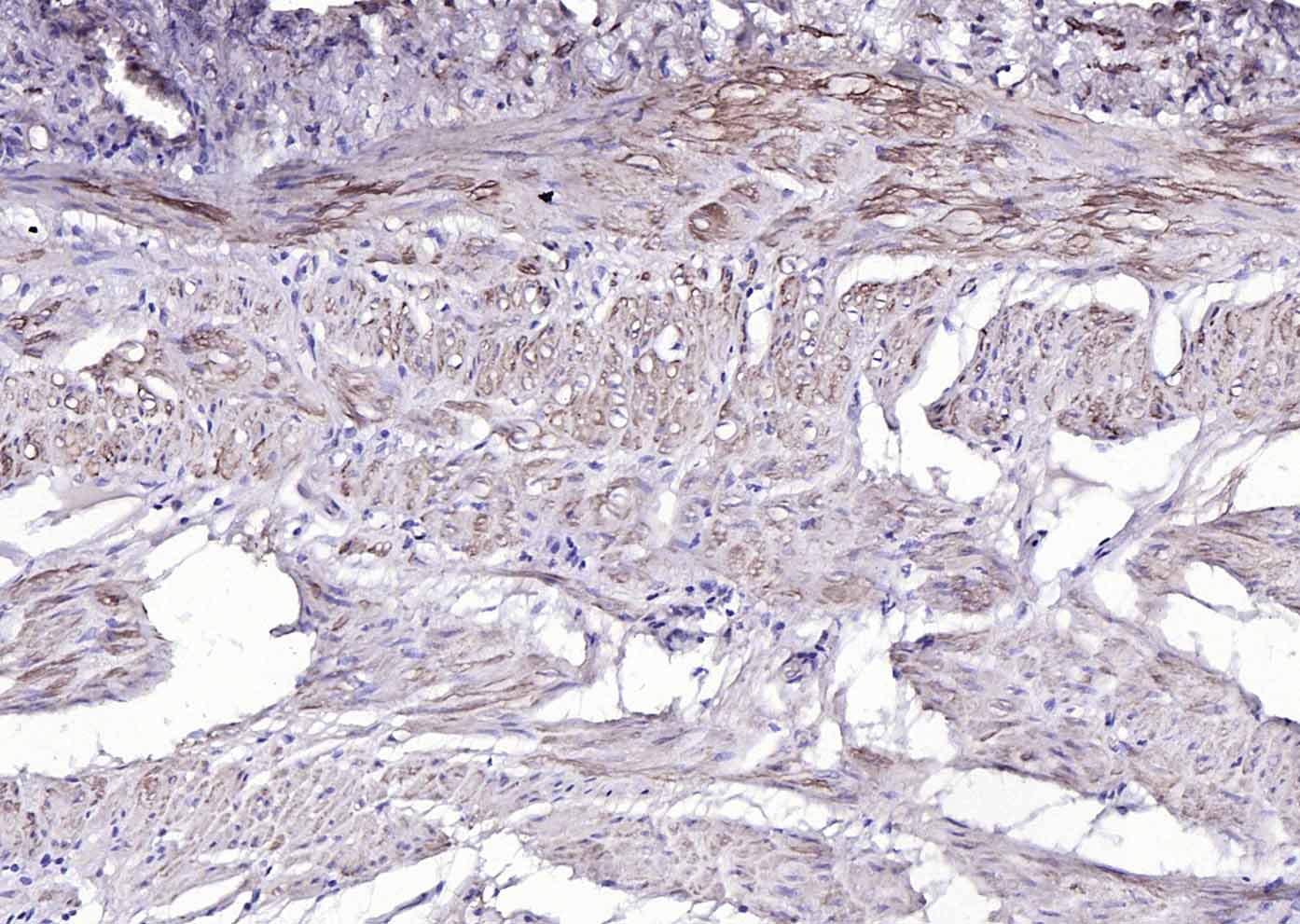
Paraformaldehyde-fixed, paraffin embedded Human Uterus; Antigen retrieval by boiling in sodium citrate buffer (pH6.0) for 15 min; The section was incubated with alpha smooth muscle Actin Polyclonal Antibody, Unconjugated (bs-0189R) at 1:200 overnight at 4°C, followed by conjugation to the bs-0295G-HRP and DAB (C-0010) staining.
Paraformaldehyde-fixed, paraffin embedded Rat Uterus; Antigen retrieval by boiling in sodium citrate buffer (pH6.0) for 15 min; The section was incubated with alpha smooth muscle Actin Polyclonal Antibody, Unconjugated (bs-0189R) at 1:200 overnight at 4°C, followed by conjugation to the bs-0295G-HRP and DAB (C-0010) staining.
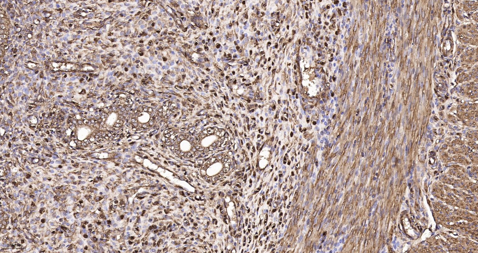
Paraformaldehyde-fixed, paraffin embedded Human Colon; Antigen retrieval by boiling in sodium citrate buffer (pH6.0) for 15 min; The section was incubated with alpha smooth muscle Actin Polyclonal Antibody, Unconjugated (bs-0189R) at 1:200 overnight at 4°C, followed by conjugation to the bs-0295G-HRP and DAB (C-0010) staining.
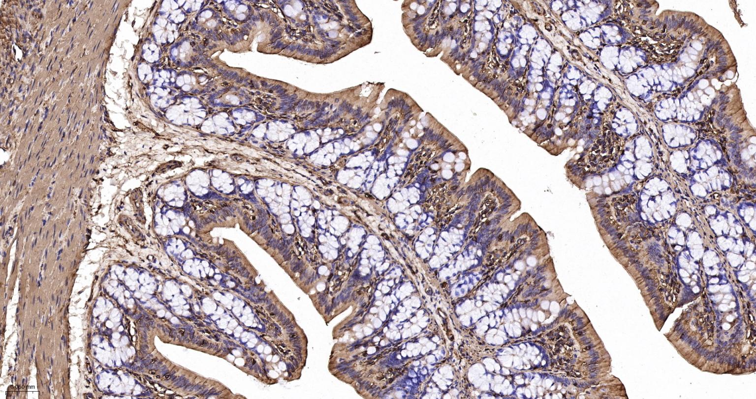
Paraformaldehyde-fixed, paraffin embedded Rat Colon; Antigen retrieval by boiling in sodium citrate buffer (pH6.0) for 15 min; The section was incubated with alpha smooth muscle Actin Polyclonal Antibody, Unconjugated (bs-0189R) at 1:200 overnight at 4°C, followed by conjugation to the bs-0295G-HRP and DAB (C-0010) staining.
Paraformaldehyde-fixed, paraffin embedded Mouse Colon; Antigen retrieval by boiling in sodium citrate buffer (pH6.0) for 15 min; The section was incubated with alpha smooth muscle Actin Polyclonal Antibody, Unconjugated (bs-0189R) at 1:200 overnight at 4°C, followed by conjugation to the bs-0295G-HRP and DAB (C-0010) staining.
Paraformaldehyde-fixed, paraffin embedded Human Cervical Cancer; Antigen retrieval by boiling in sodium citrate buffer (pH6.0) for 15 min; Antibody incubation with alpha smooth muscle Actin Polyclonal Antibody, Unconjugated (bs-0189R) at 1:200 overnight at 4°C, followed by conjugation to the SP Kit (Rabbit, SP-0023) and DAB (C-0010) staining.
Paraformaldehyde-fixed, paraffin embedded Rat Colon; Antigen retrieval by boiling in sodium citrate buffer (pH6.0) for 15 min; Antibody incubation with alpha smooth muscle Actin Polyclonal Antibody, Unconjugated (bs-0189R) at 1:200 overnight at 4°C, followed by conjugation to the SP Kit (Rabbit, SP-0023) and DAB (C-0010) staining.
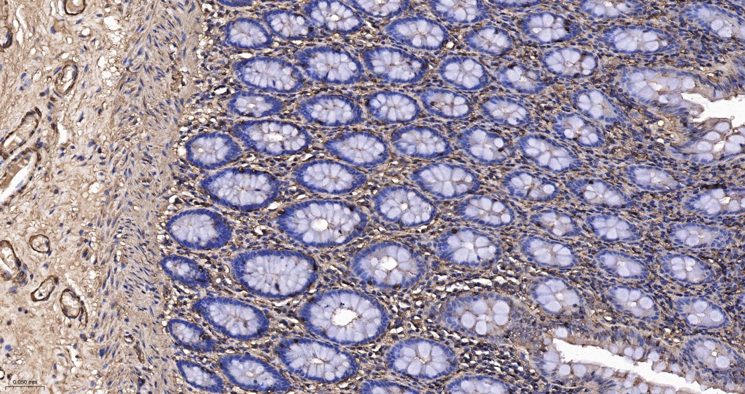
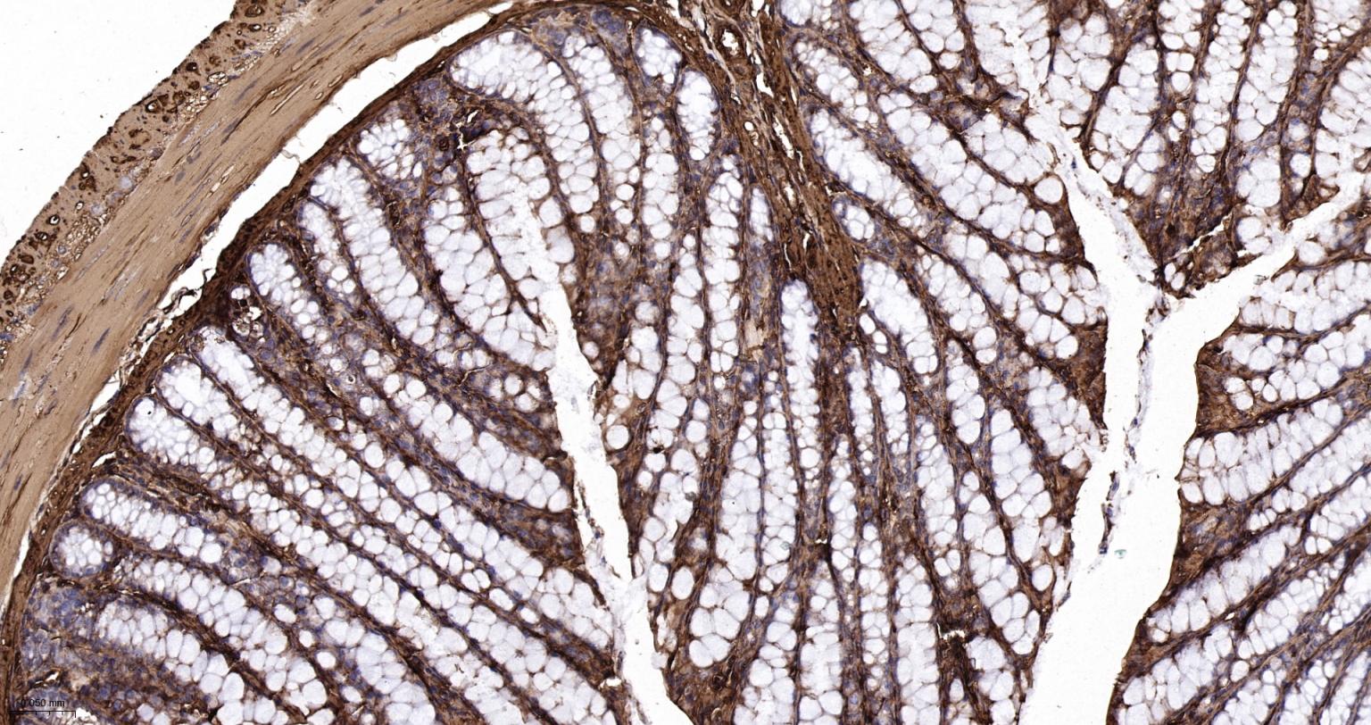
Paraformaldehyde-fixed, paraffin embedded Human Stomach; Antigen retrieval by boiling in sodium citrate buffer (pH6.0) for 15 min; Antibody incubation with alpha smooth muscle Actin Polyclonal Antibody, Unconjugated (bs-0189R) at 1:200 overnight at 4°C, followed by conjugation to the SP Kit (Rabbit, SP-0023) and DAB (C-0010) staining.
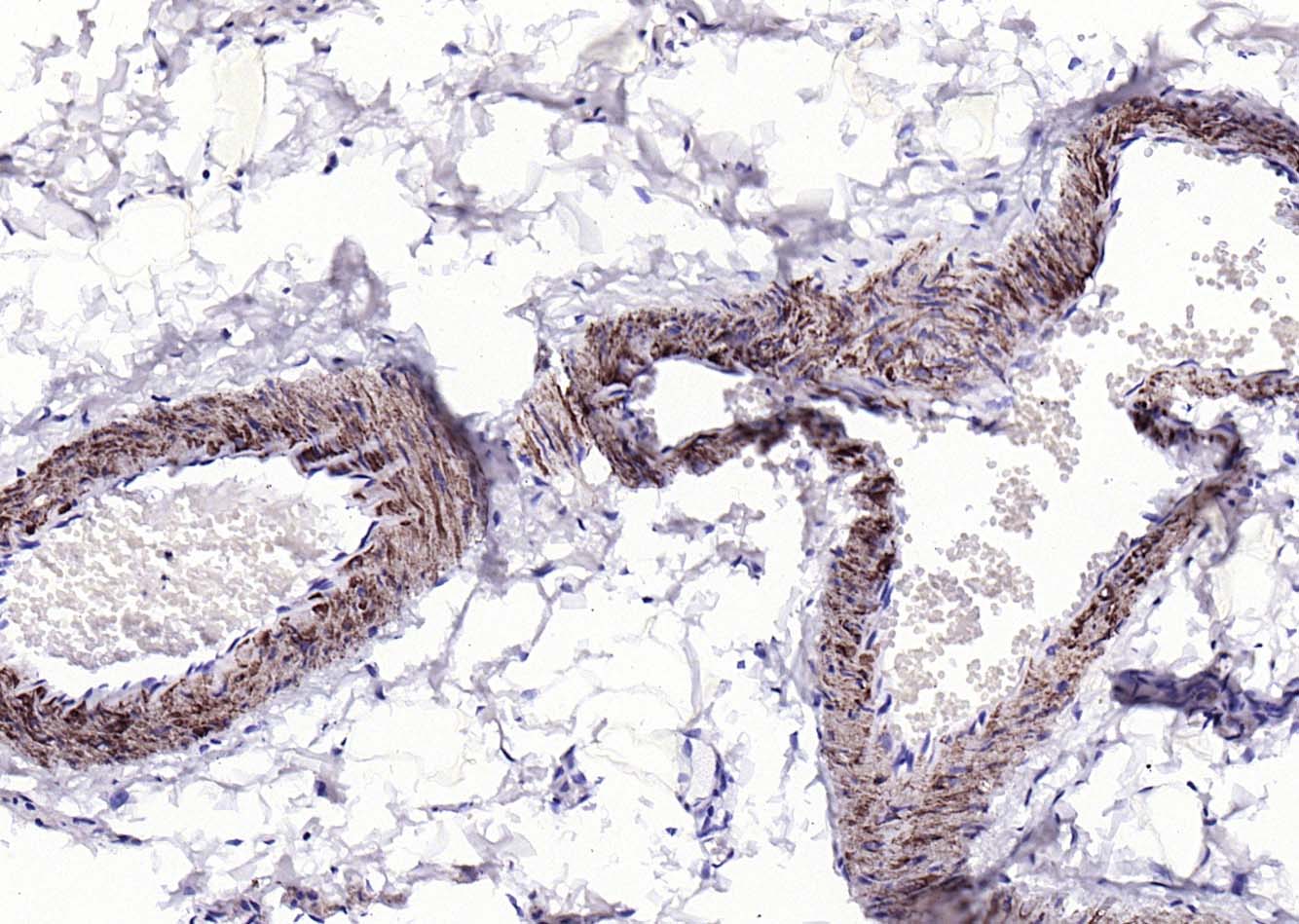
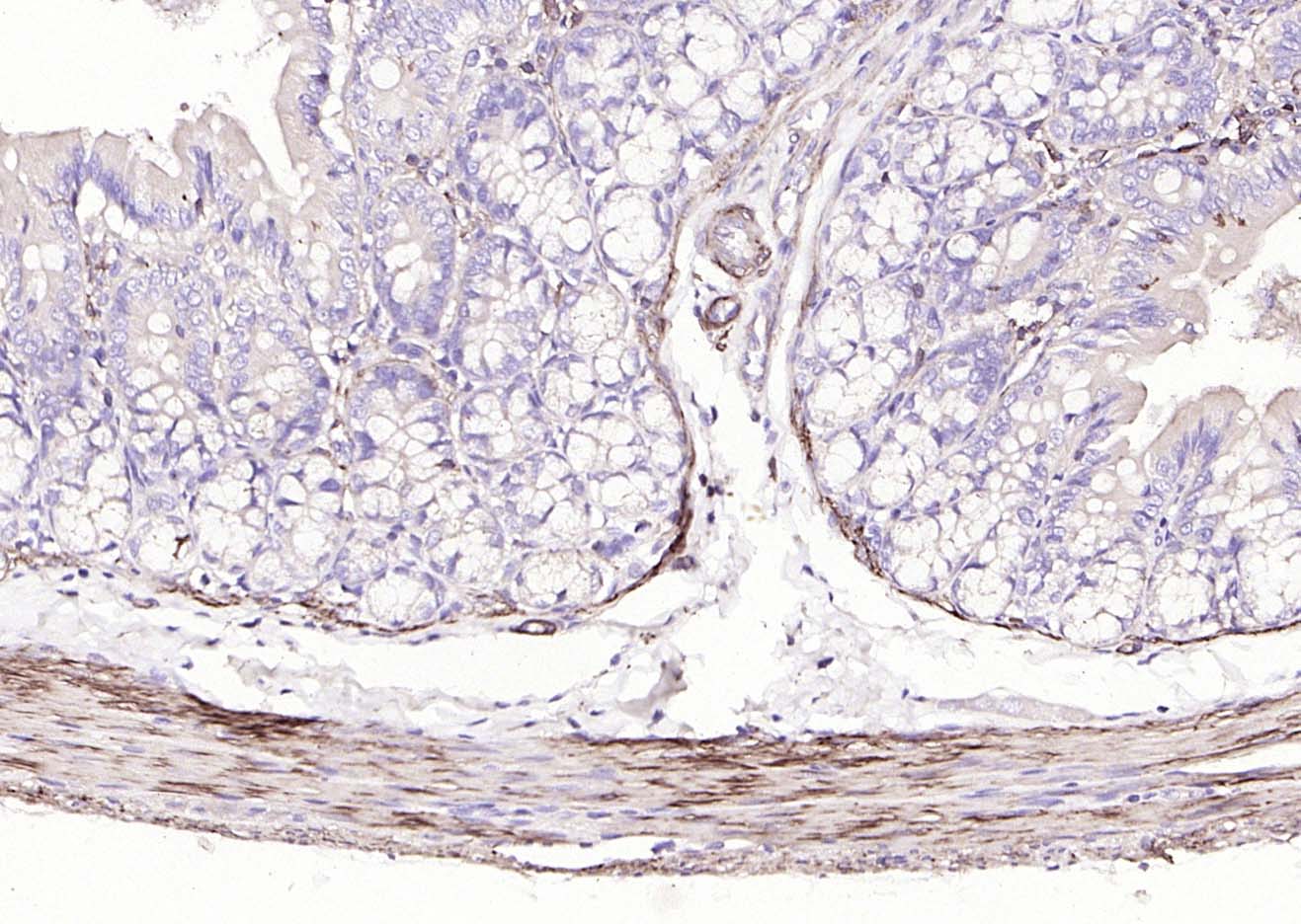
Paraformaldehyde-fixed, paraffin embedded Human Duodenum; Antigen retrieval by boiling in sodium citrate buffer (pH6.0) for 15 min; Antibody incubation with alpha smooth muscle Actin Polyclonal Antibody, Unconjugated (bs-0189R) at 1:200 overnight at 4°C, followed by conjugation to the SP Kit (Rabbit, SP-0023) and DAB (C-0010) staining.
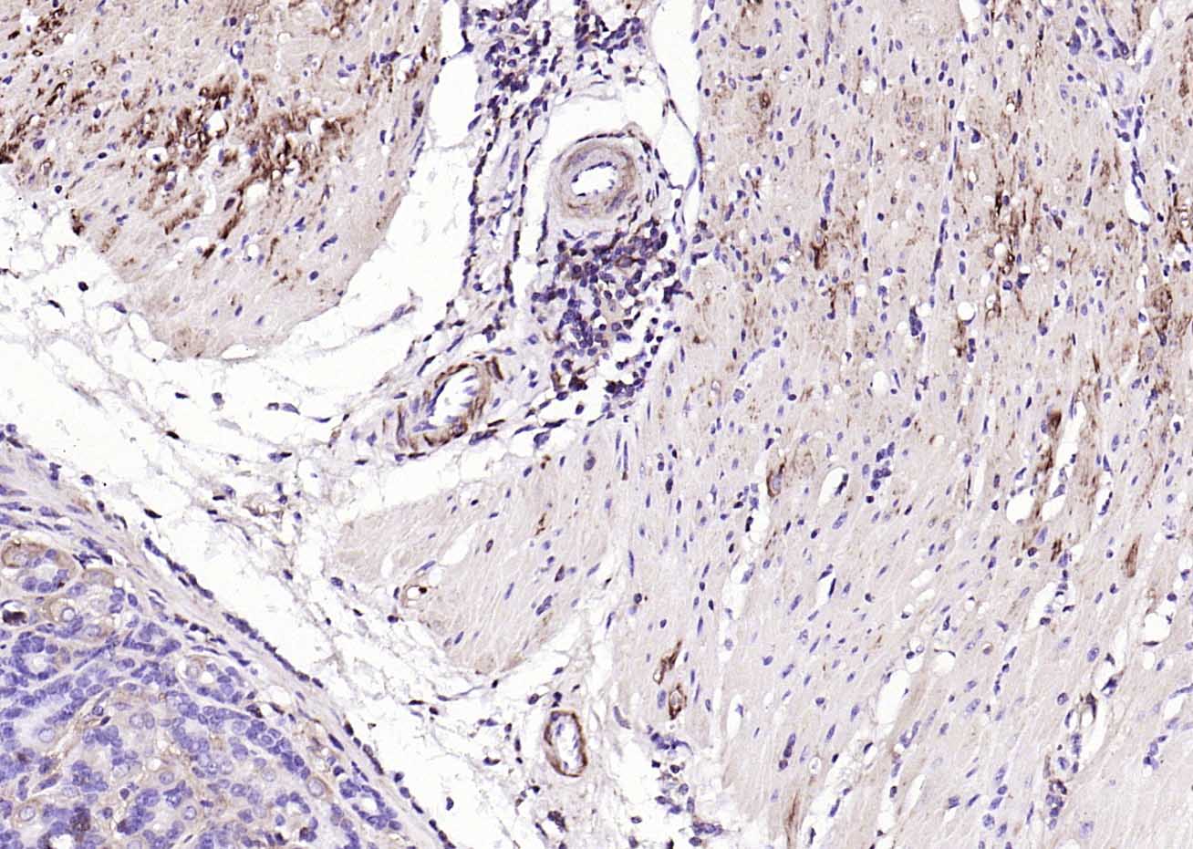
Paraformaldehyde-fixed, paraffin embedded Mouse Stomach; Antigen retrieval by boiling in sodium citrate buffer (pH6.0) for 15 min; Antibody incubation with alpha smooth muscle Actin Polyclonal Antibody, Unconjugated (bs-0189R) at 1:200 overnight at 4°C, followed by conjugation to the SP Kit (Rabbit, SP-0023) and DAB (C-0010) staining.
|
| 1、抗体溶解方法 | |
| 2、抗体修复方式 | |
| 3、常用试剂的配制 | |
| 4、免疫组化操作步骤 | |
| 5、免疫组化问题解答 | |
| 6、Western Blotting 操作步骤 | |
| 7、Western Blotting 问题解答 | |
| 8、关于肽链的设计 | |
| 9、多肽的溶解与保存 | |
| 10、酶标抗体效价测定程序 | |


