

| 产品编号 | bs-0059R |
| 英文名称 | APAF1(NT) Rabbit pAb |
| 中文名称 | 凋亡蛋白活性因子-1抗体(N端) |
| 别 名 | APAF-1; CED4; 6230400I06Rik; Apaf1l; fog; mKIAA0413; APAF_HUMAN; APAF1; KIAA0413; APAF_MOUSE; APAF_RAT; |
 |
Specific References (1) | bs-0059R has been referenced in 1 publications.
[IF=3.743] Liu F et al. Clinical and biological significances of heat shock protein 90 (Hsp90) in human nasopharyngeal carcinoma cells and anti-cancer effects of Hsp90 inhibitor. Biomed Pharmacother. 2019 Oct 18;120:109533. ICF ; Human.
|
| 研究领域 | 肿瘤 细胞生物 神经生物学 信号转导 细胞凋亡 |
| 抗体来源 | Rabbit |
| 克隆类型 | Polyclonal |
| 克 隆 号 | |
| 交叉反应 | Human,Mouse,Rat (predicted: Cow,Chicken,Dog,Horse) |
| 产品应用 | WB=1:500-2000,IHC-P=1:100-500,IHC-F=1:100-500,IF=1:100-500,Flow-Cyt=0.2ug/test,ICC/IF=1:100-500
not yet tested in other applications. optimal dilutions/concentrations should be determined by the end user. |
| 理论分子量 | 137 kDa |
| 检测分子量 | 140 |
| 细胞定位 | 细胞浆 |
| 性 状 | Liquid |
| 浓 度 | 1mg/ml |
| 免 疫 原 | KLH conjugated synthetic peptide derived from human Apaf-1: 13-80/1248 |
| 亚 型 | IgG |
| 纯化方法 | affinity purified by Protein A |
| 缓 冲 液 | 0.01M TBS (pH7.4) with 1% BSA, 0.02% Proclin300 and 50% Glycerol. |
| 保存条件 | Shipped at 4℃. Store at -20℃ for one year. Avoid repeated freeze/thaw cycles. |
| 注意事项 | This product as supplied is intended for research use only, not for use in human, therapeutic or diagnostic applications. |
| PubMed | PubMed |
| 产品介绍 |
This gene encodes a cytoplasmic protein that initiates apoptosis. This protein contains several copies of the WD-40 domain, a caspase recruitment domain (CARD), and an ATPase domain(NB-ARC). Upon binding cytochrome c and dATP, this protein forms an oligomeric apoptosome. The apoptosome binds and cleaves caspase 9 preproprotein, releasing its mature, activated form. Activated caspase 9 stimulates the subsequent caspase cascade that commits the cell to apoptosis. Alternative splicing results in several transcript variants encoding different isoforms. [provided by RefSeq, Jul 2008]. Function: Oligomeric Apaf-1 mediates the cytochrome c-dependent autocatalytic activation of pro-caspase-9 (Apaf-3), leading to the activation of caspase-3 and apoptosis. This activation requires ATP. Isoform 6 is less effective in inducing apoptosis. Subunit: Monomer. Oligomerizes upon binding of cytochrome c and dATP. Oligomeric Apaf-1 and pro-caspase-9 bind to each other via their respective NH2-terminal CARD domains and consecutively mature caspase-9 is released from the complex. Pro-caspase-3 is recruited into the Apaf-1-pro-caspase-9 complex via interaction with pro-caspase-9. Interacts with APIP. Interacts (via CARD and NACHT domains) with NAIP/BIRC1 (via NACHT domain). Subcellular Location: Cytoplasm. Tissue Specificity: Ubiquitous. Highest levels of expression in adult spleen and peripheral blood leukocytes, and in fetal brain, kidney and lung. Isoform 1 is expressed in heart, kidney and liver. Similarity: Contains 1 CARD domain. Contains 1 NB-ARC domain. Contains 13 WD repeats. SWISS: O14727 Gene ID: 317 Database links: Entrez Gene: 317 Human Entrez Gene: 11783 Mouse Omim: 602233 Human SwissProt: O14727 Human SwissProt: O88879 Mouse Unigene: 728891 Human Unigene: 220289 Mouse Unigene: 64522 Rat Apaf-1 (Apoptosis protease activating factor-1)调节细胞色素C依赖的Caspase-9原的自动催化活性,导致Caspase-3 的激活和引起凋亡。 Apaf-1在成年人的脾脏、外周血白细胞、肾脏、肺和胎儿脑、肾、肺中高水平表达。 |
| 产品图片 |
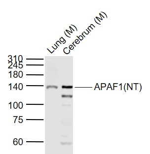
Sample:
Lane 1: Lung (Mouse) Lysate at 40 ug
Lane 2: Cerebrum (Mouse) Lysate at 40 ug
Primary: Anti-APAF1(NT) (bs-0059R) at 1/1000 dilution
Secondary: IRDye800CW Goat Anti-Rabbit IgG at 1/20000 dilution
Predicted band size: 140 kD
Observed band size: 140 kD
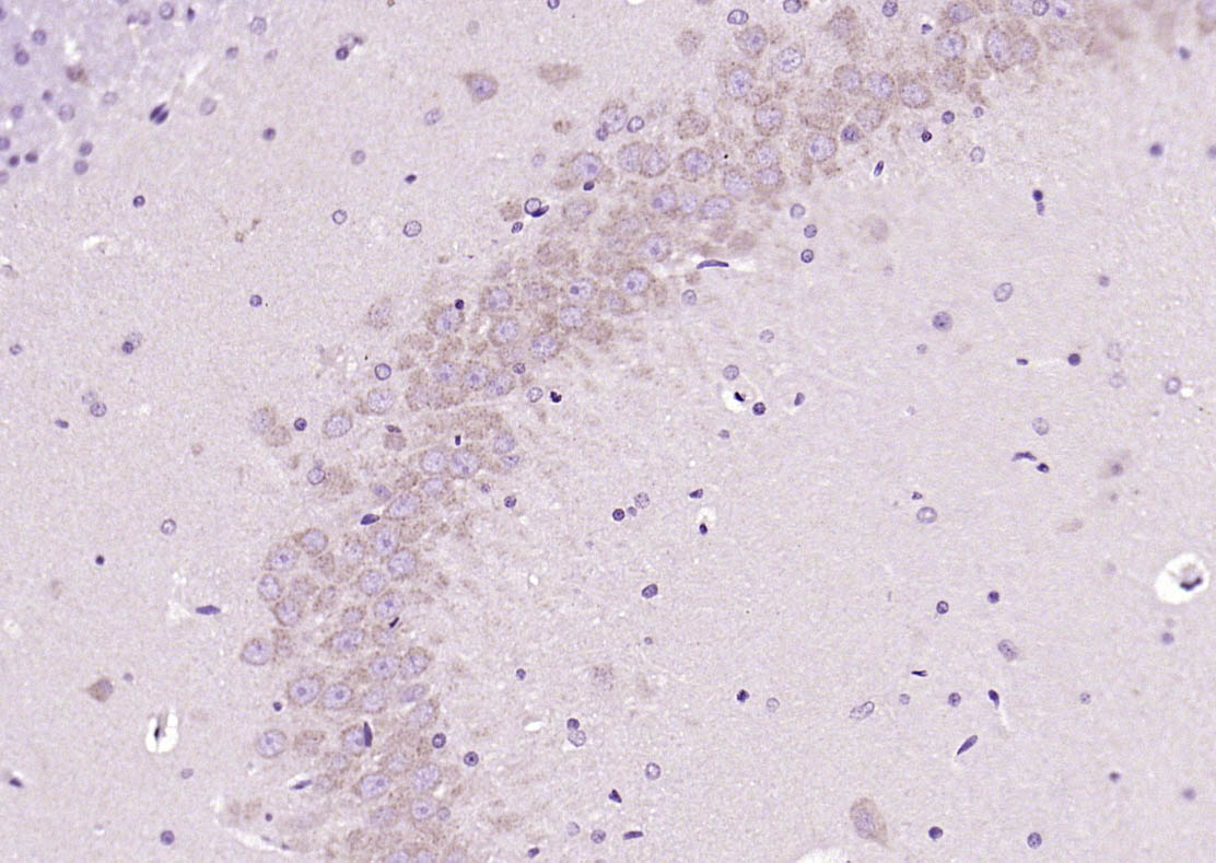
Paraformaldehyde-fixed, paraffin embedded (rat brain); Antigen retrieval by boiling in sodium citrate buffer (pH6.0) for 15min; Block endogenous peroxidase by 3% hydrogen peroxide for 20 minutes; Blocking buffer (normal goat serum) at 37°C for 30min; Antibody incubation with (APAF1(NT)) Polyclonal Antibody, Unconjugated (bs-0059R) at 1:200 overnight at 4°C, followed by operating according to SP Kit(Rabbit) (sp-0023) instructionsand DAB staining.
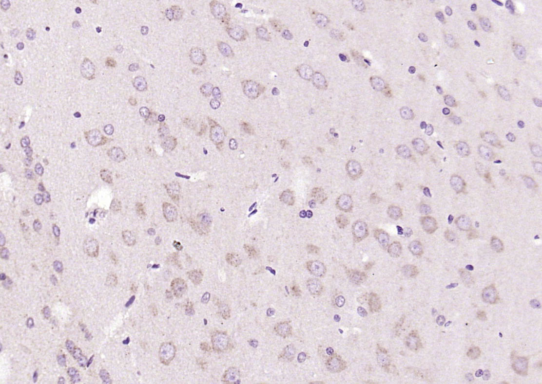
Paraformaldehyde-fixed, paraffin embedded (rat brain); Antigen retrieval by boiling in sodium citrate buffer (pH6.0) for 15min; Block endogenous peroxidase by 3% hydrogen peroxide for 20 minutes; Blocking buffer (normal goat serum) at 37°C for 30min; Antibody incubation with (APAF1(NT)) Polyclonal Antibody, Unconjugated (bs-0059R) at 1:200 overnight at 4°C, followed by operating according to SP Kit(Rabbit) (sp-0023) instructionsand DAB staining.
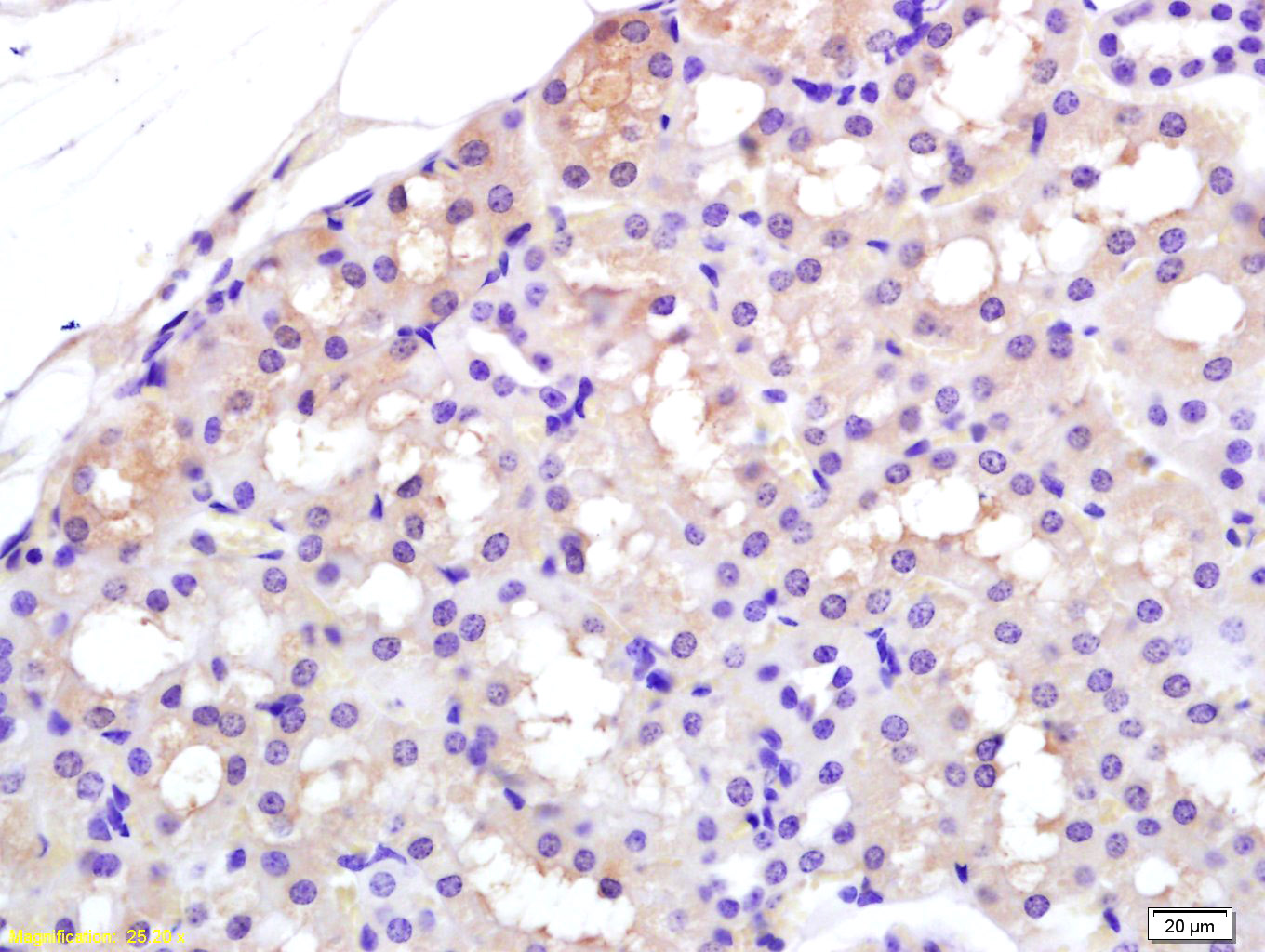
Tissue/cell: rat kidney tissue; 4% Paraformaldehyde-fixed and paraffin-embedded;
Antigen retrieval: citrate buffer ( 0.01M, pH 6.0 ), Boiling bathing for 15min; Block endogenous peroxidase by 3% Hydrogen peroxide for 30min; Blocking buffer (normal goat serum,C-0005) at 37℃ for 20 min;
Incubation: Anti-APAF1(NT) Polyclonal Antibody, Unconjugated(bs-0059R) 1:200, overnight at 4°C, followed by conjugation to the secondary antibody(SP-0023) and DAB(C-0010) staining
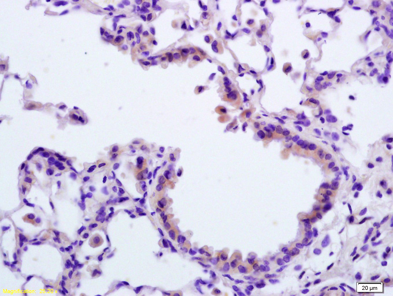
Tissue/cell: rat lung tissue; 4% Paraformaldehyde-fixed and paraffin-embedded;
Antigen retrieval: citrate buffer ( 0.01M, pH 6.0 ), Boiling bathing for 15min; Block endogenous peroxidase by 3% Hydrogen peroxide for 30min; Blocking buffer (normal goat serum,C-0005) at 37℃ for 20 min;
Incubation: Anti-APAF1(NT) Polyclonal Antibody, Unconjugated(bs-0059R) 1:200, overnight at 4°C, followed by conjugation to the secondary antibody(SP-0023) and DAB(C-0010) staining
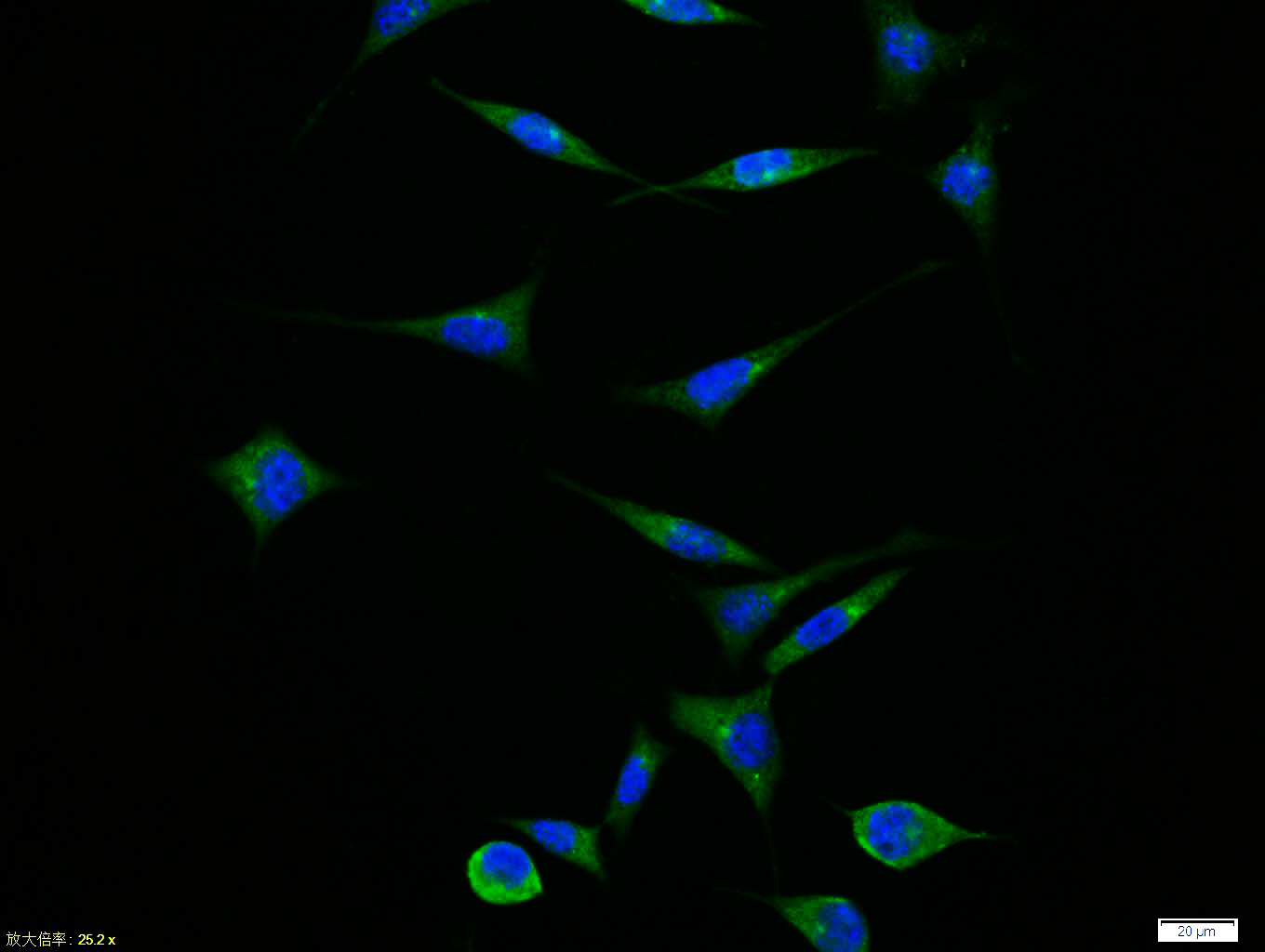
Tissue/cell:SH-SY5Y cell; 4% Paraformaldehyde-fixed; Triton X-100 at room temperature for 20 min; Blocking buffer (normal goat serum,C-0005) at 37°C for 20 min; Antibody incubation with (APAF1(NT)) polyclonal Antibody, Unconjugated (bs-0059R) 1:100, 90 minutes at 37°C; followed by a FITC conjugated Goat Anti-Rabbit IgG antibody at 37°C for 90 minutes, DAPI (blue, C02-04002) was used to stain the cell nuclei.
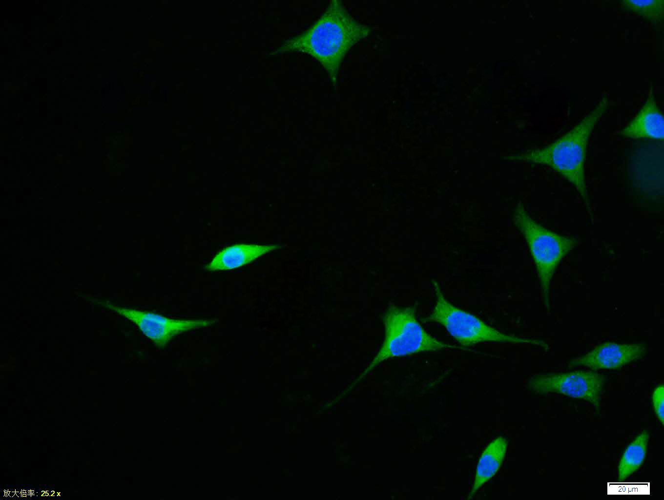
Tissue/cell:SH-SY5Y cell; 4% Paraformaldehyde-fixed; Triton X-100 at room temperature for 20 min; Blocking buffer (normal goat serum,C-0005) at 37°C for 20 min; Antibody incubation with (APAF1(NT)) polyclonal Antibody, Unconjugated (bs-0059R) 1:100, 90 minutes at 37°C; followed by a FITC conjugated Goat Anti-Rabbit IgG antibody at 37°C for 90 minutes, DAPI (blue, C02-04002) was used to stain the cell nuclei.
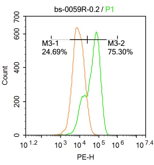
U-937 cells were fixed with 4% PFA for 10min at room temperature,permeabilized with 20% PBST for 20 min at room temperature, and incubated in 5% BSA blocking buffer for 30 min at room temperature. Cells were then stained with APAF1(NT) Antibody(bs-0059R)at 1:500 dilution in blocking buffer and incubated for 30 min at room temperature, washed twice with 2%BSA in PBS, followed by secondary antibody incubation for 40 min at room temperature. Acquisitions of 20,000 events were performed. Cells stained with primary antibody (green), and isotype control (orange).
|
| 1、抗体溶解方法 | |
| 2、抗体修复方式 | |
| 3、常用试剂的配制 | |
| 4、免疫组化操作步骤 | |
| 5、免疫组化问题解答 | |
| 6、Western Blotting 操作步骤 | |
| 7、Western Blotting 问题解答 | |
| 8、关于肽链的设计 | |
| 9、多肽的溶解与保存 | |
| 10、酶标抗体效价测定程序 | |


