

| 产品编号 | bs-0127R |
| 英文名称 | Bax Rabbit pAb |
| 中文名称 | Bax抗体 |
| 别 名 | BCL2L4; BAX_HUMAN; BAX; Bcl-2-like protein 4 (Bcl2-L-4); BAX_MOUSE; BAX_RAT; |
 |
Specific References (186) | bs-0127R has been referenced in 186 publications.
|
| 研究领域 | 细胞生物 信号转导 细胞凋亡 线粒体 |
| 抗体来源 | Rabbit |
| 克隆类型 | Polyclonal |
| 克 隆 号 | |
| 交叉反应 | Human,Mouse,Rat (predicted: Rabbit,Pig,Sheep,Cow,Dog) |
| 产品应用 | WB=1:500-2000,IHC-P=1:100-500,IHC-F=1:100-500,IF=1:100-500,Flow-Cyt=1μg /test,ICC/IF=1:50-200
not yet tested in other applications. optimal dilutions/concentrations should be determined by the end user. |
| 理论分子量 | 21 kDa |
| 检测分子量 | 20-23 |
| 细胞定位 | 细胞浆 细胞膜 线粒体 |
| 性 状 | Liquid |
| 浓 度 | 1mg/ml |
| 免 疫 原 | KLH conjugated synthetic peptide derived from human Bax: 84-175/192 |
| 亚 型 | IgG |
| 纯化方法 | affinity purified by Protein A |
| 缓 冲 液 | 0.01M TBS (pH7.4) with 1% BSA, 0.02% Proclin300 and 50% Glycerol. |
| 保存条件 | Shipped at 4℃. Store at -20℃ for one year. Avoid repeated freeze/thaw cycles. |
| 注意事项 | This product as supplied is intended for research use only, not for use in human, therapeutic or diagnostic applications. |
| PubMed | PubMed |
| 产品介绍 |
The protein encoded by this gene belongs to the BCL2 protein family. BCL2 family members form hetero- or homodimers and act as anti- or pro-apoptotic regulators that are involved in a wide variety of cellular activities. This protein forms a heterodimer with BCL2, and functions as an apoptotic activator. This protein is reported to interact with, and increase the opening of, the mitochondrial voltage-dependent anion channel (VDAC), which leads to the loss in membrane potential and the release of cytochrome c. The expression of this gene is regulated by the tumor suppressor P53 and has been shown to be involved in P53-mediated apoptosis. Multiple alternatively spliced transcript variants, which encode different isoforms, have been reported for this gene. [provided by RefSeq, Jul 2008]. Function: Accelerates programmed cell death by binding to, and antagonizing the apoptosis repressor BCL2 or its adenovirus homolog E1B 19k protein. Under stress conditions, undergoes a conformation change that causes translocation to the mitochondrion membrane, leading to the release of cytochrome c that then triggers apoptosis. Promotes activation of CASP3, and thereby apoptosis. Subunit: Homodimer. Forms higher oligomers under stress conditions. Interacts with BCL2L11. Interaction with BCL2L11 promotes BAX oligomerization and association with mitochondrial membranes, with subsequent release of cytochrome c. Forms heterodimers with BCL2, E1B 19K protein, BCL2L1 isoform Bcl-X(L), BCL2L2, MCL1 and A1. Interacts with SH3GLB1 and HN. Interacts with SFN and YWHAZ; the interaction occurs in the cytoplasm. Under stress conditions, JNK-mediated phosphorylation of SFN and YWHAZ, releases BAX to mitochondria. Isoform Sigma interacts with BCL2A1 and BCL2L1 isoform Bcl-X(L). Interacts with RNF144B, which regulates the ubiquitin-dependent stability of BAX. Interacts with CLU under stress conditions that cause a conformation change leading to BAX oligomerization and association with mitochondria. Does not interact with CLU in unstressed cells. Interacts with FAIM2/LFG2. Subcellular Location: Isoform Alpha: Mitochondrion membrane; Single-pass membrane protein. Cytoplasm. Note=Colocalizes with 14-3-3 proteins in the cytoplasm. Under stress conditions, undergoes a conformation change that causes release from JNK-phosphorylated 14-3-3 proteins and translocation to the mitochondrion membrane. Isoform Beta: Cytoplasm. Isoform Gamma: Cytoplasm. Isoform Delta: Cytoplasm (Potential). Tissue Specificity: Expressed in a wide variety of tissues. Isoform Psi is found in glial tumors. Isoform Alpha is expressed in spleen, breast, ovary, testis, colon and brain, and at low levels in skin and lung. Isoform Sigma is expressed in spleen, breast, ovary, testis, lung, colon, brain and at low levels in skin. Isoform Alpha and isoform Sigma are expressed in pro-myelocytic leukemia, histiocytic lymphoma, Burkitt's lymphoma, T-cell lymphoma, lymphoblastic leukemia, breast adenocarcinoma, ovary adenocarcinoma, prostate carcinoma, prostate adenocarcinoma, lung carcinoma, epidermoid carcinoma, small cell lung carcinoma and colon adenocarcinoma cell lines. Similarity: Belongs to the Bcl-2 family. SWISS: Q07812 Gene ID: 581 Database links: Entrez Gene: 581 Human Entrez Gene: 12028 Mouse Omim: 600040 Human SwissProt: Q07812 Human SwissProt: Q07813 Mouse Unigene: 624291 Human Unigene: 19904 Mouse Unigene: 10668 Rat 可识别分子量为21KDa的Bax蛋白,此抗体与Bax有较高特异性,且与Bc1-2及Bc1-X蛋白无交叉反应,Bax、Bc1-2和Bc1-X蛋白是凋亡调节蛋白家庭成员。与Bc1-2和Bc1-X相反,Bax蛋白的过量表达加速细胞凋亡。Bax在组织中广泛表达。 Bax与Bc1-2比值的高低可用于判断恶性肿瘤耐药及复发。此抗体用于肿瘤及细胞凋亡等方面的研究。最新的研究表明:Bax可能具有肿瘤抑制作用。 |
| 产品图片 |
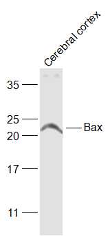
Sample:
Cerebral cortex (Rat) Lysate at 40 ug
Primary: Anti-Bax (bs-0127R) at 1/1000 dilution
Secondary: IRDye800CW Goat Anti-Rabbit IgG at 1/20000 dilution
Predicted band size: 21 kD
Observed band size: 21 kD
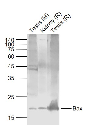
Sample:
Lane 1: Testis (Mouse) Lysate at 40 ug
Lane 2: Kidney (Rat) Lysate at 40 ug
Lane 3: Testis (Rat) Lysate at 40 ug
Primary:
Anti-Bax (bs-0127R) at 1/1000 dilution
Secondary: IRDye800CW Goat Anti-Rabbit IgG at 1/20000 dilution
Predicted band size: 21 kD
Observed band size: 21 kD
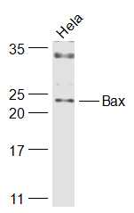
Sample:
Hela(Human) Cell Lysate at 30 ug
Primary: Anti-Bax (bs-0127R) at 1/1000 dilution
Secondary: IRDye800CW Goat Anti-Rabbit IgG at 1/20000 dilution
Predicted band size: 21 kD
Observed band size: 23 kD
Paraformaldehyde-fixed, paraffin embedded (Rat spinal cord); Antigen retrieval by boiling in sodium citrate buffer (pH6.0) for 15min; Block endogenous peroxidase by 3% hydrogen peroxide for 20 minutes; Blocking buffer (normal goat serum) at 37°C for 30min; Antibody incubation with (Bax) Polyclonal Antibody, Unconjugated (bs-0127R) at 1:400 overnight at 4°C, followed by operating according to SP Kit(Rabbit) (sp-0023) instructionsand DAB staining.
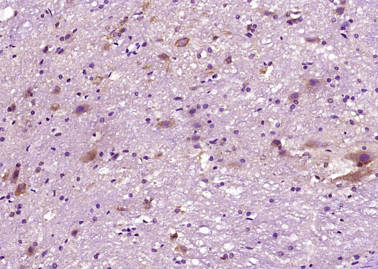
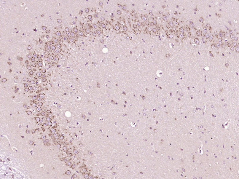
Tissue/cell: rat brain tissue; 4% Paraformaldehyde-fixed and paraffin-embedded;
Antigen retrieval: citrate buffer ( 0.01M, pH 6.0 ), Boiling bathing for 15min; Block endogenous peroxidase by 3% Hydrogen peroxide for 30min; Blocking buffer (normal goat serum,C-0005) at 37℃ for 20 min;
Incubation: Anti-Bax Polyclonal Antibody, Unconjugated(bs-0127R) 1:800, overnight at 4℃, followed by conjugation to the secondary antibody(SP-0023) and DAB(C-0010) staining
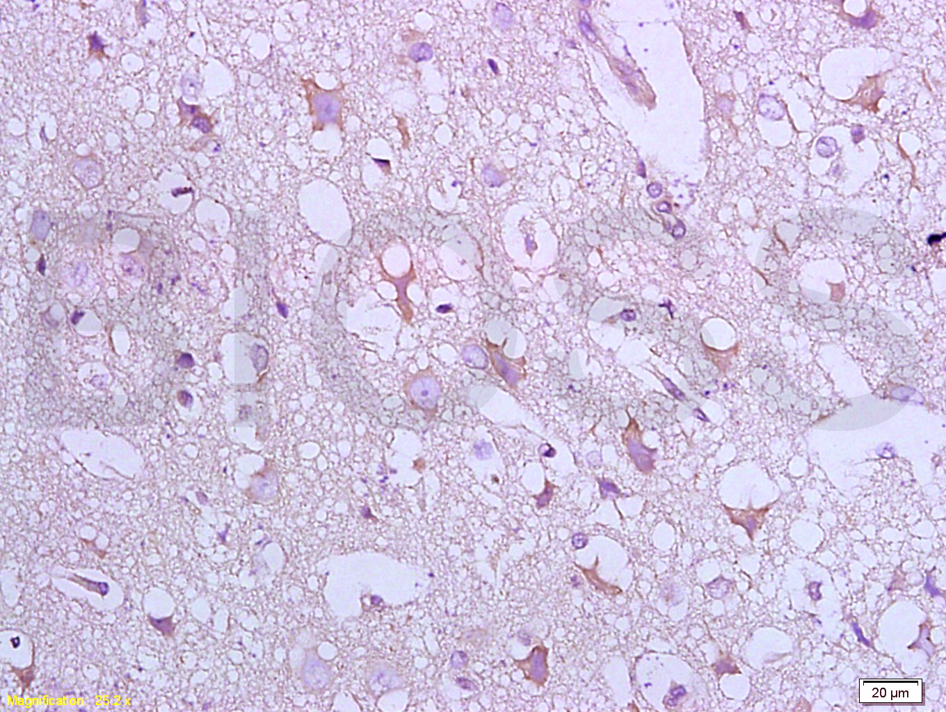
Paraformaldehyde-fixed, paraffin embedded (Rat brain); Antigen retrieval by boiling in sodium citrate buffer (pH6.0) for 15min; Block endogenous peroxidase by 3% hydrogen peroxide for 20 minutes; Blocking buffer (normal goat serum) at 37°C for 30min; Antibody incubation with (Bax) Polyclonal Antibody, Unconjugated (bs-0127R) at 1:400 overnight at 4°C, followed by operating according to SP Kit(Rabbit) (sp-0023) instructionsand DAB staining.
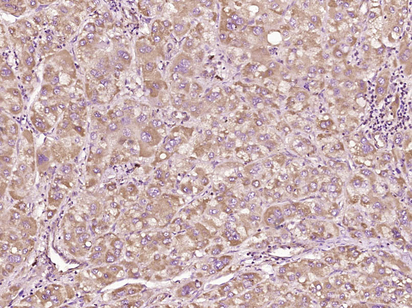
Paraformaldehyde-fixed, paraffin embedded (Human liver carcinoma); Antigen retrieval by boiling in sodium citrate buffer (pH6.0) for 15min; Block endogenous peroxidase by 3% hydrogen peroxide for 20 minutes; Blocking buffer (normal goat serum) at 37°C for 30min; Antibody incubation with (Bax) Polyclonal Antibody, Unconjugated (bs-0127R) at 1:400 overnight at 4°C, followed by operating according to SP Kit(Rabbit) (sp-0023) instructionsand DAB staining.
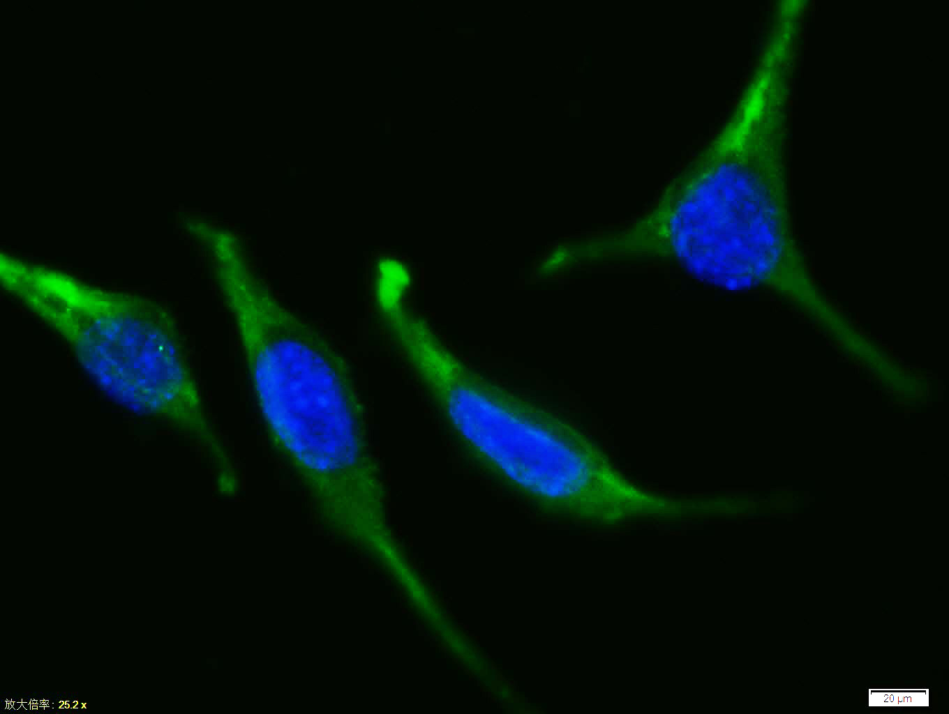
Tissue/cell:Sh-sy5y cell; 4% Paraformaldehyde-fixed; Triton X-100 at room temperature for 20 min; Blocking buffer (normal goat serum, C-0005) at 37°C for 20 min; Antibody incubation with (Bax) polyclonal Antibody, Unconjugated (bs-0127R) 1:100, 90 minutes at 37°C; followed by a FITC conjugated Goat Anti-Rabbit IgG antibody at 37°C for 90 minutes, DAPI (blue, C02-04002) was used to stain the cell nuclei.
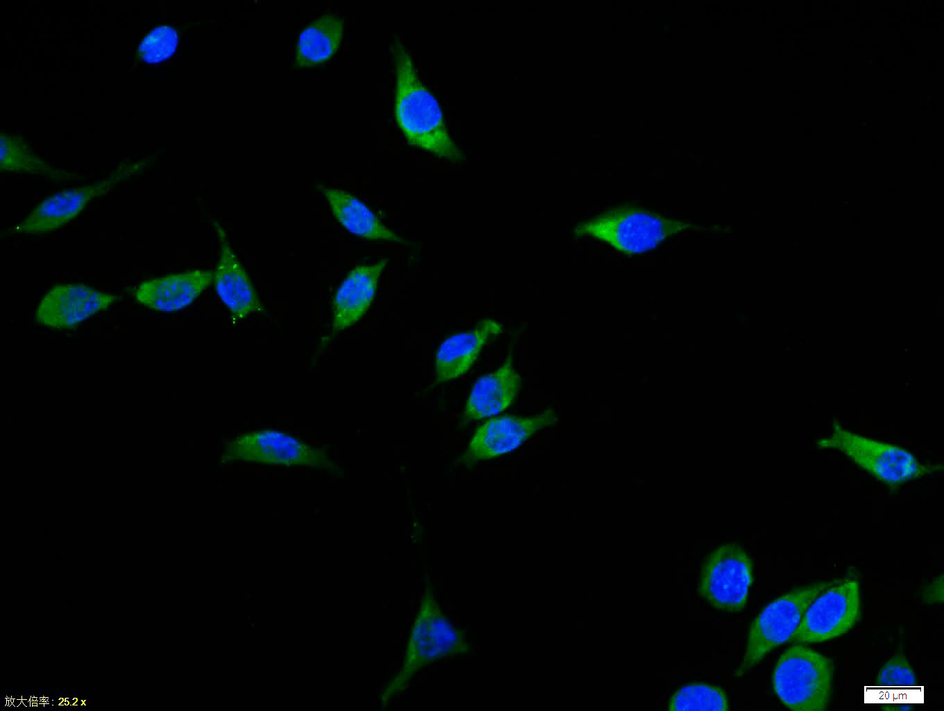
Tissue/cell:Sh-sy5y cell; 4% Paraformaldehyde-fixed; Triton X-100 at room temperature for 20 min; Blocking buffer (normal goat serum, C-0005) at 37°C for 20 min; Antibody incubation with (Bax) polyclonal Antibody, Unconjugated (bs-0127R) 1:100, 90 minutes at 37°C; followed by a FITC conjugated Goat Anti-Rabbit IgG antibody at 37°C for 90 minutes, DAPI (blue, C02-04002) was used to stain the cell nuclei.
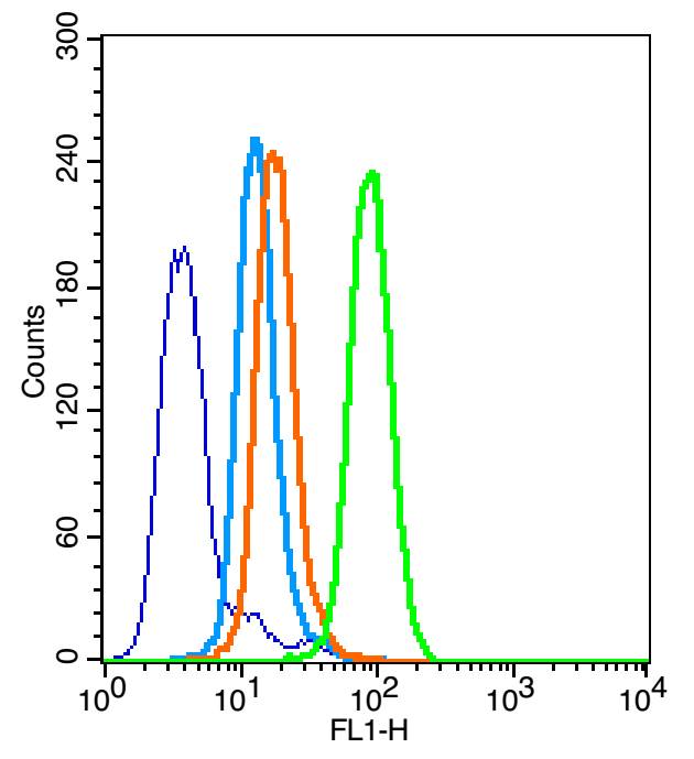
Overlay histogram showing HL 60 cells stained with bs-0127R (Green line). The cells were fixed with 90% methanol (5 min) and then permeabilized with 0.01M PBS-Tween for 20 min. The cells were then incubated in 1x PBS / 10% normal goat serum to block non-specific protein-protein interactions followed by the antibody (bs-0127R,1μg/1x10^6 cells) for 30 min at 22℃. The secondary antibody used was fluorescein isothiocyanate goat anti-rabbit IgG (H+L) (bs- 0295G-FITC , Brillant blue line) at 1/200 dilution for 30 min at 22℃. Isotype control antibody was rabbit IgG (polyclonal,bs-0295P,Orange line) (1μg/1x10^6 cells) used under the same conditions. Unlabelled sample (blue line) was also used as a control. Acquisition of 20,000 events were collected using a 20mW Argon ion laser (488nm) and 525/30 bandpass filter.
|
| 1、抗体溶解方法 | |
| 2、抗体修复方式 | |
| 3、常用试剂的配制 | |
| 4、免疫组化操作步骤 | |
| 5、免疫组化问题解答 | |
| 6、Western Blotting 操作步骤 | |
| 7、Western Blotting 问题解答 | |
| 8、关于肽链的设计 | |
| 9、多肽的溶解与保存 | |
| 10、酶标抗体效价测定程序 | |


