

| 产品编号 | bs-0032R |
| 英文名称 | Bcl-2 Rabbit pAb |
| 中文名称 | Bcl-2抗体 |
| 别 名 | Bcl-2; PPP1R50; BCL2_HUMAN; BCL2; BCL2 apoptosis regulator; B-cell CLL/lymphoma 2; BCL2, apoptosis regulator; protein phosphatase 1, regulatory subunit 50 |
 |
Specific References (162) | bs-0032R has been referenced in 162 publications.
|
| 研究领域 | 细胞生物 信号转导 细胞凋亡 细胞类型标志物 肿瘤细胞生物标志物 新陈代谢 线粒体 |
| 抗体来源 | Rabbit |
| 克隆类型 | Polyclonal |
| 交叉反应 | Human,Mouse,Rat (predicted: Chicken) |
| 产品应用 | WB=1:1000-5000,IHC-P=1:200-500,IHC-F=1:200-500,IF=1:200-500,ELISA=1:5000-10000
not yet tested in other applications. optimal dilutions/concentrations should be determined by the end user. |
| 理论分子量 | 26kDa |
| 检测分子量 | 26 |
| 细胞定位 | 细胞核 细胞浆 细胞膜 线粒体 |
| 性 状 | Liquid |
| 浓 度 | 1mg/ml |
| 免 疫 原 | KLH conjugated synthetic peptide derived from human Bcl-2: 101-160/236 |
| 亚 型 | IgG |
| 纯化方法 | affinity purified by Protein A |
| 缓 冲 液 | 0.01M TBS (pH7.4) with 1% BSA, 0.02% Proclin300 and 50% Glycerol. |
| 保存条件 | Shipped at 4℃. Store at -20℃ for one year. Avoid repeated freeze/thaw cycles. |
| 注意事项 | This product as supplied is intended for research use only, not for use in human, therapeutic or diagnostic applications. |
| PubMed | PubMed |
| 产品介绍 |
BCL2 is an integral outer mitochondrial membrane protein that blocks the apoptotic death of some cells such as lymphocytes. Constitutive expression of BCL2, such as in the case of translocation of BCL2 to Ig heavy chain locus, is thought to be the cause of follicular lymphoma. Two transcript variants (alpha and beta) produced by alternate splicing, differ in their C-terminal ends.
BCL2 suppresses apoptosis in a variety of cell systems including factor-dependent lymphohematopoietic and neural cells. It regulates cell death by controlling the mitochondrial membrane permeability. It appears to function in a feedback loop system with caspases. BCL2 inhibits caspase activity either by preventing the release of cytochrome c from the mitochondria and/or by binding to the apoptosis-activating factor (APAF1). It can form homodimers, and heterodimers with BAX, BAD, BAK and BclX(L). Heterodimerization with BAX requires intact BH1 and BH2 domains, and is necessary for anti-apoptotic activity. Also interacts with APAF1, RAF1, TP53BP2, BBC3, BCL2L1 and BNIPL. Function: Suppresses apoptosis in a variety of cell systems including factor-dependent lymphohematopoietic and neural cells. Regulates cell death by controlling the mitochondrial membrane permeability. Appears to function in a feedback loop system with caspases. Inhibits caspase activity either by preventing the release of cytochrome c from the mitochondria and/or by binding to the apoptosis-activating factor (APAF-1). Subunit: Forms homodimers, and heterodimers with BAX, BAD, BAK and Bcl-X(L). Heterodimerization with BAX requires intact BH1 and BH2 motifs, and is necessary for anti-apoptotic activity. Interacts with EI24 (By similarity). Also interacts with APAF1, BBC3, BCL2L1, BNIPL, MRPL41 and TP53BP2. Binding to FKBP8 seems to target BCL2 to the mitochondria and probably interferes with the binding of BCL2 to its targets. Interacts with BAG1 in an ATP-dependent manner. Interacts with RAF1 (the 'Ser-338' and 'Ser-339' phosphorylated form). Interacts (via the BH4 domain) with EGLN3; the interaction prevents the formation of the BAX-BCL2 complex and inhibits the anti-apoptotic activity of BCL2. Interacts with G0S2; this interaction also prevents the formation of the anti-apoptotic BAX-BCL2 complex. Subcellular Location: Mitochondrion outer membrane; Single-pass membrane protein. Nucleus membrane; Single-pass membrane protein. Endoplasmic reticulum membrane; Single-pass membrane protein. Tissue Specificity: Expressed in a variety of tissues. Post-translational modifications: Phosphorylation/dephosphorylation on Ser-70 regulates anti-apoptotic activity. Growth factor-stimulated phosphorylation on Ser-70 by PKC is required for the anti-apoptosis activity and occurs during the G2/M phase of the cell cycle. In the absence of growth factors, BCL2 appears to be phosphorylated by other protein kinases such as ERKs and stress-activated kinases. Phosphorylated by MAPK8/JNK1 at Thr-69, Ser-70 and Ser-87, wich stimulates starvation-induced autophagy. Dephosphorylated by protein phosphatase 2A (PP2A). Proteolytically cleaved by caspases during apoptosis. The cleaved protein, lacking the BH4 motif, has pro-apoptotic activity, causes the release of cytochrome c into the cytosol promoting further caspase activity. Monoubiquitinated by PARK2, leading to increase its stability. DISEASE: Note=A chromosomal aberration involving BCL2 has been found in chronic lymphatic leukemia. Translocation t(14;18)(q32;q21) with immunoglobulin gene regions. BCL2 mutations found in non-Hodgkin lymphomas carrying the chromosomal translocation could be attributed to the Ig somatic hypermutation mechanism resulting in nucleotide transitions. Similarity: Belongs to the Bcl-2 family. SWISS: P10415 Gene ID: 596 Database links: Entrez Gene: 596 Human Entrez Gene: 12043 Mouse Omim: 151430 Human SwissProt: P10415 Human SwissProt: P10417 Mouse Unigene: 150749 Human Unigene: 257460 Mouse Unigene: 9996 Rat Bcl-2基因是指B-cell lymphoma gene。人体滤泡B细胞淋巴瘤中过量表达的原癌基因。由于染色体t(14;18)易位,将Bcl-2基因置于免疫球蛋白重链的转录调控下,使其表达失控。在细胞系中其过量表达能延长细胞存活期而不诱导细胞增殖。它是哺乳动物中细胞调亡的抑制基因。参与细胞凋亡的调控。肿瘤中的Bcl-2基因可提高侵润性瘤细胞的生存能力。主要用于滤胞型淋巴瘤、毛细管性白血病及细胞凋亡等方面的研究。 目前研究认为:Bcl-2也是细胞凋亡的一种抑制因子、参与细胞凋亡调控,可以用于各种恶性肿瘤的细胞凋亡的研究。 |
| 产品图片 |
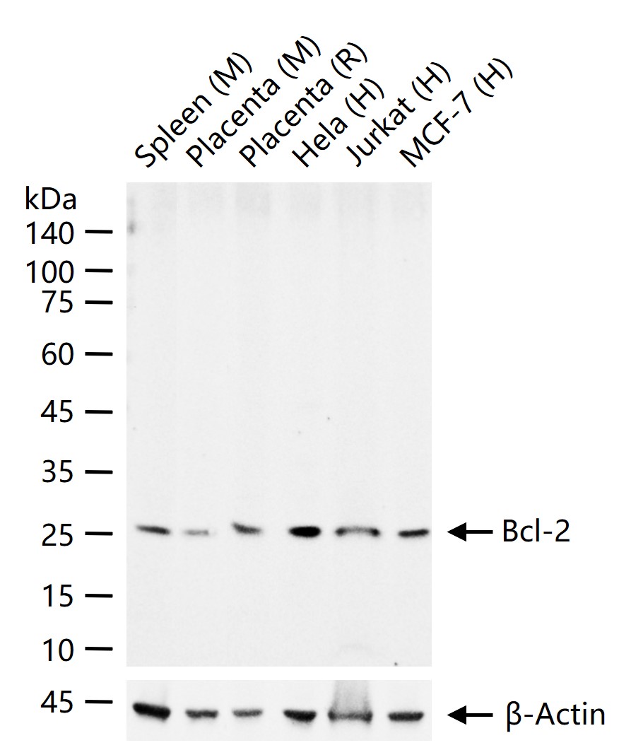
25 ug total protein per lane of various lysates (see on figure) probed with Bcl-2 polyclonal antibody, unconjugated (bs-0032R) at 1:1000 dilution and 4°C overnight incubation. Followed by conjugated secondary antibody incubation at r.t. for 60 min.
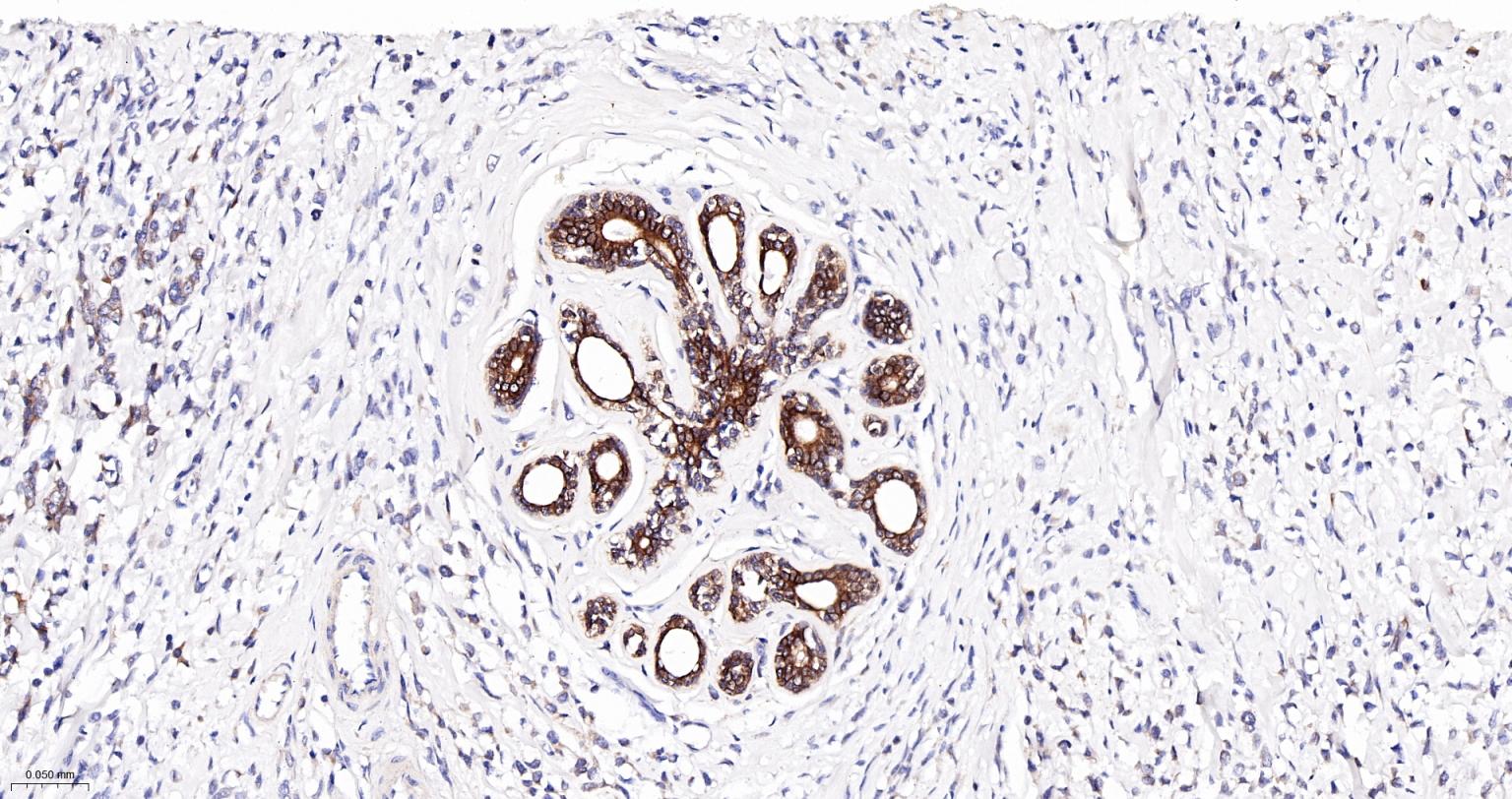
Paraformaldehyde-fixed, paraffin embedded Human Breast Cancer; Antigen retrieval by boiling in sodium citrate buffer (pH6.0) for 15 min; The section was incubated with Bcl-2 Polyclonal Antibody, Unconjugated (bs-0032R) at 1:200 overnight at 4°C, followed by conjugation to the bs-0295G-HRP and DAB (C-0010) staining.
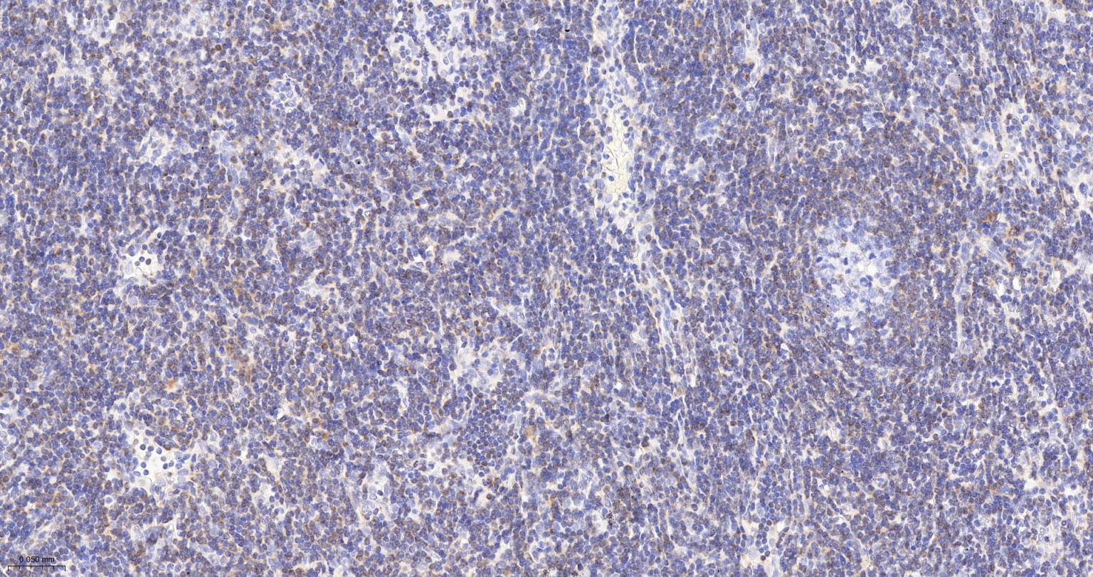
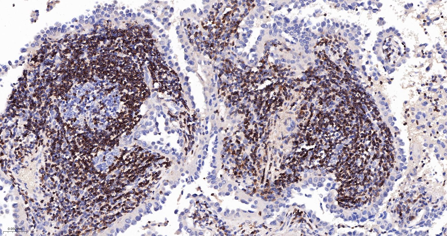
Paraformaldehyde-fixed, paraffin embedded Human Lymph; Antigen retrieval by boiling in sodium citrate buffer (pH6.0) for 15 min; The section was incubated with Bcl-2 Polyclonal Antibody, Unconjugated (bs-0032R) at 1:200 overnight at 4°C, followed by conjugation to the bs-0295G-HRP and DAB (C-0010) staining.
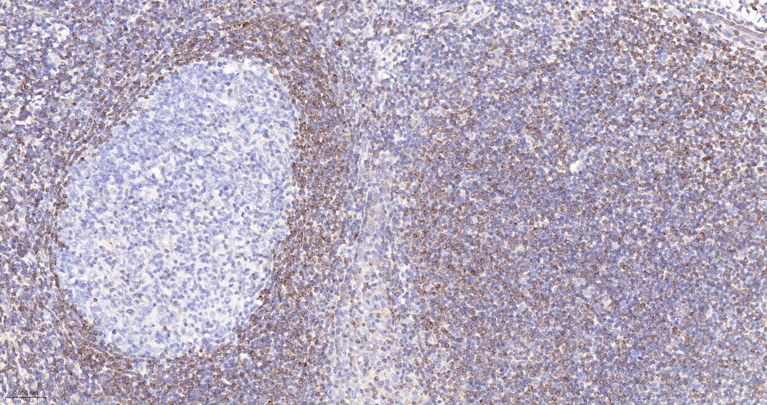
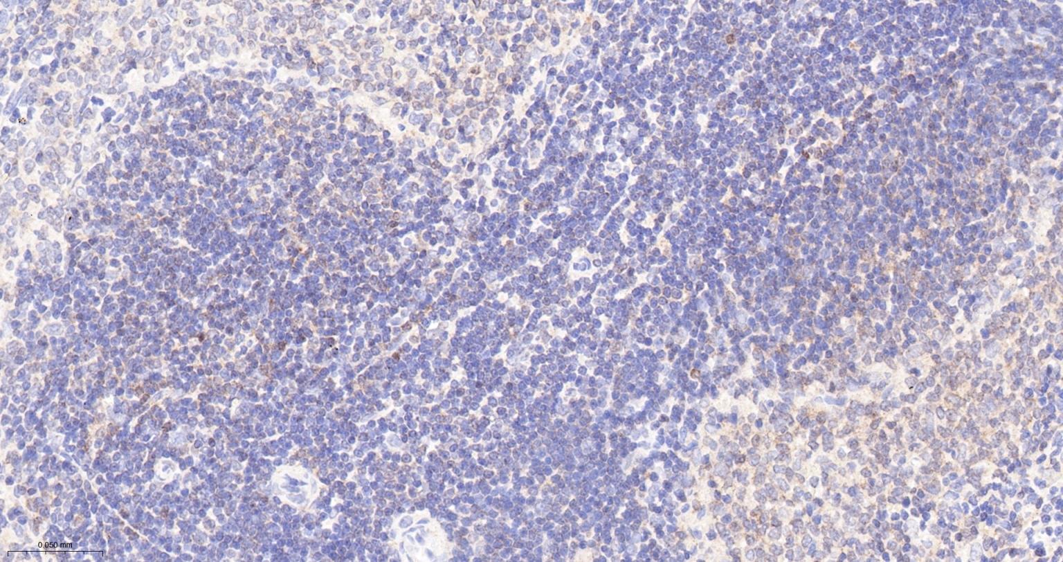
Paraformaldehyde-fixed, paraffin embedded Human Tonsil; Antigen retrieval by boiling in sodium citrate buffer (pH6.0) for 15 min; The section was incubated with Bcl-2 Polyclonal Antibody, Unconjugated (bs-0032R) at 1:200 overnight at 4°C, followed by conjugation to the bs-0295G-HRP and DAB (C-0010) staining.
Paraformaldehyde-fixed, paraffin embedded Human Lung Cancer; Antigen retrieval by boiling in sodium citrate buffer (pH6.0) for 15 min; The section was incubated with Bcl-2 Polyclonal Antibody, Unconjugated (bs-0032R) at 1:200 overnight at 4°C, followed by conjugation to the bs-0295G-HRP and DAB (C-0010) staining.
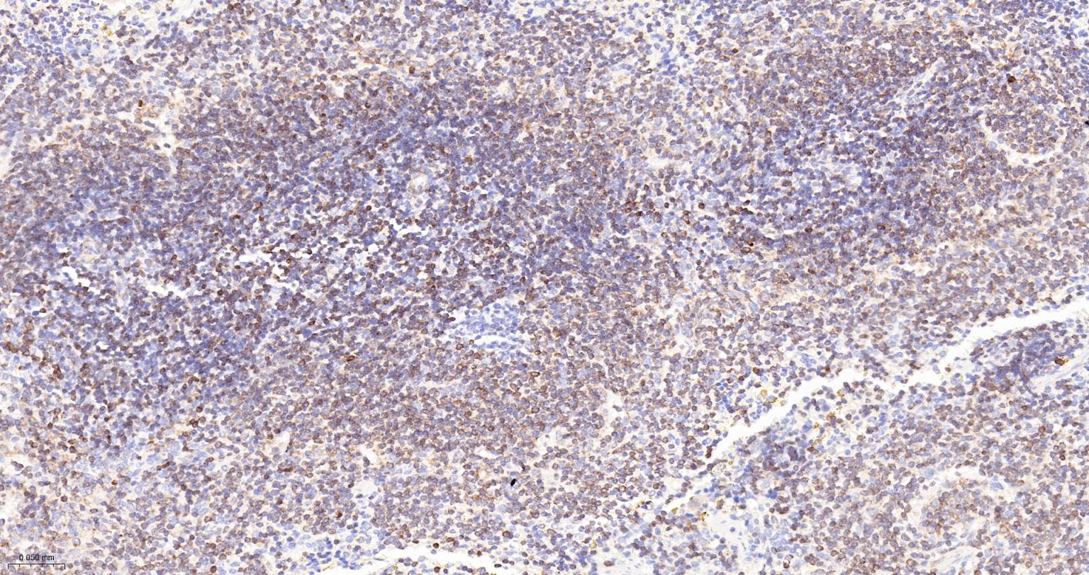
Paraformaldehyde-fixed, paraffin embedded Rat Spleen; Antigen retrieval by boiling in sodium citrate buffer (pH6.0) for 15 min; The section was incubated with Bcl-2 Polyclonal Antibody, Unconjugated (bs-0032R) at 1:200 overnight at 4°C, followed by conjugation to the bs-0295G-HRP and DAB (C-0010) staining.
Paraformaldehyde-fixed, paraffin embedded Mouse Spleen; Antigen retrieval by boiling in sodium citrate buffer (pH6.0) for 15 min; The section was incubated with Bcl-2 Polyclonal Antibody, Unconjugated (bs-0032R) at 1:200 overnight at 4°C, followed by conjugation to the bs-0295G-HRP and DAB (C-0010) staining.
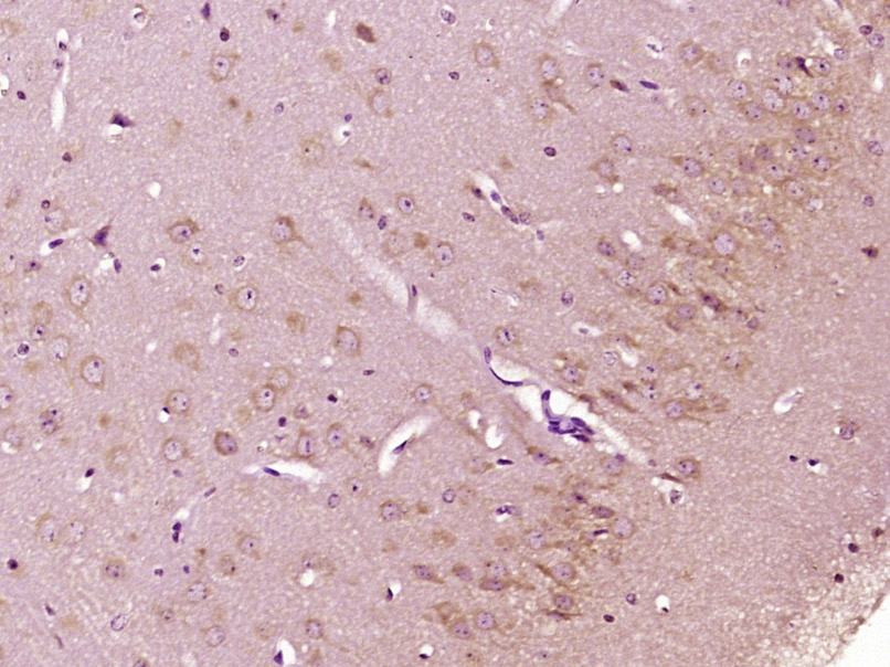
Paraformaldehyde-fixed, paraffin embedded (Mouse brain); Antigen retrieval by boiling in sodium citrate buffer (pH6.0) for 15min; Block endogenous peroxidase by 3% hydrogen peroxide for 20 minutes; Blocking buffer (normal goat serum) at 37°C for 30min; Antibody incubation with (Bcl-2) Polyclonal Antibody, Unconjugated (bs-0032R) at 1:400 overnight at 4°C, followed by operating according to SP Kit(Rabbit) (sp-0023) instructionsand DAB staining.
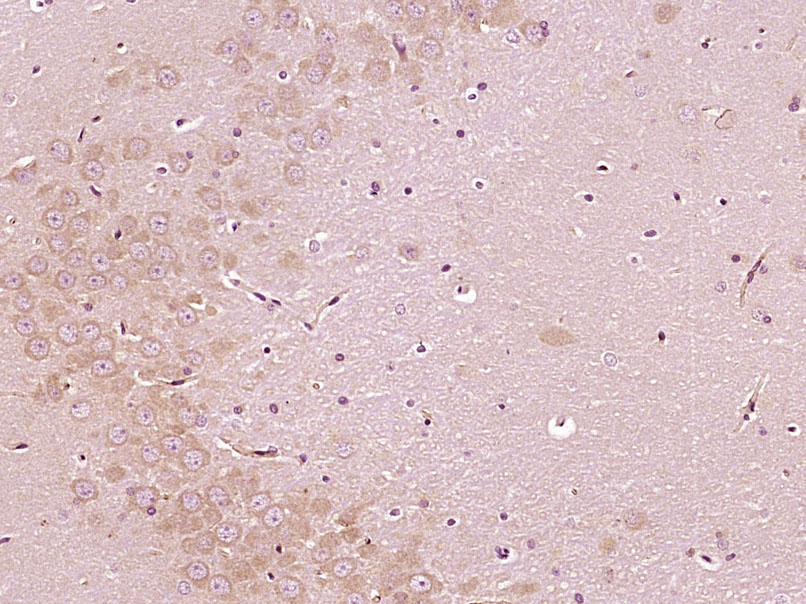
Paraformaldehyde-fixed, paraffin embedded (Rat brain); Antigen retrieval by boiling in sodium citrate buffer (pH6.0) for 15min; Block endogenous peroxidase by 3% hydrogen peroxide for 20 minutes; Blocking buffer (normal goat serum) at 37°C for 30min; Antibody incubation with (Bcl-2) Polyclonal Antibody, Unconjugated (bs-0032R) at 1:400 overnight at 4°C, followed by operating according to SP Kit(Rabbit) (sp-0023) instructionsand DAB staining.
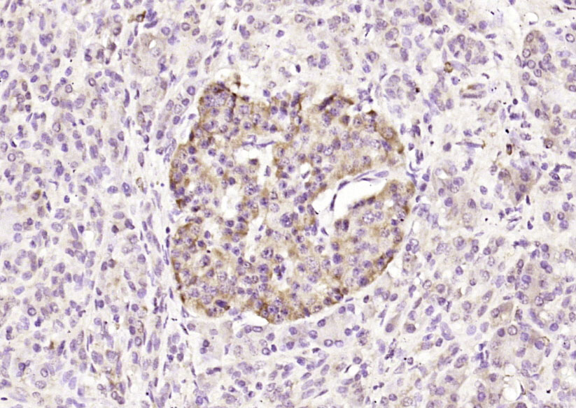
Paraformaldehyde-fixed, paraffin embedded (human pancreatic cancer); Antigen retrieval by boiling in sodium citrate buffer (pH6.0) for 15min; Block endogenous peroxidase by 3% hydrogen peroxide for 20 minutes; Blocking buffer (normal goat serum) at 37°C for 30min; Antibody incubation with (Bcl-2) Polyclonal Antibody, Unconjugated (bs-0032R) at 1:200 overnight at 4°C, followed by operating according to SP Kit(Rabbit) (sp-0023) instructionsand DAB staining.
|
| 1、抗体溶解方法 | |
| 2、抗体修复方式 | |
| 3、常用试剂的配制 | |
| 4、免疫组化操作步骤 | |
| 5、免疫组化问题解答 | |
| 6、Western Blotting 操作步骤 | |
| 7、Western Blotting 问题解答 | |
| 8、关于肽链的设计 | |
| 9、多肽的溶解与保存 | |
| 10、酶标抗体效价测定程序 | |


