

| 产品编号 | bs-0512R |
| 英文名称 | CD2AP Rabbit pAb |
| 中文名称 | 白细胞分化抗原CD2AP抗体 |
| 别 名 | CMS; METS-1; Mets1; CD2AP_HUMAN; CD2AP; Adapter protein CMS; Cas ligand with multiple SH3 domains; CD2AP_MOUSE; Mesenchyme-to-epithelium transition protein with SH3 domains 1 (METS-1); CD2 associated protein; Cas ligand with multiple Src homology (SH) 3 domains |
 |
Specific References (4) | bs-0512R has been referenced in 4 publications.
[IF=5.195] Lifeng Wei. et al. Shenqi granule upregulates CD2AP and α-actinin4 and activates autophagy through regulation of mTOR/ULK1 pathway in MPC5 cells. J ETHNOPHARMACOL. 2023 Mar;303:115942 WB ; Mouse.
[IF=3.098] Xinying Zhang. et al. Exogenous spermine attenuates diabetic kidney injury in rats by inhibiting AMPK/mTOR signaling pathway. Int J Mol Med. 2021 Mar;47(3):1-1 WB ; Rat.
[IF=2.518] Derya Karabulut. et al. A different perspective on the filtration barrier after kidney stone formation: An immunohistochemical and biochemical study. 2020 Nov 05 IHC ; Rat.
[IF=2.064] Jiang J et al. Erzhi Formula Extracts Reverse Renal Injury in Diabetic Nephropathy Rats by Protecting the Renal Podocytes.
Evid Based Complement Alternat Med. 2018 Aug 23;2018:1741924. WB ; Rat.
|
| 研究领域 | 信号转导 转运蛋白 结合蛋白 |
| 抗体来源 | Rabbit |
| 克隆类型 | Polyclonal |
| 交叉反应 | Mouse,Rat (predicted: Human,Rabbit,Pig,Sheep,Cow,Chicken,Dog,GuineaPig,Horse) |
| 产品应用 | WB=1:500-2000,IHC-P=1:100-500,IHC-F=1:100-500,IF=1:100-500
not yet tested in other applications. optimal dilutions/concentrations should be determined by the end user. |
| 理论分子量 | 71kDa |
| 检测分子量 | 77 |
| 细胞定位 | 细胞浆 |
| 性 状 | Liquid |
| 浓 度 | 1mg/ml |
| 免 疫 原 | KLH conjugated synthetic peptide derived from human CD2AP: 561-639/639 |
| 亚 型 | IgG |
| 纯化方法 | affinity purified by Protein A |
| 缓 冲 液 | 0.01M TBS (pH7.4) with 1% BSA, 0.02% Proclin300 and 50% Glycerol. |
| 保存条件 | Shipped at 4℃. Store at -20℃ for one year. Avoid repeated freeze/thaw cycles. |
| 注意事项 | This product as supplied is intended for research use only, not for use in human, therapeutic or diagnostic applications. |
| PubMed | PubMed |
| 产品介绍 |
This gene encodes a scaffolding molecule that regulates the actin cytoskeleton. The protein directly interacts with filamentous actin and a variety of cell membrane proteins through multiple actin binding sites, SH3 domains, and a proline-rich region containing binding sites for SH3 domains. The cytoplasmic protein localizes to membrane ruffles, lipid rafts, and the leading edges of cells. It is implicated in dynamic actin remodeling and membrane trafficking that occurs during receptor endocytosis and cytokinesis. Haploinsufficiency of this gene is implicated in susceptibility to glomerular disease. [provided by RefSeq, Jul 2008]. Function: Seems to act as an adapter protein between membrane proteins and the actin cytoskeleton. May play a role in receptor clustering and cytoskeletal polarity in the junction between T-cell and antigen-presenting cell. May anchor the podocyte slit diaphragm to the actin cytoskeleton in renal glomerolus. Also required for cytokinesis. Subunit: Self-associates. Homodimer (Potential). Interacts with F-actin, PKD2, NPHS1 and NPHS2. Interacts with WTIP. Interacts with DDN; interaction is direct. Interacts (via SH3 2 domain) with CBL (via phosphorylated C-terminus). Interacts with BCAR1/p130Cas (via SH3 domain). Interacts with MVB12A and ARHGAP17. Interacts with ANLN, CD2 and CBLB. Interacts with PDCD6IP and TSG101. Interacts with RIN3. Subcellular Location: Cytoplasm, cytoskeleton. Cell projection, ruffle. Note=Colocalizes with F-actin and BCAR1/p130Cas in membrane ruffles. Located at podocyte slit diaphragm between podocyte foot processes. During late anaphase and telophase, concentrates in the vicinity of the midzone microtubules and in the midbody in late telophase. Tissue Specificity: Widely expressed in fetal and adult tissues. Post-translational modifications: Phosphorylated on tyrosine residues; probably by c-Abl, Fyn and c-Src. DISEASE: Focal segmental glomerulosclerosis 3 (FSGS3) [MIM:607832]: A renal pathology defined by the presence of segmental sclerosis in glomeruli and resulting in proteinuria, reduced glomerular filtration rate and progressive decline in renal function. Renal insufficiency often progresses to end-stage renal disease, a highly morbid state requiring either dialysis therapy or kidney transplantation. Note=Disease susceptibility is associated with variations affecting the gene represented in this entry. Similarity: Contains 3 SH3 domains. SWISS: Q9Y5K6 Gene ID: 23607 Database links: Entrez Gene: 23607 Human Entrez Gene: 12488 Mouse Omim: 604241 Human SwissProt: Q9Y5K6 Human SwissProt: Q9JLQ0 Mouse Unigene: 485518 Human Unigene: 218637 Mouse Unigene: 212220 Rat CD2AP可能作为细胞裂孔隔膜分子与细胞骨架的连接蛋白,在细胞分化即增殖的过程中发挥重要作用。 目前主用用于肾脏功能方面的研究,D2AP不仅参与T细胞的活化,而且对肾脏功能起着至关重要的作用,其异常表达可能是引起人类肾脏疾病的诱因之一。 |
| 产品图片 |
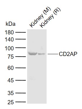
Sample:
Lane 1: Mouse Kidney tissue lysates
Lane 2: Rat Kidney tissue lysates
Primary: Anti-CD2AP (bs-0512R) at 1/1000 dilution
Secondary: IRDye800CW Goat Anti-Rabbit IgG at 1/20000 dilution
Predicted band size: 71 kDa
Observed band size: 77 kDa
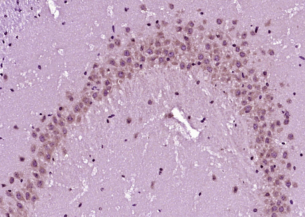
Paraformaldehyde-fixed, paraffin embedded (Mouse brain); Antigen retrieval by boiling in sodium citrate buffer (pH6.0) for 15min; Block endogenous peroxidase by 3% hydrogen peroxide for 20 minutes; Blocking buffer (normal goat serum) at 37°C for 30min; Antibody incubation with (CD2AP) Polyclonal Antibody, Unconjugated (bs-0512R) at 1:400 overnight at 4°C, followed by operating according to SP Kit(Rabbit) (sp-0023) instructionsand DAB staining.
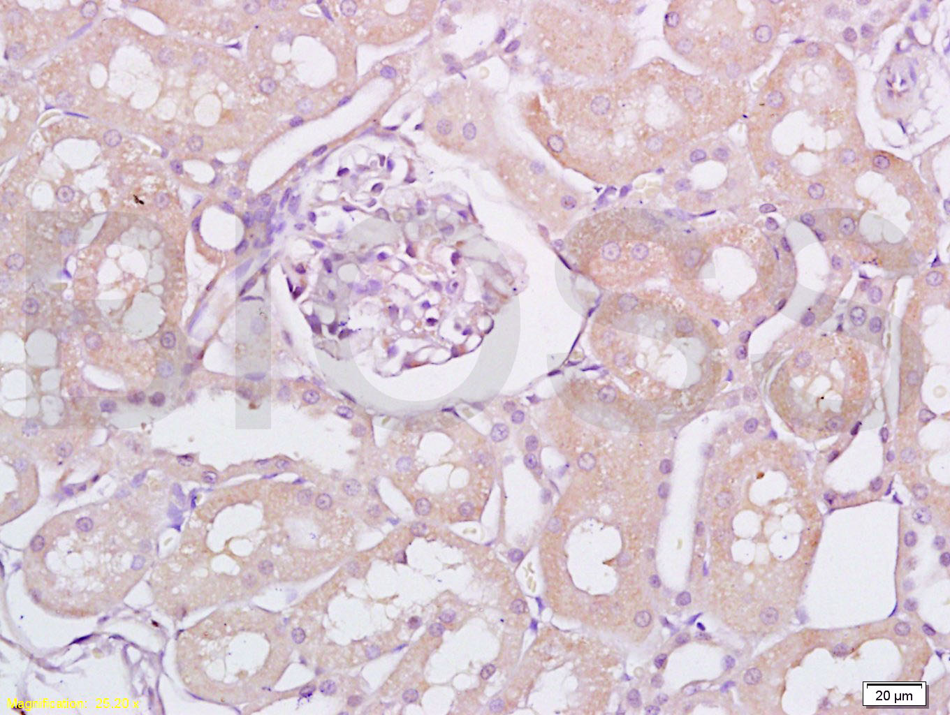
Tissue/cell: rat kidney tissue; 4% Paraformaldehyde-fixed and paraffin-embedded;
Antigen retrieval: citrate buffer ( 0.01M, pH 6.0 ), Boiling bathing for 15min; Block endogenous peroxidase by 3% Hydrogen peroxide for 30min; Blocking buffer (normal goat serum,C-0005) at 37℃ for 20 min;
Incubation: Anti-CD2AP Polyclonal Antibody, Unconjugated(bs-0512R) 1:200, overnight at 4°C, followed by conjugation to the secondary antibody(SP-0023) and DAB(C-0010) staining
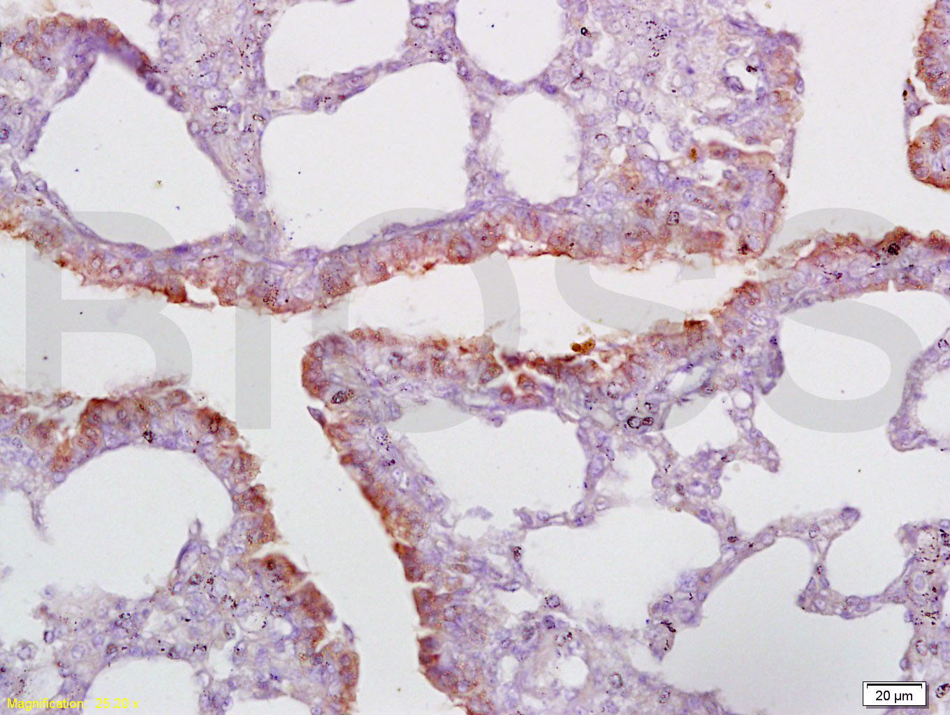
Tissue/cell: rat lung tissue; 4% Paraformaldehyde-fixed and paraffin-embedded;
Antigen retrieval: citrate buffer ( 0.01M, pH 6.0 ), Boiling bathing for 15min; Block endogenous peroxidase by 3% Hydrogen peroxide for 30min; Blocking buffer (normal goat serum,C-0005) at 37℃ for 20 min;
Incubation: Anti-CD2AP Polyclonal Antibody, Unconjugated(bs-0512R) 1:200, overnight at 4°C, followed by conjugation to the secondary antibody(SP-0023) and DAB(C-0010) staining
|
| 1、抗体溶解方法 | |
| 2、抗体修复方式 | |
| 3、常用试剂的配制 | |
| 4、免疫组化操作步骤 | |
| 5、免疫组化问题解答 | |
| 6、Western Blotting 操作步骤 | |
| 7、Western Blotting 问题解答 | |
| 8、关于肽链的设计 | |
| 9、多肽的溶解与保存 | |
| 10、酶标抗体效价测定程序 | |


