

| 产品编号 | bs-0569R |
| 英文名称 | CDK7 Rabbit pAb |
| 中文名称 | 周期素依赖性激酶7抗体 |
| 别 名 | CAK; CAK1; CDKN7; HCAK; MO15; STK1; p39MO15; CDK7_HUMAN; CDK7; 39 kDa protein kinase (p39 Mo15); CDK-activating kinase 1; Cell division protein kinase 7; Serine/threonine-protein kinase 1; TFIIH basal transcription factor complex kinase subunit; 2.7.11.22; 2.7.11.23; cyclin dependent kinase 7; cyclin-dependent kinase 7 (homolog of Xenopus MO15 cdk-activating kinase); cyclin-dependent kinase 7 (MO15 homolog, Xenopus laevis, cdk-activating kinase) |
| 研究领域 | 肿瘤 细胞生物 染色质和核信号 信号转导 细胞周期蛋白 激酶和磷酸酶 |
| 抗体来源 | Rabbit |
| 克隆类型 | Polyclonal |
| 交叉反应 | Human,Mouse,Rat (predicted: Cow) |
| 产品应用 | WB=1:500-2000,IHC-P=1:100-500,IHC-F=1:100-500,IF=1:100-500,ICC/IF=1:100-500
not yet tested in other applications. optimal dilutions/concentrations should be determined by the end user. |
| 理论分子量 | 40kDa |
| 检测分子量 | 40 |
| 细胞定位 | 细胞核 细胞浆 |
| 性 状 | Liquid |
| 浓 度 | 1mg/ml |
| 免 疫 原 | KLH conjugated synthetic peptide derived from human CDK7: 1-80/346 |
| 亚 型 | IgG |
| 纯化方法 | affinity purified by Protein A |
| 缓 冲 液 | 0.01M TBS (pH7.4) with 1% BSA, 0.02% Proclin300 and 50% Glycerol. |
| 保存条件 | Shipped at 4℃. Store at -20℃ for one year. Avoid repeated freeze/thaw cycles. |
| 注意事项 | This product as supplied is intended for research use only, not for use in human, therapeutic or diagnostic applications. |
| PubMed | PubMed |
| 产品介绍 |
The protein encoded by this gene is a member of the cyclin-dependent protein kinase (CDK) family. CDK family members are highly similar to the gene products of Saccharomyces cerevisiae cdc28, and Schizosaccharomyces pombe cdc2, and are known to be important regulators of cell cycle progression. This protein forms a trimeric complex with cyclin H and MAT1, which functions as a Cdk-activating kinase (CAK). It is an essential component of the transcription factor TFIIH, that is involved in transcription initiation and DNA repair. This protein is thought to serve as a direct link between the regulation of transcription and the cell cycle. [provided by RefSeq, Jul 2008] Function: Serine/threonine kinase involved in cell cycle control and in RNA polymerase II-mediated RNA transcription. Cyclin-dependent kinases (CDKs) are activated by the binding to a cyclin and mediate the progression through the cell cycle. Each different complex controls a specific transition between 2 subsequent phases in the cell cycle. Required for both activation and complex formation of CDK1/cyclin-B during G2-M transition, and for activation of CDK2/cyclins during G1-S transition (but not complex formation). CDK7 is the catalytic subunit of the CDK-activating kinase (CAK) complex. Phosphorylates SPT5/SUPT5H, SF1/NR5A1, POLR2A, p53/TP53, CDK1, CDK2, CDK4, CDK6 and CDK11B/CDK11. CAK activates the cyclin-associated kinases CDK1, CDK2, CDK4 and CDK6 by threonine phosphorylation, thus regulating cell cycle progression. CAK complexed to the core-TFIIH basal transcription factor activates RNA polymerase II by serine phosphorylation of the repetitive C-terminus domain (CTD) of its large subunit (POLR2A), allowing its escape from the promoter and elongation of the transcripts. Phosphorylation of POLR2A in complex with DNA promotes transcription initiation by triggering dissociation from DNA. Its expression and activity are constant throughout the cell cycle. Upon DNA damage, triggers p53/TP53 activation by phosphorylation, but is inactivated in turn by p53/TP53; this feedback loop may lead to an arrest of the cell cycle and of the transcription, helping in cell recovery, or to apoptosis. Required for DNA-bound peptides-mediated transcription and cellular growth inhibition. Subunit: Associates primarily with cyclin-H (CCNH) and MAT1 to form the CAK complex. CAK can further associate with the core-TFIIH to form the TFIIH basal transcription factor; this complex is sensitive to UV light. The CAK complex binds to p53/TP53 in response to DNA damage. Interacts with CDK2, SF1/NR5A1, PUF60 and PRKCI. Subcellular Location: Nucleus. Cytoplasm. Cytoplasm, perinuclear region. Note=Colocalizes with PRKCI in the cytoplasm and nucleus. Translocates from the nucleus to cytoplasm and perinuclear region in response to DNA-bound peptides. Tissue Specificity: Ubiquitous. Post-translational modifications: Phosphorylation of Ser-164 during mitosis inactivates the enzyme. Phosphorylation of Thr-170 is required for activity. Phosphorylated at Ser-164 and Thr-170 by CDK2. Similarity: Belongs to the protein kinase superfamily. CMGC Ser/Thr protein kinase family. CDC2/CDKX subfamily. Contains 1 protein kinase domain. SWISS: P50613 Gene ID: 1022 Database links: Entrez Gene: 1022 Human Entrez Gene: 12572 Mouse Omim: 601955 Human SwissProt: P50613 Human SwissProt: Q03147 Mouse Unigene: 184298 Human Unigene: 259718 Mouse Unigene: 98896 Rat |
| 产品图片 |
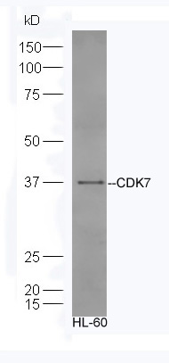
Sample:HL-60 Cell lysate;
Primary:Anti-CDK7 (bs-0569R) at 1:300;
Secondary: HRP conjugated Goat-Anti-rabbit IgG(bs-0295G-HRP) at 1: 5000;
Predicted band size:40 kD
Observed band size:40 kD
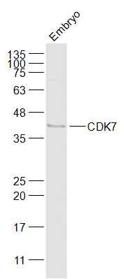
Sample:
Embryo (Mouse) Lysate at 40 ug
Primary: Anti-CDK7 (bs-0569R) at 1/1000 dilution
Secondary: IRDye800CW Goat Anti-Rabbit IgG at 1/20000 dilution
Predicted band size: 40 kD
Observed band size: 40 kD
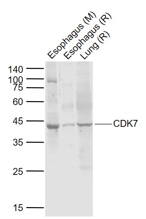
Sample:
Lane 1: Esophagus (Mouse) Lysate at 40 ug
Lane 2: Esophagus (Rat) Lysate at 40 ug
Lane 3: Lung (Rat) Lysate at 40 ug
Primary: Anti-CDK7 (bs-0569R) at 1/1000 dilution
Secondary: IRDye800CW Goat Anti-Rabbit IgG at 1/20000 dilution
Predicted band size: 40 kD
Observed band size: 40 kD

Paraformaldehyde-fixed, paraffin embedded (Human breast carcinoma); Antigen retrieval by boiling in sodium citrate buffer (pH6.0) for 15min; Block endogenous peroxidase by 3% hydrogen peroxide for 20 minutes; Blocking buffer (normal goat serum) at 37°C for 30min; Antibody incubation with (CDK7) Polyclonal Antibody, Unconjugated (bs-0569R) at 1:400 overnight at 4°C, followed by operating according to SP Kit(Rabbit) (sp-0023) instructionsand DAB staining.
Paraformaldehyde-fixed, paraffin embedded (rat lung); Antigen retrieval by boiling in sodium citrate buffer (pH6.0) for 15min; Block endogenous peroxidase by 3% hydrogen peroxide for 20 minutes; Blocking buffer (normal goat serum) at 37°C for 30min; Antibody incubation with (CDK7) Polyclonal Antibody, Unconjugated (bs-0569R) at 1:200 overnight at 4°C, followed by operating according to SP Kit(Rabbit) (sp-0023) instructionsand DAB staining.
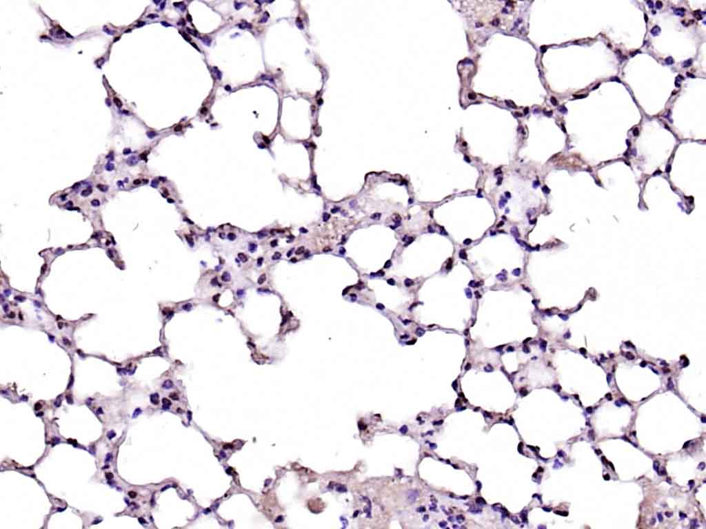
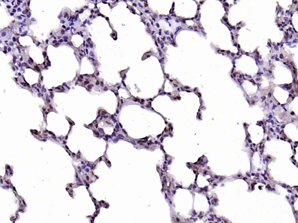
Paraformaldehyde-fixed, paraffin embedded (mouse lung); Antigen retrieval by boiling in sodium citrate buffer (pH6.0) for 15min; Block endogenous peroxidase by 3% hydrogen peroxide for 20 minutes; Blocking buffer (normal goat serum) at 37°C for 30min; Antibody incubation with (CDK7) Polyclonal Antibody, Unconjugated (bs-0569R) at 1:200 overnight at 4°C, followed by operating according to SP Kit(Rabbit) (sp-0023) instructionsand DAB staining.
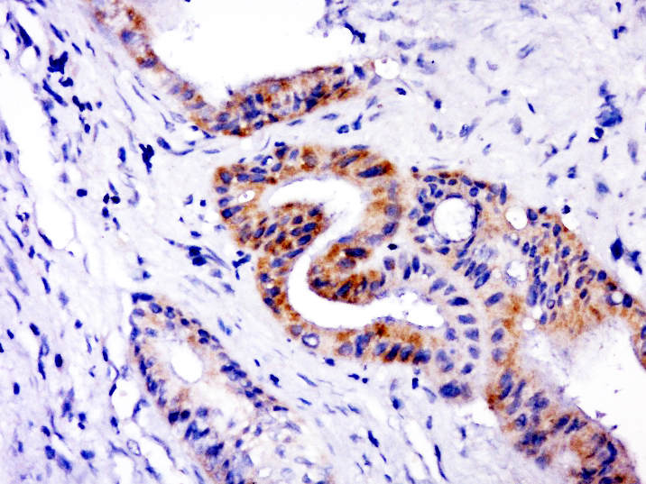
Paraformaldehyde-fixed, paraffin embedded (human cervical carcinoma); Antigen retrieval by boiling in sodium citrate buffer (pH6.0) for 15min; Block endogenous peroxidase by 3% hydrogen peroxide for 20 minutes; Blocking buffer (normal goat serum) at 37°C for 30min; Antibody incubation with (CDK7) Polyclonal Antibody, Unconjugated (bs-0569R) at 1:400 overnight at 4°C, followed by a conjugated secondary (sp-0023) for 20 minutes and DAB staining.
Hela cell; 4% Paraformaldehyde-fixed; Triton X-100 at room temperature for 20 min; Blocking buffer (normal goat serum, C-0005) at 37°C for 20 min; Antibody incubation with (CDK7) polyclonal Antibody, Unconjugated (bs-0569R) 1:100, 90 minutes at 37°C; followed by a conjugated Goat Anti-Rabbit IgG antibody at 37°C for 90 minutes, DAPI (blue, C02-04002) was used to stain the cell nuclei.

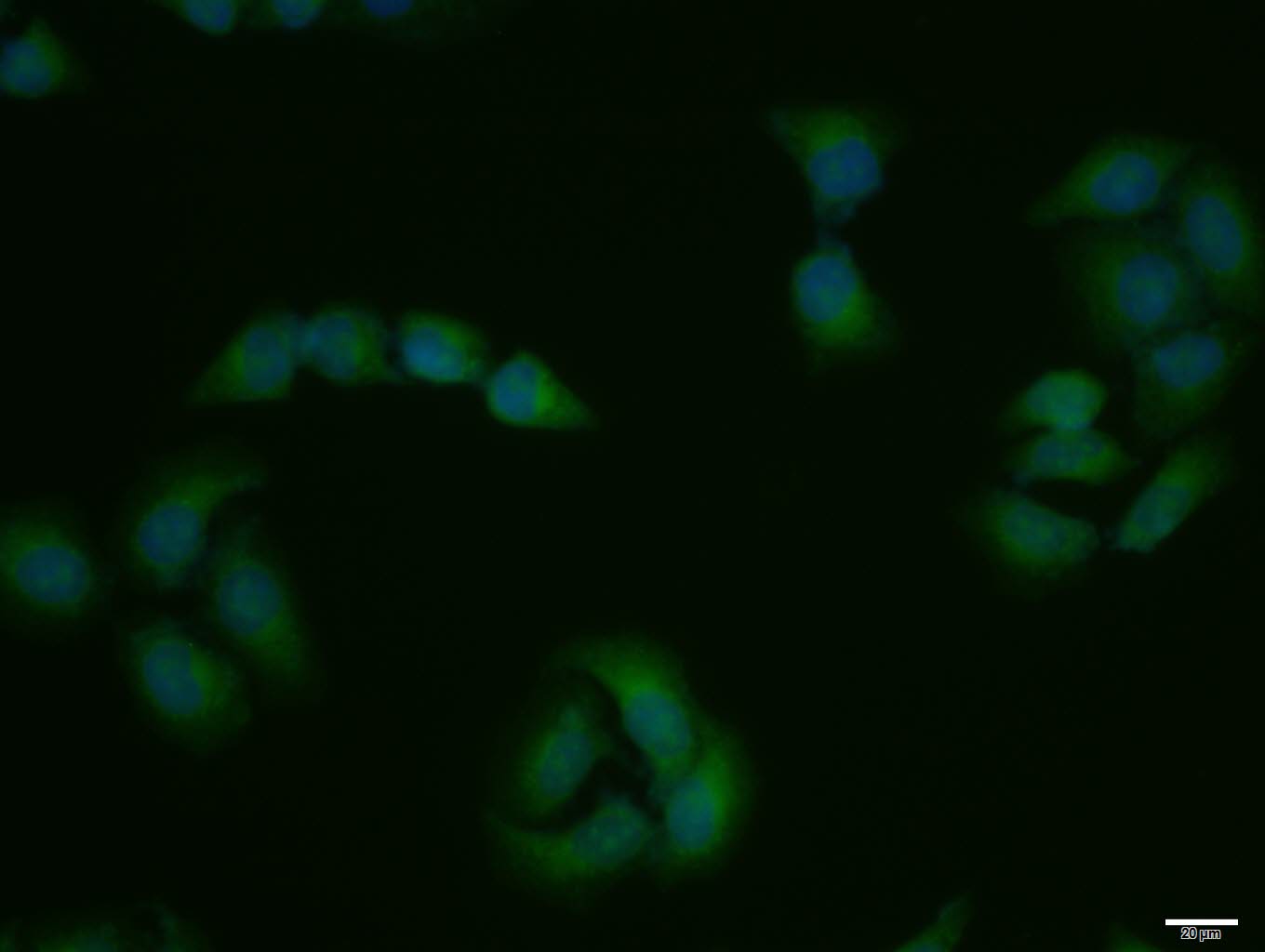
HepG2 cell; 4% Paraformaldehyde-fixed; Triton X-100 at room temperature for 20 min; Blocking buffer (normal goat serum, C-0005) at 37°C for 20 min; Antibody incubation with (CDK7) polyclonal Antibody, Unconjugated (bs-0569R) 1:100, 90 minutes at 37°C; followed by a conjugated Goat Anti-Rabbit IgG antibody at 37°C for 90 minutes, DAPI (blue, C02-04002) was used to stain the cell nuclei.
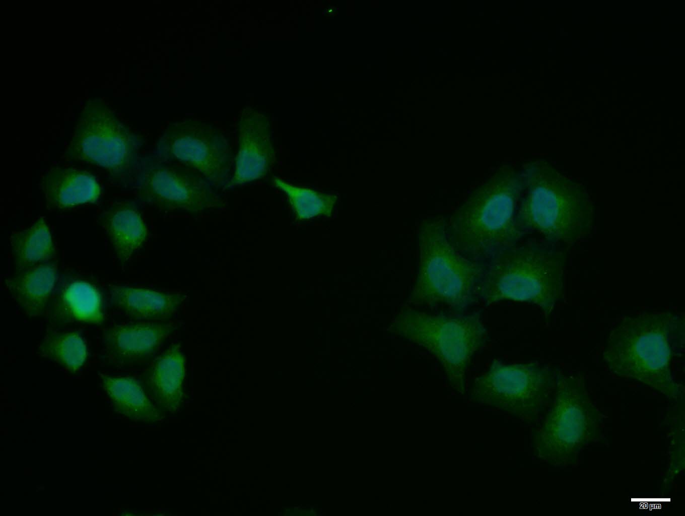
Hela cell; 4% Paraformaldehyde-fixed; Triton X-100 at room temperature for 20 min; Blocking buffer (normal goat serum, C-0005) at 37°C for 20 min; Antibody incubation with (CDK7) polyclonal Antibody, Unconjugated (bs-0569R) 1:100, 90 minutes at 37°C; followed by a conjugated Goat Anti-Rabbit IgG antibody at 37°C for 90 minutes, DAPI (blue, C02-04002) was used to stain the cell nuclei.
|
| 1、抗体溶解方法 | |
| 2、抗体修复方式 | |
| 3、常用试剂的配制 | |
| 4、免疫组化操作步骤 | |
| 5、免疫组化问题解答 | |
| 6、Western Blotting 操作步骤 | |
| 7、Western Blotting 问题解答 | |
| 8、关于肽链的设计 | |
| 9、多肽的溶解与保存 | |
| 10、酶标抗体效价测定程序 | |


