

| 产品编号 | bs-0623R |
| 英文名称 | Cyclin D1 Rabbit pAb |
| 中文名称 | 周期素D1抗体 |
| 别 名 | BCL1; D11S287E; PRAD1; U21B31; CycD1; Cyl-1; bcl-1; cD1; CCND1_HUMAN; CCND1; B-cell lymphoma 1 protein (BCL-1); BCL-1 oncogene; PRAD1 oncogene; CCND1_MOUSE; cyclin D1; cyclin D1 (PRAD1: parathyroid adenomatosis 1); parathyroid adenomatosis 1; B-cell CLL/lymphoma 1; G1/S-specific cyclin D1 |
 |
Specific References (72) | bs-0623R has been referenced in 72 publications.
|
| 研究领域 | 肿瘤 细胞生物 染色质和核信号 细胞周期蛋白 表观遗传学 |
| 抗体来源 | Rabbit |
| 克隆类型 | Polyclonal |
| 交叉反应 | Human,Mouse,Rat |
| 产品应用 | WB=1:500-2000,IHC-P=1:50-200,IHC-F=1:50-200,IF=1:50-200,Flow-Cyt=1:50-100,ICC/IF=1:50-200
not yet tested in other applications. optimal dilutions/concentrations should be determined by the end user. |
| 理论分子量 | 32kDa |
| 检测分子量 | 34 |
| 细胞定位 | 细胞核 细胞浆 细胞膜 |
| 性 状 | Liquid |
| 浓 度 | 1mg/ml |
| 免 疫 原 | KLH conjugated synthetic peptide derived from human Cyclin D1: 61-110/295 |
| 亚 型 | IgG |
| 纯化方法 | affinity purified by Protein A |
| 缓 冲 液 | 0.01M TBS (pH7.4) with 1% BSA, 0.02% Proclin300 and 50% Glycerol. |
| 保存条件 | Shipped at 4℃. Store at -20℃ for one year. Avoid repeated freeze/thaw cycles. |
| 注意事项 | This product as supplied is intended for research use only, not for use in human, therapeutic or diagnostic applications. |
| PubMed | PubMed |
| 产品介绍 |
The protein encoded by this gene belongs to the highly conserved cyclin family, whose members are characterized by a dramatic periodicity in protein abundance throughout the cell cycle. Cyclins function as regulators of CDK kinases. Different cyclins exhibit distinct expression and degradation patterns which contribute to the temporal coordination of each mitotic event. This cyclin forms a complex with and functions as a regulatory subunit of CDK4 or CDK6, whose activity is required for cell cycle G1/S transition. This protein has been shown to interact with tumor suppressor protein Rb and the expression of this gene is regulated positively by Rb. Mutations, amplification and overexpression of this gene, which alters cell cycle progression, are observed frequently in a variety of tumors and may contribute to tumorigenesis. [provided by RefSeq, Jul 2008]. Function: Regulatory component of the cyclin D1-CDK4 (DC) complexthat phosphorylates and inhibits members of the retinoblastoma (RB)protein family including RB1 and regulates the cell-cycle duringG(1)/S transition. Phosphorylation of RB1 allows dissociation ofthe transcription factor E2F from the RB/E2F complex and thesubsequent transcription of E2F target genes which are responsiblefor the progression through the G(1) phase. Hypophosphorylates RB1in early G(1) phase. Cyclin D-CDK4 complexes are major integratorsof various mitogenenic and antimitogenic signals. Also substratefor SMAD3, phosphorylating SMAD3 in a cell-cycle-dependent mannerand repressing its transcriptional activity. Component of theternary complex, cyclin D1/CDK4/CDKN1B, required for nucleartranslocation and activity of the cyclin D-CDK4 complex. Subunit: Interacts with FBXO4. Interacts witheither CDK4 or CDK6 protein kinase to form a serine/threoninekinase holoenzyme complex. The cyclin subunit imparts substratespecificity to the complex. Component of the ternary complexCCND1/CDK4/CDKN1B required for nuclear translocation and modulationof CDK4-mediated kinase activity. Interacts directly with CDKN1B.Interacts with UHRF2; the interaction ubiquitinates CCND1 andappears to occur independently of phosphorylation. Can form similarcomplexes with either CDKN1A or CDKN2A. Interacts with USP2. Subcellular Location: Nucleus. Cytoplasm. Membrane. Note=CyclinD-CDK4 complexes accumulate at the nuclear membrane and are thentranslocated to the nucleus through interaction with KIP/CIP familymembers. Post-translational modifications: Phosphorylation at Thr-286 by MAP kinases is required forubiquitination and degradation following DNA damage. It probablyplays an essential role for recognition by the FBXO31 component ofSCF (SKP1-cullin-F-box) protein ligase complex. Ubiquitinated, primarily as 'Lys-48'-linkedpolyubiquitination. Ubiquitinated by a SCF (SKP1-CUL1-F-boxprotein) ubiquitin-protein ligase complex containing FBXO4 andCRYAB. Following DNA damage it is ubiquitinated by some SCF(SKP1-cullin-F-box) protein ligase complex containing FBXO31.SCF-type ubiquitination is dependent on Thr-286 phosphorylation (Bysimilarity). Ubiquitinated also by UHRF2 apparently in aphosphorylation-independent manner. Ubiquitination leads to itsdegradation and G1 arrest. Deubiquitinated by USP2; leading to itsstabilization. DISEASE: Note=A chromosomal aberration involving CCND1 may be acause of B-lymphocytic malignancy, particularly mantle-celllymphoma (MCL). Translocation t(11;14)(q13;q32) with immunoglobulingene regions. Activation of CCND1 may be oncogenic by directlyaltering progression through the cell cycle. Note=A chromosomal aberration involving CCND1 may be acause of parathyroid adenomas. Translocation t(11;11)(q13;p15) withthe parathyroid hormone (PTH) enhancer. Defects in CCND1 are a cause of multiple myeloma (MM)[MIM:254500]. MM is a malignant tumor of plasma cells usuallyarising in the bone marrow and characterized by diffuse involvementof the skeletal system, hyperglobulinemia, Bence-Jones proteinuriaand anemia. Complications of multiple myeloma are bone pain,hypercalcemia, renal failure and spinal cord compression. Theaberrant antibodies that are produced lead to impaired humoralimmunity and patients have a high prevalence of infection.Amyloidosis may develop in some patients. Multiple myeloma is partof a spectrum of diseases ranging from monoclonal gammopathy ofunknown significance (MGUS) to plasma cell leukemia. Note=Achromosomal aberration involving CCND1 is found in multiplemyeloma. Translocation t(11;14)(q13;q32) with the IgH locus. Similarity: Belongs to the cyclin family. Cyclin D subfamily. SWISS: P24385 Gene ID: 595 Database links: Entrez Gene: 595 Human Entrez Gene: 12443 Mouse Omim: 168461 Human SwissProt: P24385 Human SwissProt: P25322 Mouse Unigene: 523852 Human Unigene: 667996 Human Unigene: 273049 Mouse Unigene: 22279 Rat 细胞周期素D1蛋白(Cyclin D1)是细胞周期中的重要调控因子,它作用于细胞周期的G1→S期调控点,为G1期的限速步骤。 细胞周期蛋白D1-Cyclin D1的过度表达使细胞周期G1期缩短,细胞生长对有丝分裂原和粘附信号的需求降低,最终引发肿瘤的发生。该抗原的氨基酸序列抗原决定簇-结合位点,我们选在了细胞质的粗面内质网。越来越多的研究表明。CyclinD1在正常细胞周期及肿瘤的调节中起着重要作用。周期素D1/Bcl-1属于细胞周期调控蛋白家族成员之一,在细胞周期从G1进入S期中起到重要作用。周期素D1的过度表达与癌症的早发、肿瘤的进展和转移相关。该抗体可用于细胞周期方面的研究, |
| 产品图片 |
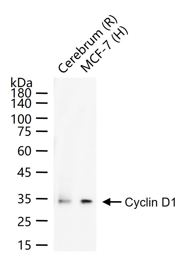
25 ug total protein per lane of various lysates (see on figure) probed with Cyclin D1 polyclonal antibody, unconjugated (bs-0623R) at 1:2000 dilution and 4°C overnight incubation. Followed by conjugated secondary antibody incubation at r.t. for 60 min.
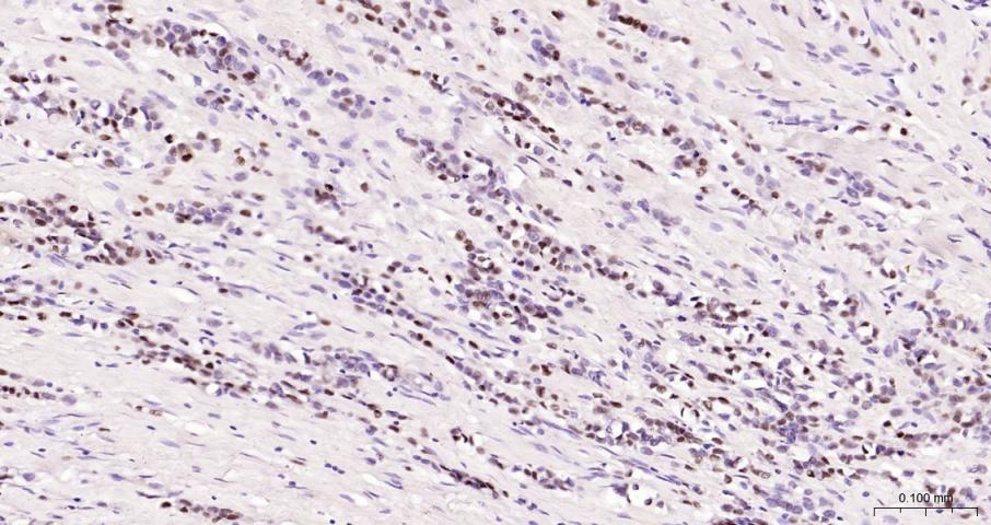
Paraformaldehyde-fixed, paraffin embedded Human Breast Cancer; Antigen retrieval by boiling in sodium citrate buffer (pH6.0) for 15 min; Antibody incubation with Cyclin D1 Polyclonal Antibody, Unconjugated (bs-0623R) at 1:200 overnight at 4°C, followed by conjugation to the bs-0295G-HRP and DAB (C-0010) staining.
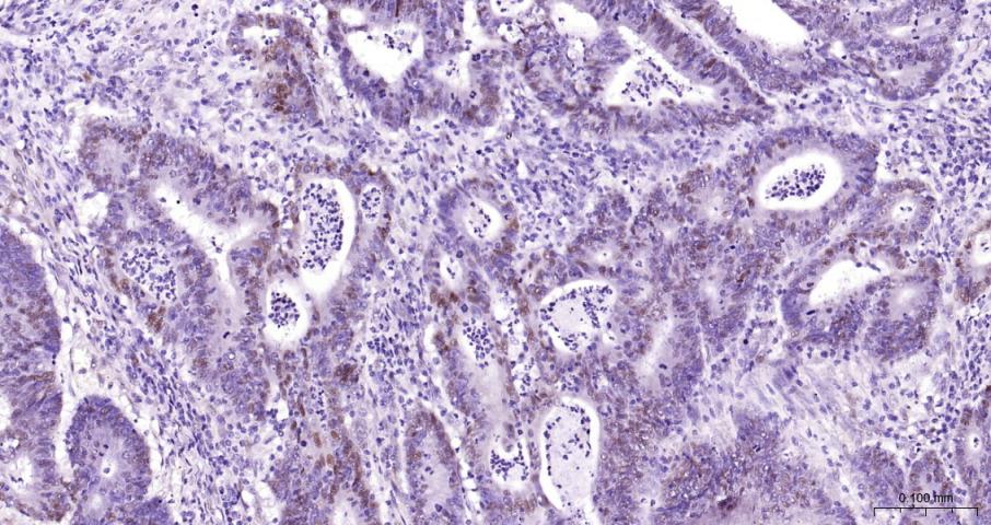
Paraformaldehyde-fixed, paraffin embedded Human Colon Cancer; Antigen retrieval by boiling in sodium citrate buffer (pH6.0) for 15 min; Antibody incubation with Cyclin D1 Polyclonal Antibody, Unconjugated (bs-0623R) at 1:200 overnight at 4°C, followed by conjugation to the bs-0295G-HRP and DAB (C-0010) staining.
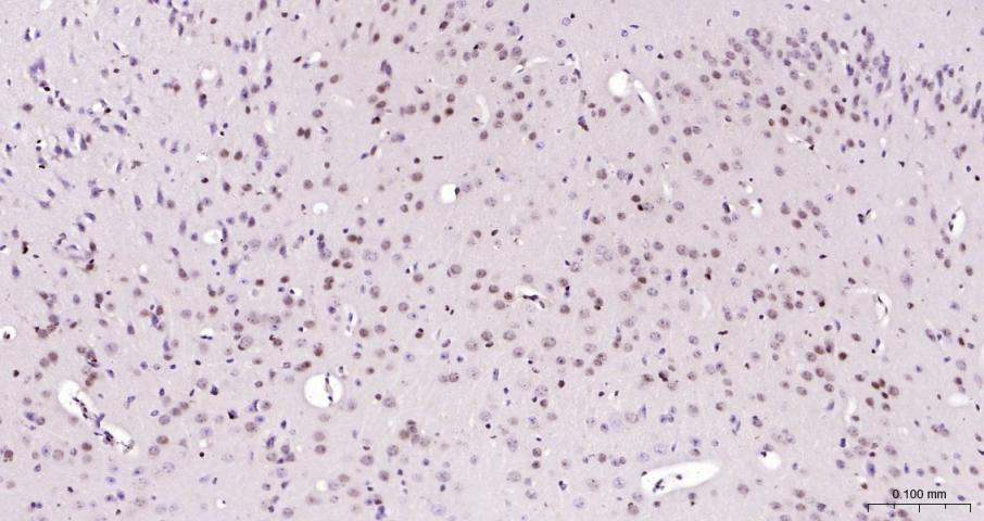
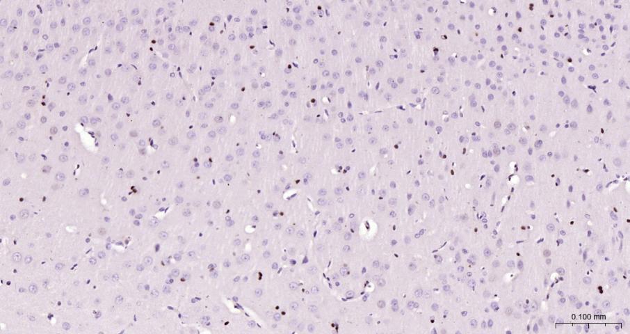
Paraformaldehyde-fixed, paraffin embedded Mouse Cerebrum; Antigen retrieval by boiling in sodium citrate buffer (pH6.0) for 15 min; Antibody incubation with Cyclin D1 Polyclonal Antibody, Unconjugated (bs-0623R) at 1:200 overnight at 4°C, followed by conjugation to the bs-0295G-HRP and DAB (C-0010) staining.
Paraformaldehyde-fixed, paraffin embedded Rat Cerebrum; Antigen retrieval by boiling in sodium citrate buffer (pH6.0) for 15 min; Antibody incubation with Cyclin D1 Polyclonal Antibody, Unconjugated (bs-0623R) at 1:200 overnight at 4°C, followed by conjugation to the bs-0295G-HRP and DAB (C-0010) staining.
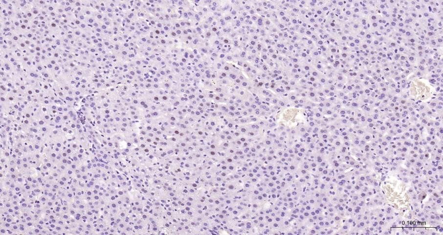
Paraformaldehyde-fixed, paraffin embedded Rat Liver; Antigen retrieval by boiling in sodium citrate buffer (pH6.0) for 15 min; Antibody incubation with Cyclin D1 Polyclonal Antibody, Unconjugated (bs-0623R) at 1:200 overnight at 4°C, followed by conjugation to the bs-0295G-HRP and DAB (C-0010) staining.
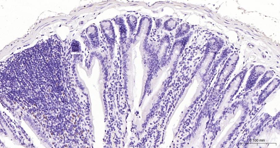
Paraformaldehyde-fixed, paraffin embedded Mouse Small Intestine; Antigen retrieval by boiling in sodium citrate buffer (pH6.0) for 15 min; Antibody incubation with Cyclin D1 Polyclonal Antibody, Unconjugated (bs-0623R) at 1:200 overnight at 4°C, followed by conjugation to the bs-0295G-HRP and DAB (C-0010) staining.
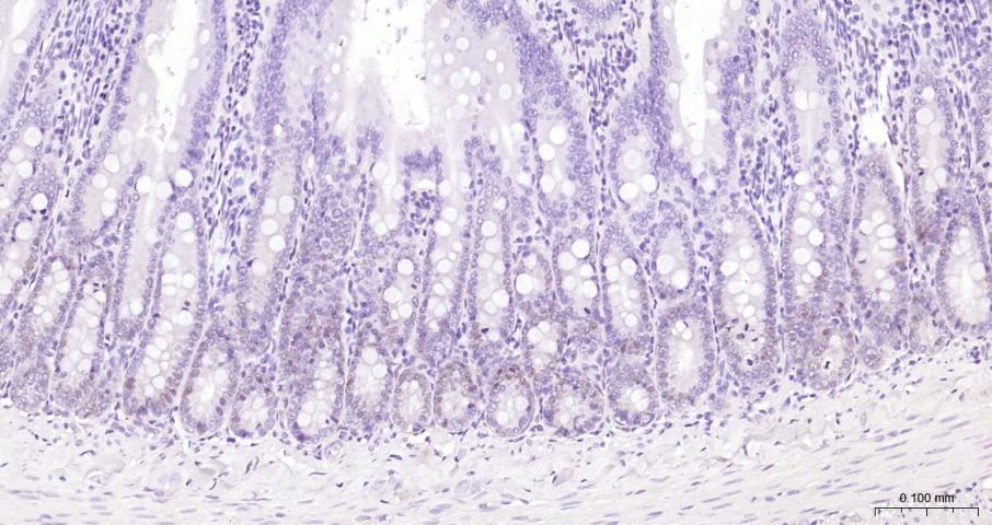
Paraformaldehyde-fixed, paraffin embedded Rat Small Intestine; Antigen retrieval by boiling in sodium citrate buffer (pH6.0) for 15 min; Antibody incubation with Cyclin D1 Polyclonal Antibody, Unconjugated (bs-0623R) at 1:200 overnight at 4°C, followed by conjugation to the bs-0295G-HRP and DAB (C-0010) staining.
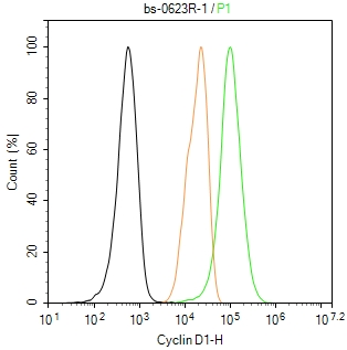
The SH-SY5Y (H) cells were fixed with 4% PFA (10 min at r.t.) and then permeabilized with 90% ice-cold methanol for 20 min at -20℃,the cells then were incubated in 5%BSA to block non-specific protein-protein interactions (30 min at r.t.), followed by secondary antibody incubation for 40 min at room temperature. Primary Antibody (green):Rabbit Anti-Cyclin D1 antibody (bs-0623R,1:100); Isotype Control (orange): Rabbit IgG (bs-0295P). Blank control (black): PBS. Acquisition of 20,000 events was performed.
|
| 1、抗体溶解方法 | |
| 2、抗体修复方式 | |
| 3、常用试剂的配制 | |
| 4、免疫组化操作步骤 | |
| 5、免疫组化问题解答 | |
| 6、Western Blotting 操作步骤 | |
| 7、Western Blotting 问题解答 | |
| 8、关于肽链的设计 | |
| 9、多肽的溶解与保存 | |
| 10、酶标抗体效价测定程序 | |


