

| 产品编号 | bs-0573R |
| 英文名称 | Cyclin E1 Rabbit pAb |
| 中文名称 | 周期素E抗体 |
| 别 名 | CCNE; pCCNE1; CycE1; CYCLE; CCNE1_HUMAN; CCNE1; CCNE1_MOUSE; CCNE1_RAT; |
 |
Specific References (13) | bs-0573R has been referenced in 13 publications.
|
| 研究领域 | 细胞生物 细胞周期蛋白 |
| 抗体来源 | Rabbit |
| 克隆类型 | Polyclonal |
| 克 隆 号 | |
| 交叉反应 | Human,Mouse,Rat |
| 产品应用 | WB=1:500-2000,IHC-P=1:100-500,IHC-F=1:100-500,IF=1:100-500,Flow-Cyt=1μg/Test
not yet tested in other applications. optimal dilutions/concentrations should be determined by the end user. |
| 理论分子量 | 45 kDa |
| 检测分子量 | 50 |
| 细胞定位 | 细胞核 |
| 性 状 | Liquid |
| 浓 度 | 1mg/ml |
| 免 疫 原 | KLH conjugated synthetic peptide derived from rat Cyclin E: 375-411/411 |
| 亚 型 | IgG |
| 纯化方法 | affinity purified by Protein A |
| 缓 冲 液 | 0.01M TBS (pH7.4) with 1% BSA, 0.02% Proclin300 and 50% Glycerol. |
| 保存条件 | Shipped at 4℃. Store at -20℃ for one year. Avoid repeated freeze/thaw cycles. |
| 注意事项 | This product as supplied is intended for research use only, not for use in human, therapeutic or diagnostic applications. |
| PubMed | PubMed |
| 产品介绍 |
The protein encoded by this gene belongs to the highly conserved cyclin family, whose members are characterized by a dramatic periodicity in protein abundance through the cell cycle. Cyclins function as regulators of CDK kinases. Different cyclins exhibit distinct expression and degradation patterns which contribute to the temporal coordination of each mitotic event. This cyclin forms a complex with and functions as a regulatory subunit of CDK2, whose activity is required for cell cycle G1/S transition. This protein accumulates at the G1-S phase boundary and is degraded as cells progress through S phase. Overexpression of this gene has been observed in many tumors, which results in chromosome instability, and thus may contribute to tumorigenesis. This protein was found to associate with, and be involved in, the phosphorylation of NPAT protein (nuclear protein mapped to the ATM locus), which participates in cell-cycle regulated histone gene expression and plays a critical role in promoting cell-cycle progression in the absence of pRB. Two alternatively spliced transcript variants of this gene, which encode distinct isoforms, have been described. Two additional splice variants were reported but detailed nucleotide sequence information is not yet available. Transcript Variant: This variant (1) contains a different 5' end region, which includes an upstream in-frame translation start codon, when compared to variant 2. The encoded protein has a 15 aa longer N-terminus, as compared to isoform 2. Subunit: Interacts with a member of the CDK2/CDK protein kinases to form a serine/threonine kinase holoenzyme complex. The cyclin subunit imparts substrate specificity to the complex. Found in a complex with CDK2, CABLES1 and CCNA1 (By similarity). Part of a complex consisting of UHRF2, CDK2 and CCNE1. Interacts directly with UHRF2; the interaction ubiquitinates CCNE1 and appears to occur independently of CCNE1 phosphorylation. Subcellular Location: Nucleus. Tissue Specificity: Highly expressed in testis and placenta. Low levels in bronchial epithelial cells. Post-translational modifications: Phosphorylation of Thr-395 by GSK3 and of Ser-399 by CDK2 accelerates degradation via the ubiquitin proteasome pathway. Phosphorylated upon DNA damage, probably by ATM or ATR. Similarity: Belongs to the cyclin family. Cyclin E subfamily. SWISS: P39949 Gene ID: 25729 Database links: Entrez Gene: 898 Human Entrez Gene: 12447 Mouse Omim: 123837 Human SwissProt: P24864 Human SwissProt: Q61457 Mouse Unigene: 244723 Human Unigene: 16110 Mouse Unigene: 15455 Rat 细胞周期素E是调控细胞G-1→S期转变的关键因素。由于在多种肿瘤中的不适当表达,细胞周期素E现在已明确为原癌基因。 |
| 产品图片 |
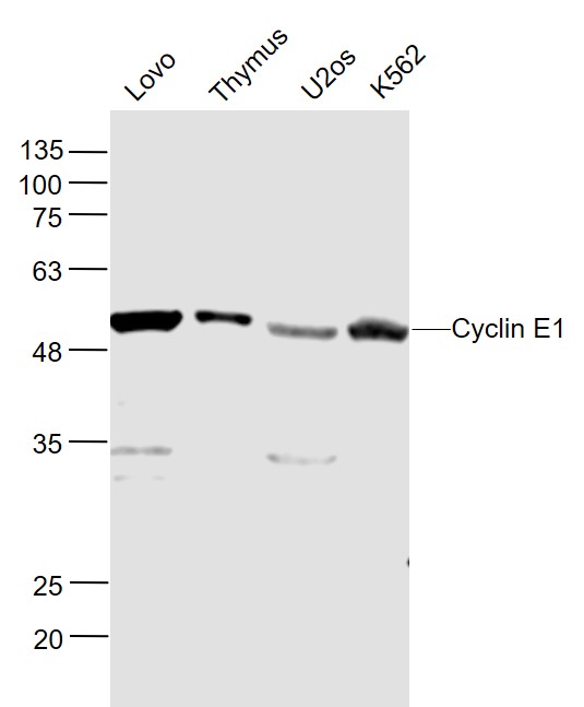
Sample:
Lovo (Human) Cell Lysate at 30 ug
Thymus (Mouse) Lysate at 40 ug
U2os (Human) Cell Lysate at 30 ug
K562 (Human) Cell Lysate at 30 ug
Primary: Anti- Cyclin E1 (bs-0573R) at 1/1000 dilution
Secondary: IRDye800CW Goat Anti-Rabbit IgG at 1/20000 dilution
Predicted band size: 45 kD
Observed band size: 50 kD
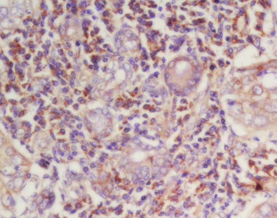
Tissue/cell: human laryngocarcinoma; 4% Paraformaldehyde-fixed and paraffin-embedded;
Antigen retrieval: citrate buffer ( 0.01M, pH 6.0 ), Boiling bathing for 15min; Block endogenous peroxidase by 3% Hydrogen peroxide for 30min; Blocking buffer (normal goat serum,C-0005) at 37℃ for 20 min;
Incubation: Anti-Cyclin-E Polyclonal Antibody, Unconjugated(bs-0573R) 1:200, overnight at 4°C, followed by conjugation to the secondary antibody(SP-0023) and DAB(C-0010) staining

Tissue/cell: rat testis tissue;4% Paraformaldehyde-fixed and paraffin-embedded;
Antigen retrieval: citrate buffer ( 0.01M, pH 6.0 ), Boiling bathing for 15min; Blocking buffer (normal goat serum,C-0005) at 37℃ for 20 min;
Incubation: Anti-Cyclin E Polyclonal Antibody, Unconjugated(bs-0573R) 1:200, overnight at 4°C; The secondary antibody was Goat Anti-Rabbit IgG, Cy3 conjugated(bs-0295G-Cy3)used at 1:200 dilution for 40 minutes at 37°C.

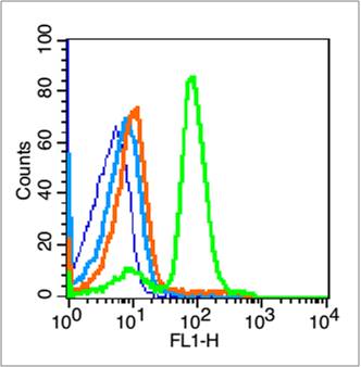
Cell: NIH/3T3
Concentration:1:100
Host/Isotype:Rabbit/IgG
Flow cytometric analysis of primary antibody (Cat#: bs-0573R) on NIH/3T3(green) compared with Rabbit IgG isotype control in the absence of primary antibody (blue) followed by Alexa Fluor 488-conjugated goat anti-rabbit IgG(H+L) secondary antibody .
Blank control (blue line): Mouse spleen cells (blue).
Primary Antibody (green line): Rabbit Anti-Cyclin E1 antibody (bs-0573R)
Dilution: 1μg /10^6 cells;
Isotype Control Antibody (orange line): Rabbit IgG .
Secondary Antibody (white blue line): Goat anti-rabbit IgG-FITC
Dilution: 1μg /test.
Protocol
The cells were fixed with 70% ethanol (overninght at 4℃) and then permeabilized with 0.1% PBS-Tween for 20 min at room temperature. Cells stained with Primary Antibody for 30 min at room temperature. The cells were then incubated in 1 X PBS/2%BSA/10% goat serum to block non-specific protein-protein interactions followed by the antibody for 15 min at room temperature. The secondary antibody used for 40 min at room temperature. Acquisition of 20,000 events was performed.
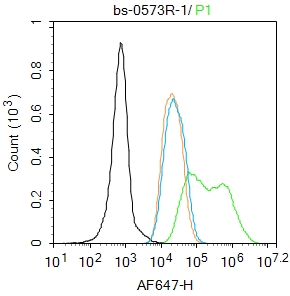
Blank control: MCF7.
Primary Antibody (green line): Rabbit Anti-Cyclin E1 antibody (bs-0573R)
Dilution: 2μg /10^6 cells;
Isotype Control Antibody (orange line): Rabbit IgG .
Secondary Antibody : Goat anti-rabbit IgG-AF647
Dilution: 1μg /test.
Protocol
The cells were fixed with 4% PFA (10min at room temperature)and then permeabilized with 90% ice-cold methanol for 20 min at-20℃.The cells were then incubated in 5%BSA to block non-specific protein-protein interactions for 30 min at room temperature .Cells stained with Primary Antibody for 30 min at room temperature. The secondary antibody used for 40 min at room temperature. Acquisition of 20,000 events was performed.
|
| 1、抗体溶解方法 | |
| 2、抗体修复方式 | |
| 3、常用试剂的配制 | |
| 4、免疫组化操作步骤 | |
| 5、免疫组化问题解答 | |
| 6、Western Blotting 操作步骤 | |
| 7、Western Blotting 问题解答 | |
| 8、关于肽链的设计 | |
| 9、多肽的溶解与保存 | |
| 10、酶标抗体效价测定程序 | |


