

| 产品编号 | bs-1020R |
| 英文名称 | ERK1 Rabbit pAb |
| 中文名称 | 丝裂原活化蛋白激酶1抗体 |
| 别 名 | MK03_HUMAN; MAPK3; MAP kinase 3; MAPK 3; ERT2; Extracellular signal-regulated kinase 1 (ERK-1); Insulin-stimulated MAP2 kinase; MAP kinase isoform p44 (p44-MAPK); Microtubule-associated protein 2 kinase; p44-ERK1; 2.7.11.24; ERK1; PRKM3; MK01_HUMAN; MAPK1 |
 |
Specific References (7) | bs-1020R has been referenced in 7 publications.
|
| 研究领域 | 细胞生物 神经生物学 信号转导 干细胞 激酶和磷酸酶 Alzheimer's |
| 抗体来源 | Rabbit |
| 克隆类型 | Polyclonal |
| 克 隆 号 | |
| 交叉反应 | Human,Mouse,Rat |
| 产品应用 | IHC-P=1:100-500,IHC-F=1:100-500,IF=1:100-500,Flow-Cyt=1μg/Test,ICC/IF=1:100-500
not yet tested in other applications. optimal dilutions/concentrations should be determined by the end user. |
| 理论分子量 | 43 kDa |
| 细胞定位 | 细胞核 细胞浆 |
| 性 状 | Liquid |
| 浓 度 | 1mg/ml |
| 免 疫 原 | KLH conjugated synthetic peptide derived from human MAPK1: 101-200/380 |
| 亚 型 | IgG |
| 纯化方法 | affinity purified by Protein A |
| 缓 冲 液 | 0.01M TBS (pH7.4) with 1% BSA, 0.02% Proclin300 and 50% Glycerol. |
| 保存条件 | Shipped at 4℃. Store at -20℃ for one year. Avoid repeated freeze/thaw cycles. |
| 注意事项 | This product as supplied is intended for research use only, not for use in human, therapeutic or diagnostic applications. |
| PubMed | PubMed |
| 产品介绍 |
The protein encoded by this gene is a member of the MAP kinase family. MAP kinases, also known as extracellular signal-regulated kinases (ERKs), act in a signaling cascade that regulates various cellular processes such as proliferation, differentiation, and cell cycle progression in response to a variety of extracellular signals. This kinase is activated by upstream kinases, resulting in its translocation to the nucleus where it phosphorylates nuclear targets. Alternatively spliced transcript variants encoding different protein isoforms have been described. [provided by RefSeq, Jul 2008] Function: Serine/threonine kinase which acts as an essential component of the MAP kinase signal transduction pathway. MAPK1/ERK2 and MAPK3/ERK1 are the 2 MAPKs which play an important role in the MAPK/ERK cascade. They participate also in a signaling cascade initiated by activated KIT and KITLG/SCF. Depending on the cellular context, the MAPK/ERK cascade mediates diverse biological functions such as cell growth, adhesion, survival and differentiation through the regulation of transcription, translation, cytoskeletal rearrangements. The MAPK/ERK cascade plays also a role in initiation and regulation of meiosis, mitosis, and postmitotic functions in differentiated cells by phosphorylating a number of transcription factors. About 160 substrates have already been discovered for ERKs. Many of these substrates are localized in the nucleus, and seem to participate in the regulation of transcription upon stimulation. However, other substrates are found in the cytosol as well as in other cellular organelles, and those are responsible for processes such as translation, mitosis and apoptosis. Moreover, the MAPK/ERK cascade is also involved in the regulation of the endosomal dynamics, including lysosome processing and endosome cycling through the perinuclear recycling compartment (PNRC); as well as in the fragmentation of the Golgi apparatus during mitosis. The substrates include transcription factors (such as ATF2, BCL6, ELK1, ERF, FOS, HSF4 or SPZ1), cytoskeletal elements (such as CANX, CTTN, GJA1, MAP2, MAPT, PXN, SORBS3 or STMN1), regulators of apoptosis (such as BAD, BTG2, CASP9, DAPK1, IER3, MCL1 or PPARG), regulators of translation (such as EIF4EBP1) and a variety of other signaling-related molecules (like ARHGEF2, FRS2 or GRB10). Protein kinases (such as RAF1, RPS6KA1/RSK1, RPS6KA3/RSK2, RPS6KA2/RSK3, RPS6KA6/RSK4, SYK, MKNK1/MNK1, MKNK2/MNK2, RPS6KA5/MSK1, RPS6KA4/MSK2, MAPKAPK3 or MAPKAPK5) and phosphatases (such as DUSP1, DUSP4, DUSP6 or DUSP16) are other substrates which enable the propagation the MAPK/ERK signal to additional cytosolic and nuclear targets, thereby extending the specificity of the cascade. Subunit: Binds both upstream activators and downstream substrates in multimolecular complexes. Found in a complex with at least BRAF, HRAS1, MAP2K1/MEK1, MAPK3 and RGS14. Binds to HIV-1 Nef through its SH3 domain. This interaction inhibits its tyrosine-kinase activity. Interacts with ADAM15, ARRB2, CANX, DAPK1 (via death domain), HSF4, IER3, MAP2K1/MEK1, MORG1, NISCH, and SGK1. Interacts with PEA15 and MKNK2. MKNK2 isoform 1 binding prevents from dephosphorylation and inactivation. Interacts with TPR. Subcellular Location: Cytoplasm. Nucleus. Note=Autophosphorylation at Thr-207 promotes nuclear localization. Post-translational modifications: Phosphorylated upon KIT and FLT3 signaling. Dually phosphorylated on Thr-202 and Tyr-204, which activates the enzyme. Ligand-activated ALK induces tyrosine phosphorylation. Dephosphorylated by PTPRJ at Tyr-204. Similarity: Belongs to the protein kinase superfamily. CMGC Ser/Thr protein kinase family. MAP kinase subfamily. Contains 1 protein kinase domain. SWISS: P28482 Gene ID: 5595 Database links: Entrez Gene: 5595 Human Entrez Gene: 26417 Mouse Omim: 601795 Human SwissProt: P27361 Human SwissProt: Q63844 Mouse Unigene: 861 Human Unigene: 8385 Mouse Unigene: 2592 Rat |
| 产品图片 |
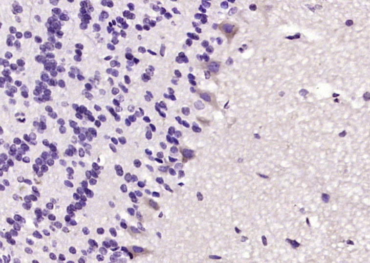
Paraformaldehyde-fixed, paraffin embedded (rat brain); Antigen retrieval by boiling in sodium citrate buffer (pH6.0) for 15min; Block endogenous peroxidase by 3% hydrogen peroxide for 20 minutes; Blocking buffer (normal goat serum) at 37°C for 30min; Antibody incubation with (ERK1) Polyclonal Antibody, Unconjugated (bs-1020R) at 1:200 overnight at 4°C, followed by operating according to SP Kit(Rabbit) (sp-0023) instructionsand DAB staining.
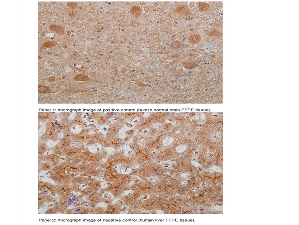
Images provided by the Independent Validation Program (badge number 029731) Formalin-fixed and paraffin embedded human brain (panel 1) and human liver (panel 2) labeled with Rabbit Anti-ERK1 Polyclonal Antibody (bs-1020R) at 1:250 overnight at room temperature followed by conjugation to secondary antibody.
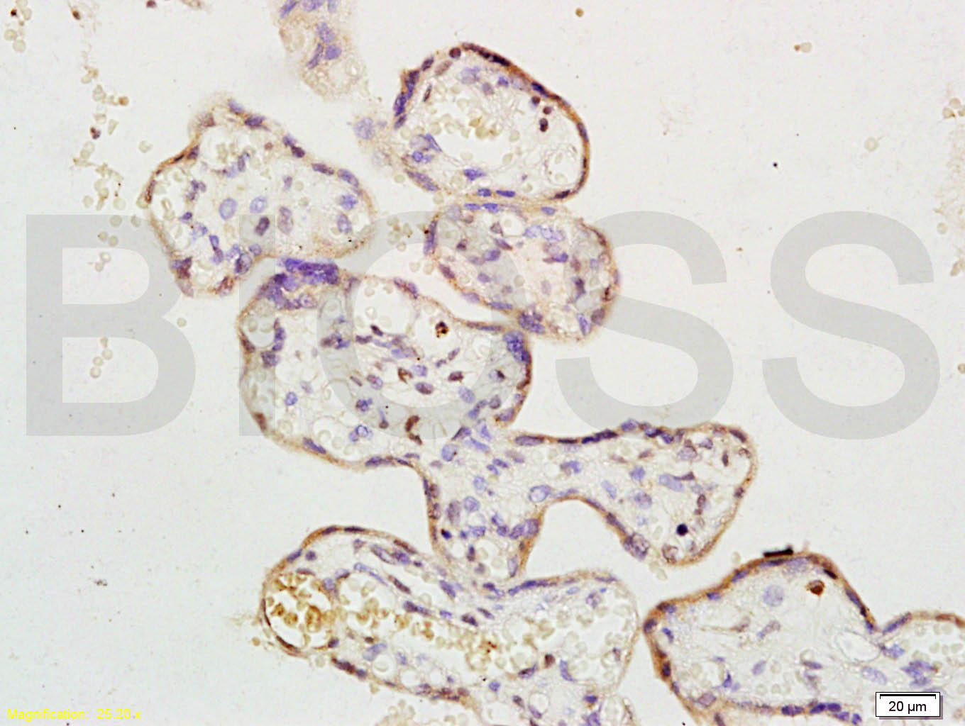
Tissue/cell: human placenta tissue; 4% Paraformaldehyde-fixed and paraffin-embedded;
Antigen retrieval: citrate buffer ( 0.01M, pH 6.0 ), Boiling bathing for 15min; Block endogenous peroxidase by 3% Hydrogen peroxide for 30min; Blocking buffer (normal goat serum,C-0005) at 37℃ for 20 min;
Incubation: Anti-ERK1 Polyclonal Antibody, Unconjugated(bs-1020R) 1:200, overnight at 4°C, followed by conjugation to the secondary antibody(SP-0023) and DAB(C-0010) staining
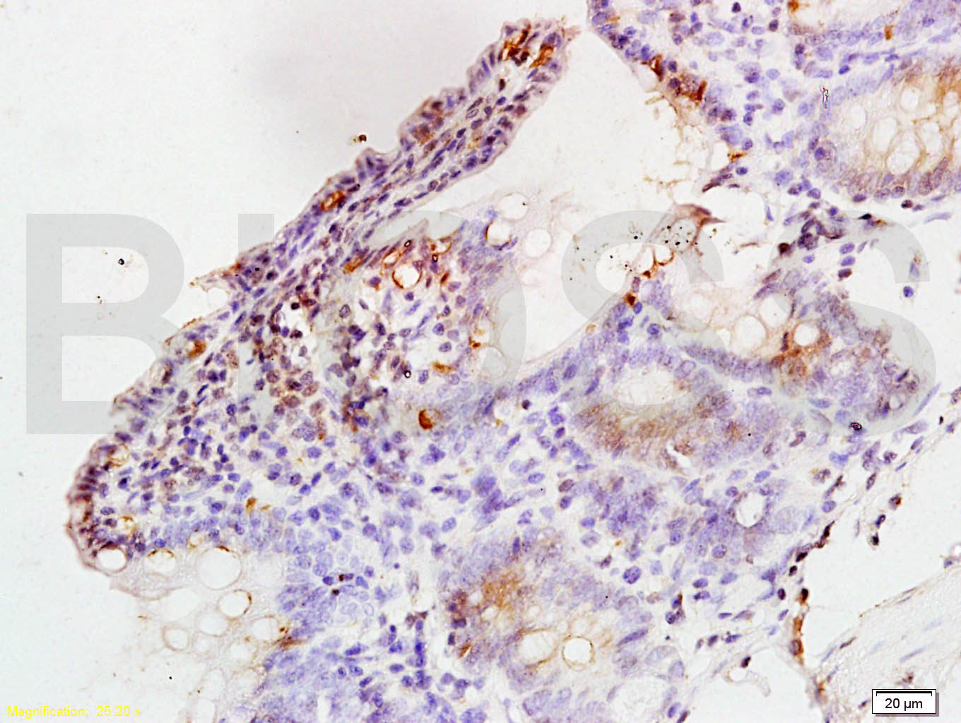
Tissue/cell: rat colon tissue; 4% Paraformaldehyde-fixed and paraffin-embedded;
Antigen retrieval: citrate buffer ( 0.01M, pH 6.0 ), Boiling bathing for 15min; Block endogenous peroxidase by 3% Hydrogen peroxide for 30min; Blocking buffer (normal goat serum,C-0005) at 37℃ for 20 min;
Incubation: Anti-ERK1 Polyclonal Antibody, Unconjugated(bs-1020R) 1:200, overnight at 4°C, followed by conjugation to the secondary antibody(SP-0023) and DAB(C-0010) staining
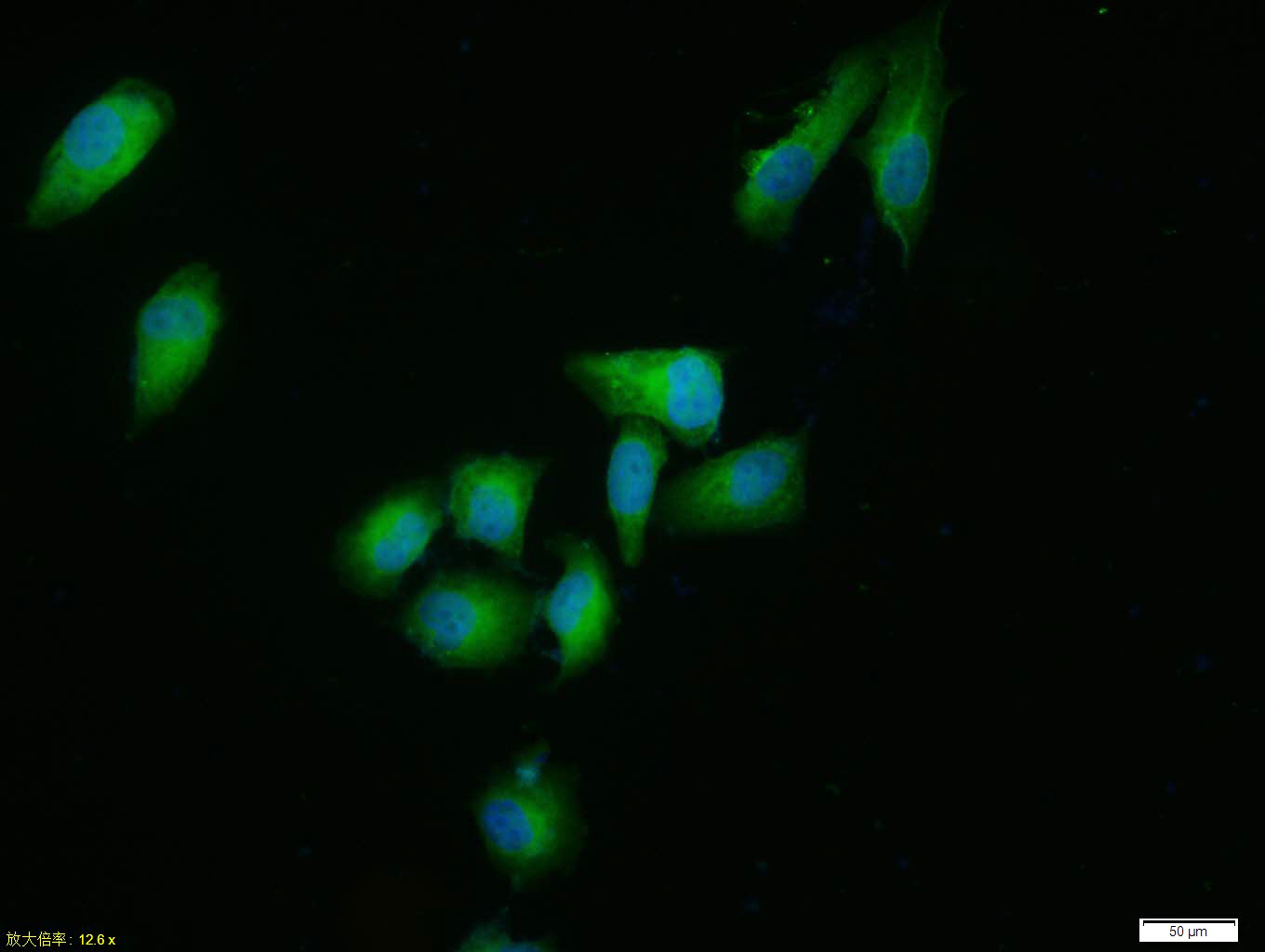
Tissue/cell: HUVEC cell; 4% Paraformaldehyde-fixed; Triton X-100 at room temperature for 20 min; Blocking buffer (normal goat serum, C-0005) at 37°C for 20 min; Antibody incubation with (ERK1) polyclonal Antibody, Unconjugated (bs-1020R) 1:100, 90 minutes at 37°C; followed by a FITC conjugated Goat Anti-Rabbit IgG antibody at 37°C for 90 minutes, DAPI (blue, C02-04002) was used to stain the cell nuclei.
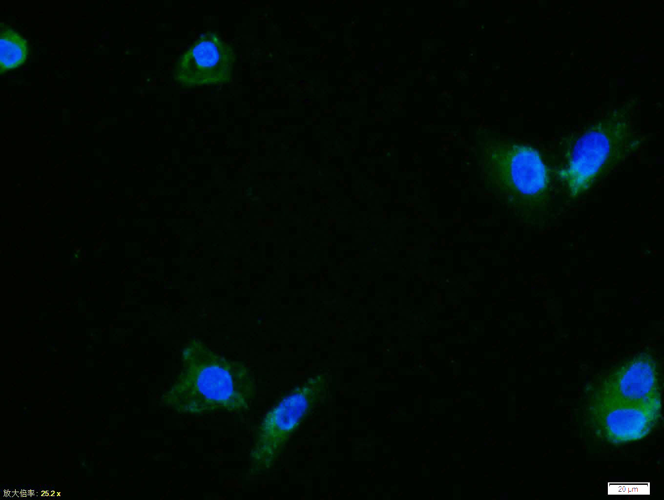
Tissue/cell: Hela cell; 4% Paraformaldehyde-fixed; Triton X-100 at room temperature for 20 min; Blocking buffer (normal goat serum, C-0005) at 37°C for 20 min; Antibody incubation with (ERK1) polyclonal Antibody, Unconjugated (bs-1020R) 1:100, 90 minutes at 37°C; followed by a FITC conjugated Goat Anti-Rabbit IgG antibody at 37°C for 90 minutes, DAPI (blue, C02-04002) was used to stain the cell nuclei.
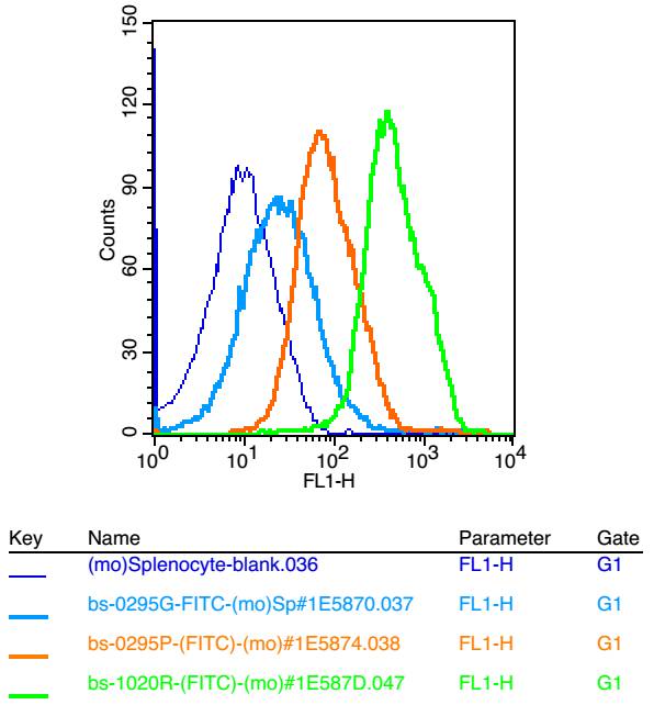
Blank control: mouse splenocytes(blue)
Isotype Control Antibody: Rabbit IgG(orange) ;
Secondary Antibody: Goat anti-rabbit IgG-FITC(white blue),
Dilution: 1:100 in 1 X PBS containing 0.5% BSA ;
Primary Antibody Dilution: 1μl in 100 μL1X PBS containing 0.5% BSA(green).
|
| 1、抗体溶解方法 | |
| 2、抗体修复方式 | |
| 3、常用试剂的配制 | |
| 4、免疫组化操作步骤 | |
| 5、免疫组化问题解答 | |
| 6、Western Blotting 操作步骤 | |
| 7、Western Blotting 问题解答 | |
| 8、关于肽链的设计 | |
| 9、多肽的溶解与保存 | |
| 10、酶标抗体效价测定程序 | |


