

| 产品编号 | bs-0501R |
| 英文名称 | JNK1 + JNK3 Rabbit pAb |
| 中文名称 | 氨基末端激酶1/3抗体 |
| 别 名 | JNK; JNK-46; JNK1; JNK1A2; JNK21B1/2; PRKM8; SAPK1; SAPK1c; MK08_HUMAN; MAPK8; MAP kinase 8; MAPK 8; Stress-activated protein kinase 1c (SAPK1c); Stress-activated protein kinase JNK1; c-Jun N-terminal kinase 1; 2.7.11.24; MK08_MOUSE; MK08_RAT; SAPK gamma; |
 |
Specific References (6) | bs-0501R has been referenced in 6 publications.
[IF=9.473] Shuting Wei. et al. Particle matters induce airway epithelial barrier dysfunction in vivo and in vitro: from a more realistic inhalation scenario. ENVIRON SCI-NANO. 2022 Jun;: WB ; Human.
[IF=7.963] Meiqiong Wu. et al. Suppression of NADPH oxidase 4 inhibits PM2.5-induced cardiac fibrosis through ROS-P38 MAPK pathway. SCI TOTAL ENVIRON. 2022 Apr;:155558 WB ; Mouse,Rat.
[IF=5.285] Huawei Liu. et al. Integrated multi-omics reveals the beneficial role of chlorogenic acid in improving the growth performance and immune function of immunologically-stressed broilers. ANIM NUTR. 2023 May;: WB ; Chicken.
[IF=3.31] Król, Magdalena, et al. "Macrophages Mediate a Switch between Canonical and Non-Canonical Wnt Pathways in Canine Mammary Tumors." PloS one 9.1 (2014): e83995. WB ; Dog.
[IF=3.234] Zhuang S et al. Rhein ameliorates lipopolysaccharide-induced intestinal barrier injury via modulation of Nrf2 and MAPKs.(2019)Life Sci. Life Sci. Jan 1;216:168-175. WB ; Rat.
[IF=2.027] Li QH et al. Effect of heat stress on mitogen-activated protein kinases in the hypothalamic− pituitary− gonadal axis of developing Wenchang chicks. Poultry Science.2019. WB ; chick.
|
| 研究领域 | 肿瘤 细胞生物 免疫学 信号转导 转录调节因子 激酶和磷酸酶 |
| 抗体来源 | Rabbit |
| 克隆类型 | Polyclonal |
| 克 隆 号 | |
| 交叉反应 | Human,Mouse,Rat (predicted: Rabbit,Pig,Cow,Chicken,Dog) |
| 产品应用 | WB=1:500-2000,IHC-P=1:100-500,IHC-F=1:100-500,IF=1:100-500,Flow-Cyt=1μg/Test,ICC/IF=1:100-500
not yet tested in other applications. optimal dilutions/concentrations should be determined by the end user. |
| 理论分子量 | 42 kDa |
| 检测分子量 | 42 |
| 细胞定位 | 细胞核 细胞浆 |
| 性 状 | Liquid |
| 浓 度 | 1mg/ml |
| 免 疫 原 | KLH conjugated synthetic peptide derived from human JNK1: 201-300/427 |
| 亚 型 | IgG |
| 纯化方法 | affinity purified by Protein A |
| 缓 冲 液 | 0.01M TBS (pH7.4) with 1% BSA, 0.02% Proclin300 and 50% Glycerol. |
| 保存条件 | Shipped at 4℃. Store at -20℃ for one year. Avoid repeated freeze/thaw cycles. |
| 注意事项 | This product as supplied is intended for research use only, not for use in human, therapeutic or diagnostic applications. |
| PubMed | PubMed |
| 产品介绍 |
JNK1(MAPK8) is a member of the MAP kinase family. MAP kinases act as an integration point for multiple biochemical signals, and are involved in a wide variety of cellular processes such as proliferation, differentiation, transcription regulation and development. This kinase is activated by various cell stimuli, and targets specific transcription factors, and thus mediates immediate-early gene expression in response to cell stimuli. The activation of this kinase by tumor-necrosis factor alpha (TNF-alpha) is found to be required for TNF-alpha induced apoptosis. This kinase is also involved in UV radiation induced apoptosis, which is thought to be related to cytochrome c-mediated cell death pathway. Studies of the mouse counterpart of this gene suggested that this kinase play a key role in T cell proliferation, apoptosis and differentiation. Four alternatively spliced transcript variants encoding distinct isoforms have been reported.
JNK1 is activated by threonine and tyrosine phosphorylation by either of two dual specificity kinases, MAP2K4 and MAP2K7.
The JNK pathway is critically involved in diabetes and levels are abnormally elevated in obesity. The cell-permeable JNK inhibitory peptide may have promise as a therapeutic agent for diabetes. Function: Serine/threonine-protein kinase involved in various processes such as cell proliferation, differentiation, migration, transformation and programmed cell death. Extracellular stimuli such as proinflammatory cytokines or physical stress stimulate the stress-activated protein kinase/c-Jun N-terminal kinase (SAP/JNK) signaling pathway. In this cascade, two dual specificity kinases MAP2K4/MKK4 and MAP2K7/MKK7 phosphorylate and activate MAPK8/JNK1. In turn, MAPK8/JNK1 phosphorylates a number of transcription factors, primarily components of AP-1 such as JUN, JDP2 and ATF2 and thus regulates AP-1 transcriptional activity. Phosphorylates the replication licensing factor CDT1, inhibiting the interaction between CDT1 and the histone H4 acetylase HBO1 to replication origins. Loss of this interaction abrogates the acetylation required for replication initiation. Promotes stressed cell apoptosis by phosphorylating key regulatory factors including p53/TP53 and Yes-associates protein YAP1. In T-cells, MAPK8 and MAPK9 are required for polarized differentiation of T-helper cells into Th1 cells. Contributes to the survival of erythroid cells by phosphorylating the antagonist of cell death BAD upon EPO stimulation. Mediates starvation-induced BCL2 phosphorylation, BCL2 dissociation from BECN1, and thus activation of autophagy. Phosphorylates STMN2 and hence regulates microtubule dynamics, controlling neurite elongation in cortical neurons. In the developing brain, through its cytoplasmic activity on STMN2, negatively regulates the rate of exit from multipolar stage and of radial migration from the ventricular zone. Phosphorylates several other substrates including heat shock factor protein 4 (HSF4), the deacetylase SIRT1, ELK1, or the E3 ligase ITCH. Subunit: Binds to at least four scaffolding proteins, MAPK8IP1/JIP-1, MAPK8IP2/JIP-2, MAPK8IP3/JIP-3/JSAP1 and SPAG9/MAPK8IP4/JIP-4. These proteins also bind other components of the JNK signaling pathway. Forms a complex with MAPK8IP1 and RGNEF. Interacts with TP53 and WWOX. Interacts with JAMP. Interacts with NFATC4. Interacts with MECOM; regulates JNK signaling. Interacts with PIN1; this interaction mediates MAPK8 conformational changes leading to the binding of MAPK8 to its substrates. Interacts (phosphorylated form) with NFE2; the interaction phosphorylates NFE2 in undifferentiated cells. Subcellular Location: Cytoplasm. Nucleus. Post-translational modifications: Phosphorylated by TAOK2. Dually phosphorylated on Thr-183 and Tyr-185 by MAP2K7 and MAP2K4, which activates the enzyme. Similarity: Belongs to the protein kinase superfamily. CMGC Ser/Thr protein kinase family. MAP kinase subfamily. Contains 1 protein kinase domain. SWISS: P45983 Gene ID: 5599 Database links: Entrez Gene: 5599 Human Entrez Gene: 5601 Human Entrez Gene: 26419 Mouse Entrez Gene: 26420 Mouse Omim: 601158 Human Omim: 602896 Human SwissProt: P45983 Human SwissProt: P45984 Human SwissProt: Q91Y86 Mouse SwissProt: Q9WTU6 Mouse Unigene: 138211 Human Unigene: 348446 Human Unigene: 21495 Mouse Unigene: 68933 Mouse |
| 产品图片 |
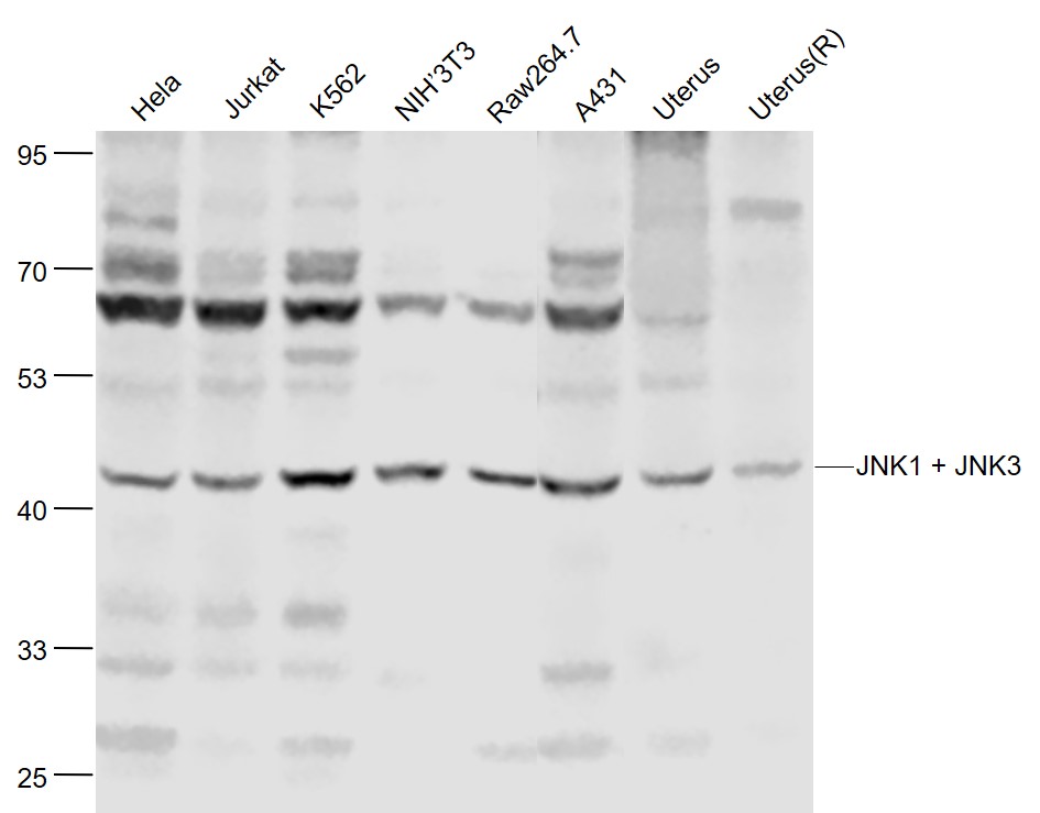
Sample:
Hela(Human) Cell Lysate at 30 ug
Jurkat(Human) Cell Lysate at 30 ug
K562(Human) Cell Lysate at 30 ug
NIH/3T3(Mouse) Cell Lysate at 30 ug
Raw264.7(Mouse) Cell Lysate at 30 ug
A431(Human) Cell Lysate at 30 ug
Uterus(Mouse) Lysate at 40 ug
Uterus(Rat) Lysate at 40 ug
Primary: Anti-JNK1 + JNK3 (bs-0501R) at 1/1000 dilution
Secondary: IRDye800CW Goat Anti-Rabbit IgG at 1/20000 dilution
Predicted band size: 42 kD
Observed band size: 42 kD
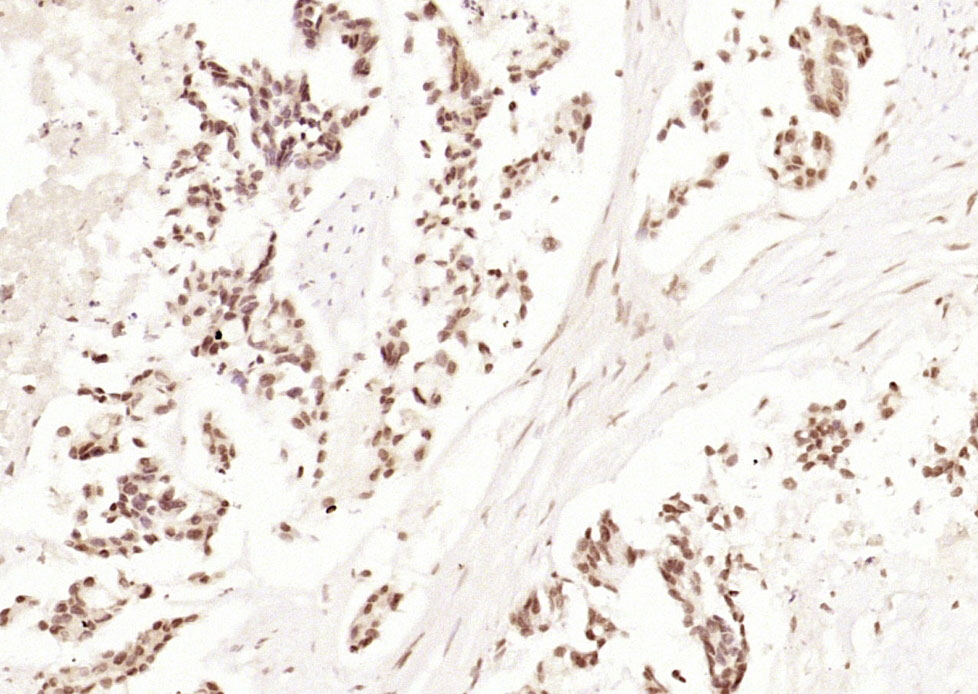
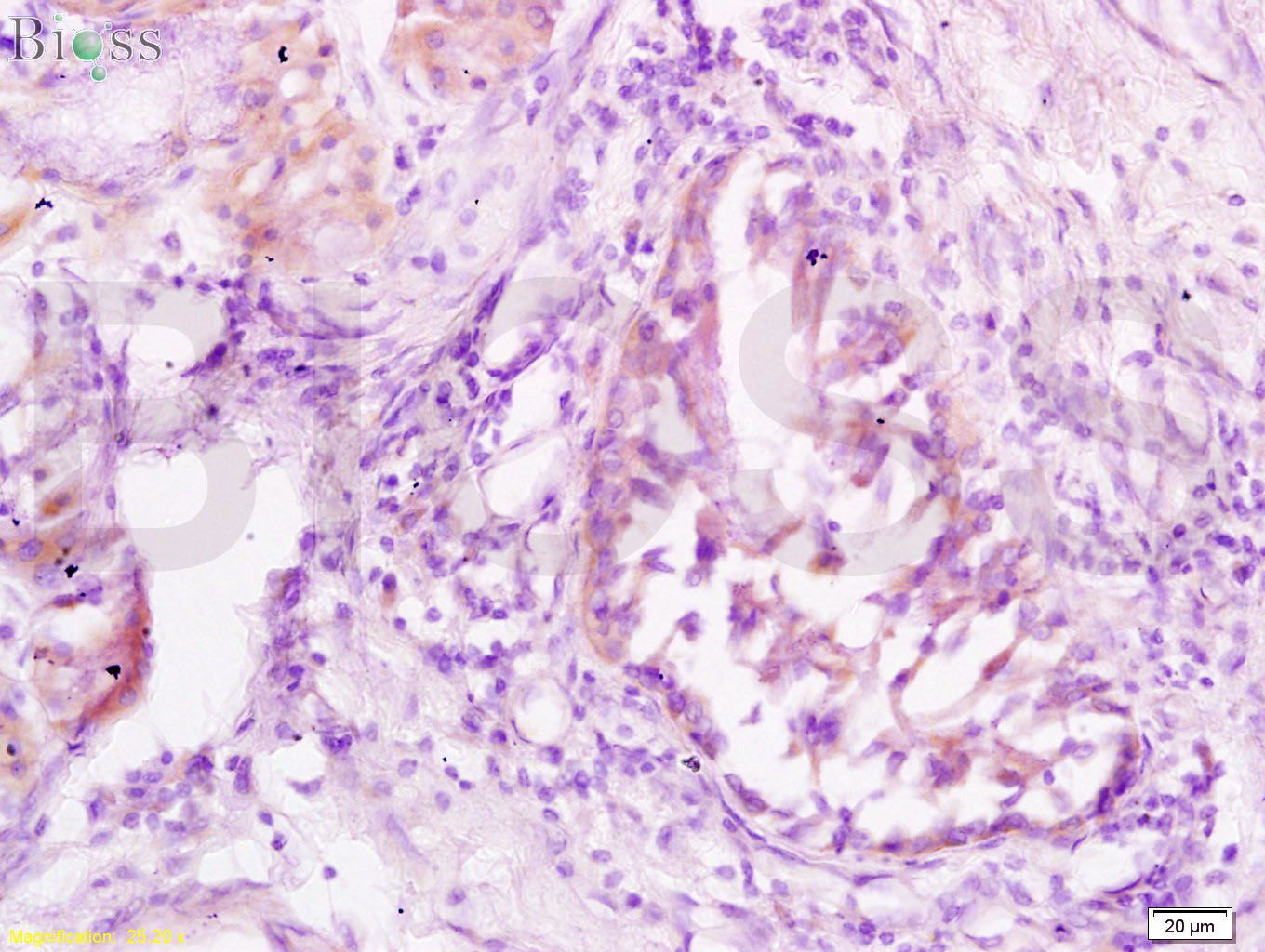
Paraformaldehyde-fixed, paraffin embedded (human gastric carcinoma); Antigen retrieval by boiling in sodium citrate buffer (pH6.0) for 15min; Block endogenous peroxidase by 3% hydrogen peroxide for 20 minutes; Blocking buffer (normal goat serum) at 37°C for 30min; Antibody incubation with (JNK1 + JNK3) Polyclonal Antibody, Unconjugated (bs-0501R) at 1:200 overnight at 4°C, followed by operating according to SP Kit(Rabbit) (sp-0023) instructionsand DAB staining.
Paraformaldehyde-fixed, paraffin embedded (rat uterus); Antigen retrieval by boiling in sodium citrate buffer (pH6.0) for 15min; Block endogenous peroxidase by 3% hydrogen peroxide for 20 minutes; Blocking buffer (normal goat serum) at 37°C for 30min; Antibody incubation with (JNK1 + JNK3) Polyclonal Antibody, Unconjugated (bs-0501R) at 1:200 overnight at 4°C, followed by operating according to SP Kit(Rabbit) (sp-0023) instructionsand DAB staining.
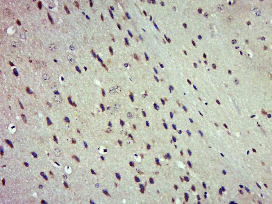
Paraformaldehyde-fixed, paraffin embedded (Mouse brain); Antigen retrieval by boiling in sodium citrate buffer (pH6.0) for 15min; Block endogenous peroxidase by 3% hydrogen peroxide for 20 minutes; Blocking buffer (normal goat serum) at 37°C for 30min; Antibody incubation with (JNK1 + JNK3) Polyclonal Antibody, Unconjugated (bs-0501R) at 1:500 overnight at 4°C, followed by a conjugated secondary (sp-0023) for 20 minutes and DAB staining.
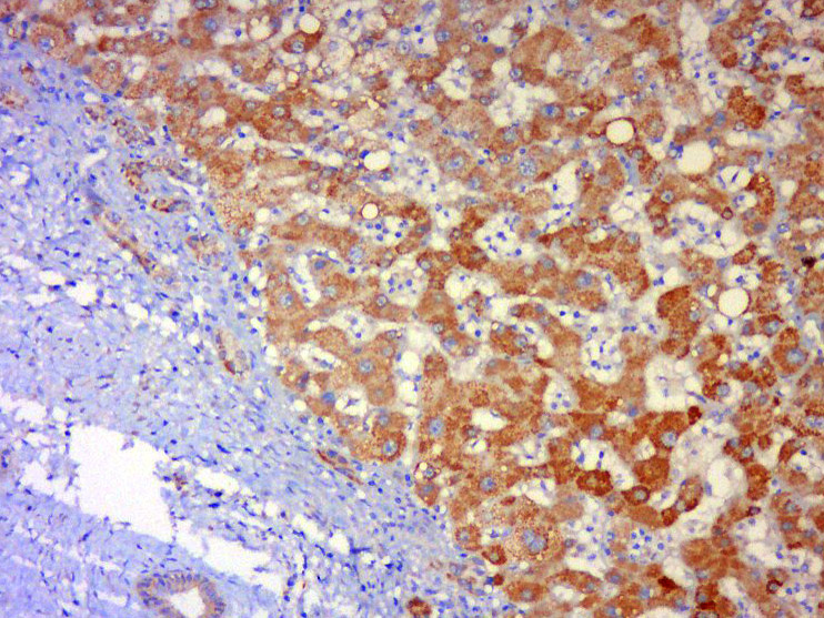
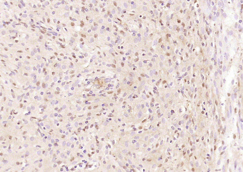
Tissue/cell: human liver carcinoma; 4% Paraformaldehyde-fixed and paraffin-embedded;
Antigen retrieval: citrate buffer ( 0.01M, pH 6.0 ), Boiling bathing for 15min; Block endogenous peroxidase by 3% Hydrogen peroxide for 30min; Blocking buffer (normal goat serum,C-0005) at 37℃ for 20 min;
Incubation: Anti-JNK1+ JNK3 Polyclonal Antibody, Unconjugated(bs-0501R) 1:500, overnight at 4°C, followed by conjugation to the secondary antibody(SP-0023) and DAB(C-0010) staining
Tissue/cell: human lung carcinoma; 4% Paraformaldehyde-fixed and paraffin-embedded;
Antigen retrieval: citrate buffer ( 0.01M, pH 6.0 ), Boiling bathing for 15min; Block endogenous peroxidase by 3% Hydrogen peroxide for 30min; Blocking buffer (normal goat serum,C-0005) at 37℃ for 20 min;
Incubation: Anti-JNK1/3 Polyclonal Antibody, Unconjugated(bs-0501R) 1:200, overnight at 4°C, followed by conjugation to the secondary antibody(SP-0023) and DAB(C-0010) staining
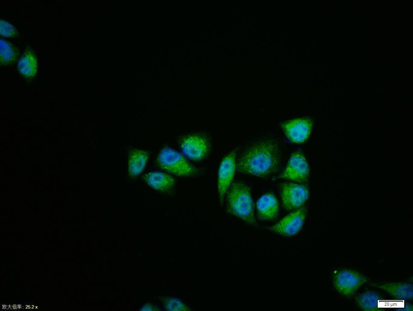
Hela cell; 4% Paraformaldehyde-fixed; Triton X-100 at room temperature for 20 min; Blocking buffer (normal goat serum, C-0005) at 37°C for 20 min; Antibody incubation with (JNK1 + JNK3) polyclonal Antibody, Unconjugated (bs-0501R) 1:100, 90 minutes at 37°C; followed by a conjugated Goat Anti-Rabbit IgG antibody at 37°C for 90 minutes, DAPI (blue, C02-04002) was used to stain the cell nuclei.
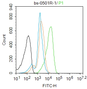
Blank control: K562.
Primary Antibody (green line): Rabbit Anti-JNK1 + JNK3 antibody (bs-0501R)
Dilution: 1μg /10^6 cells;
Isotype Control Antibody (orange line): Rabbit IgG .
Secondary Antibody : Goat anti-rabbit IgG-FITC
Dilution: 1μg /test.
Protocol
The cells were fixed with 4% PFA (10min at room temperature)and then permeabilized with 90% ice-cold methanol for 20 min at-20℃. The cells were then incubated in 5%BSA to block non-specific protein-protein interactions for 30 min at room temperature .Cells stained with Primary Antibody for 30 min at room temperature. The secondary antibody used for 40 min at room temperature. Acquisition of 20,000 events was performed.
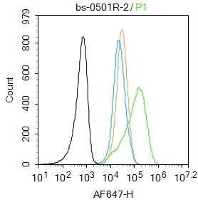
Blank control: Jurkat.
Primary Antibody (green line): Rabbit Anti-JNK1 + JNK3 antibody (bs-0501R)
Dilution: 2μg /10^6 cells;
Isotype Control Antibody (orange line): Rabbit IgG .
Secondary Antibody : Goat anti-rabbit IgG-AF647
Dilution: 1μg /test.
Protocol
The cells were fixed with 4% PFA (10min at room temperature)and then permeabilized with 90% ice-cold methanol for 20 min at-20℃. The cells were then incubated in 5%BSA to block non-specific protein-protein interactions for 30 min at room temperature .Cells stained with Primary Antibody for 30 min at room temperature. The secondary antibody used for 40 min at room temperature. Acquisition of 20,000 events was performed.
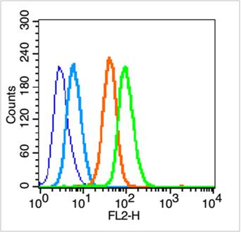
Blank control (blue line): Hep G2 (blue).
Primary Antibody (green line): Rabbit Anti-JNK1 + JNK3 antibody (bs-0501R)
Dilution: 1μg /10^6 cells;
Isotype Control Antibody (orange line): Rabbit IgG .
Secondary Antibody (white blue line): Goat anti-rabbit IgG-PE
Dilution: 1μg /test.
Protocol
The cells were fixed with 70% ethanol (Overnight at 4℃) and then permeabilized with 90% methanol for 20 min at -20℃. Cells stained with Primary Antibody for 30 min at room temperature. The cells were then incubated in 1 X PBS/2%BSA/10% goat serum to block non-specific protein-protein interactions followed by the antibody for 15 min at room temperature. The secondary antibody used for 40 min at room temperature. Acquisition of 20,000 events was performed.
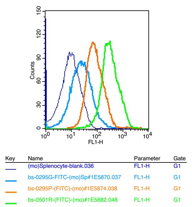
Blank control: mouse splenocytes(blue)
Isotype Control Antibody: Rabbit IgG(orange) ;
Secondary Antibody: Goat anti-rabbit IgG-FITC(white blue),
Dilution: 1:100 in 1 X PBS containing 0.5% BSA ;
Primary Antibody Dilution: 1μl in 100 μL1X PBS containing 0.5% BSA(green).
|
| 1、抗体溶解方法 | |
| 2、抗体修复方式 | |
| 3、常用试剂的配制 | |
| 4、免疫组化操作步骤 | |
| 5、免疫组化问题解答 | |
| 6、Western Blotting 操作步骤 | |
| 7、Western Blotting 问题解答 | |
| 8、关于肽链的设计 | |
| 9、多肽的溶解与保存 | |
| 10、酶标抗体效价测定程序 | |


