

| 产品编号 | bs-0465R |
| 英文名称 | NFKB p65 Rabbit pAb |
| 中文名称 | 细胞核因子/k基因结合核因子抗体 |
| 别 名 | AIF3BL3; CMCU; NFKB3; p65; p65 NF-kappa B; p65 NFkB; NFkB; nos2; TF65_HUMAN; RELA; Nuclear factor NF-kappa-B p65 subunit; Nuclear factor of kappa light polypeptide gene enhancer in B-cells 3; TF65_MOUSE; RELA proto-oncogene, NF-kB subunit; v-rel avian reticuloendotheliosis viral oncogene homolog A |
 |
Specific References (190) | bs-0465R has been referenced in 190 publications.
|
| 研究领域 | 细胞生物 免疫学 神经生物学 信号转导 细胞凋亡 转录调节因子 |
| 抗体来源 | Rabbit |
| 克隆类型 | Polyclonal |
| 交叉反应 | Human,Mouse (predicted: Rat,Rabbit,Pig,Sheep,Cow,Zebrafish,Chicken,Dog,Horse) |
| 产品应用 | Flow-Cyt=1μg/Test,ICC/IF=1:50-200
not yet tested in other applications. optimal dilutions/concentrations should be determined by the end user. |
| 理论分子量 | 61kDa |
| 细胞定位 | 细胞核 细胞浆 |
| 性 状 | Liquid |
| 浓 度 | 1mg/ml |
| 免 疫 原 | KLH conjugated synthetic peptide derived from human NFKBp65: 51-100/551 |
| 亚 型 | IgG |
| 纯化方法 | affinity purified by Protein A |
| 缓 冲 液 | 0.01M TBS (pH7.4) with 1% BSA, 0.02% Proclin300 and 50% Glycerol. |
| 保存条件 | Shipped at 4℃. Store at -20℃ for one year. Avoid repeated freeze/thaw cycles. |
| 注意事项 | This product as supplied is intended for research use only, not for use in human, therapeutic or diagnostic applications. |
| PubMed | PubMed |
| 产品介绍 |
NF-kappa-B is a ubiquitous transcription factor involved in several biological processes. It is held in the cytoplasm in an inactive state by specific inhibitors. Upon degradation of the inhibitor, NF-kappa-B moves to the nucleus and activates transcription of specific genes. NF-kappa-B is composed of NFKB1 or NFKB2 bound to either REL, RELA, or RELB. The most abundant form of NF-kappa-B is NFKB1 complexed with the product of this gene, RELA. Four transcript variants encoding different isoforms have been found for this gene. [provided by RefSeq, Sep 2011]. Function: NF-kappa-B is a pleiotropic transcription factor present in almost all cell types and is the endpoint of a series of signal transduction events that are initiated by a vast array of stimuli related to many biological processes such as inflammation, immunity, differentiation, cell growth, tumorigenesis and apoptosis. NF-kappa-B is a homo- or heterodimeric complex formed by the Rel-like domain-containing proteins RELA/p65, RELB, NFKB1/p105, NFKB1/p50, REL and NFKB2/p52 and the heterodimeric p65-p50 complex appears to be most abundant one. The dimers bind at kappa-B sites in the DNA of their target genes and the individual dimers have distinct preferences for different kappa-B sites that they can bind with distinguishable affinity and specificity. Different dimer combinations act as transcriptional activators or repressors, respectively. NF-kappa-B is controlled by various mechanisms of post-translational modification and subcellular compartmentalization as well as by interactions with other cofactors or corepressors. NF-kappa-B complexes are held in the cytoplasm in an inactive state complexed with members of the NF-kappa-B inhibitor (I-kappa-B) family. In a conventional activation pathway, I-kappa-B is phosphorylated by I-kappa-B kinases (IKKs) in response to different activators, subsequently degraded thus liberating the active NF-kappa-B complex which translocates to the nucleus. NF-kappa-B heterodimeric p65-p50 and p65-c-Rel complexes are transcriptional activators. The NF-kappa-B p65-p65 complex appears to be involved in invasin-mediated activation of IL-8 expression. The inhibitory effect of I-kappa-B upon NF-kappa-B the cytoplasm is exerted primarily through the interaction with p65. p65 shows a weak DNA-binding site which could contribute directly to DNA binding in the NF-kappa-B complex. Associates with chromatin at the NF-kappa-B promoter region via association with DDX1. Subunit: Component of the NF-kappa-B p65-p50 complex. Component of the NF-kappa-B p65-c-Rel complex. Homodimer; component of the NF-kappa-B p65-p65 complex. Component of the NF-kappa-B p65-p52 complex. May interact with ETHE1. Binds AES and TLE1. Interacts with TP53BP2. Binds to and is phosphorylated by the activated form of either RPS6KA4 or RPS6KA5. Interacts with ING4 and this interaction may be indirect. Interacts with CARM1, USP48 and UNC5CL. Interacts with IRAK1BP1. Interacts with NFKBID. Interacts with NFKBIA. Interacts with GSK3B. Interacts with NFKBIB. Interacts with NFKBIE. Interacts with NFKBIZ. Interacts with EHMT1 (via ANK repeats). Part of a 70-90 kDa complex at least consisting of CHUK, IKBKB, NFKBIA, RELA, IKBKAP and MAP3K14. Interacts with HDAC3; HDAC3 mediates the deacetylation of RELA. Interacts with HDAC1; the interaction requires non-phosphorylated RELA. Interacts with CBP; the interaction requires phosphorylated RELA. Interacts (phosphorylated at 'Thr-254') with PIN1; the interaction inhibits p65 binding to NFKBIA. Interacts with SOCS1. Interacts with UXT. Interacts with MTDH and PHF11. Interacts with ARRB2. Interacts with human respiratory syncytial virus (HRSV) protein M2-1. Interacts with NFKBIA (when phosphorylated), the interaction is direct; phosphorylated NFKBIA is part of a SCF(BTRC)-like complex lacking CUL1. Interacts with RNF25. Interacts (via C-terminus) with DDX1. Interacts with UFL1 and COMMD1. Interacts with BRMS1; this promotes deacetylation of 'Lys-310'. Interacts with NOTCH2. Directly interacts with MEN1; this interaction represses NFKB-mediated transactivation. Interacts with AKIP1, which promotes the phosphorylation and nuclear retention of RELA. Interacts (via the RHD) with GFI1; the interaction, after bacterial lipopolysaccharide (LPS) stimulation, inhibits the transcriptional activity by interfering with the DNA-binding activity to target gene promoter DNA. Subcellular Location: Nucleus. Cytoplasm. Note=Colocalized with DDX1 in the nucleus upon TNF-alpha induction. Nuclear, but also found in the cytoplasm in an inactive form complexed to an inhibitor (I-kappa-B). Colocalizes with GFI1 in the nucleus after LPS stimulation. Post-translational modifications: Ubiquitinated, leading to its proteasomal degradation. Degradation is required for termination of NF-kappa-B response. Monomethylated at Lys-310 by SETD6. Monomethylation at Lys-310 is recognized by the ANK repeats of EHMT1 and promotes the formation of repressed chromatin at target genes, leading to down-regulation of NF-kappa-B transcription factor activity. Phosphorylation at Ser-311 disrupts the interaction with EHMT1 without preventing monomethylation at Lys-310 and relieves the repression of target genes. Phosphorylation at Ser-311 disrupts the interaction with EHMT1 and promotes transcription factor activity. Phosphorylation on Ser-536 stimulates acetylation on Lys-310 and interaction with CBP; the phosphorylated and acetylated forms show enhanced transcriptional activity. Phosphorylation at Ser-276 by RPS6KA4 and RPS6KA5 promotes its transactivation and transcriptional activities. Reversibly acetylated; the acetylation seems to be mediated by CBP, the deacetylation by HDAC3 and SIRT2. Acetylation at Lys-122 enhances DNA binding and impairs association with NFKBIA. Acetylation at Lys-310 is required for full transcriptional activity in the absence of effects on DNA binding and NFKBIA association. Acetylation can also lower DNA-binding and results in nuclear export. Interaction with BRMS1 promotes deacetylation of Lys-310. Lys-310 is deacetylated by SIRT2. S-nitrosylation of Cys-38 inactivates the enzyme activity. Sulfhydration at Cys-38 mediates the anti-apoptotic activity by promoting the interaction with RPS3 and activating the transcription factor activity. Sumoylation by PIAS3 negatively regulates DNA-bound activated NF-kappa-B. Similarity: Contains 1 RHD (Rel-like) domain. SWISS: Q04206 Gene ID: 5970 Database links: Entrez Gene: 5970 Human Entrez Gene: 19697 Mouse Omim: 164014 Human SwissProt: Q04206 Human SwissProt: Q04207 Mouse Unigene: 502875 Human Unigene: 249966 Mouse Unigene: 19480 Rat 转录调节因子(Transcriptin Regulators) NF-κBp65是一种重要的转录因子,NF-kBp65可激活参与炎症、细胞增殖、细胞凋亡等基因的调节,影响着细胞的凋亡,同时影响着肿瘤细胞对细胞毒性药物及离子辐射的敏感性。ras基因诱导的致癌突变作用需NFkB的活化,提示NFkB在致癌发生方面可能起一定作用;另有文献报道,在乳腺癌、非小细胞性肺癌、甲状腺癌、T或B淋巴细胞白血病及病毒诱变导致的肿瘤等人类肿瘤中,NFkB活化或表达。 经研究认为:NFKBp65蛋白在静息状态下以结合态的方式存在于胞浆中,当NFKBp65蛋白被激活后解离进入细胞核。(NF-κB与其抑制蛋白IκB家族成员结合,以无活性的复合物形式存在于胞浆中,当细胞受到各种刺激后,NF- κB与IκB 解离,从而进入细胞核,与相应的靶序列结合,调节基因的表达) NF-кB可以保护细胞免受肿瘤坏死因子以及电离辐射等引起的凋亡作用,而抑制NFkB的表达可以增加TNF等引起的细胞凋亡,以及增加化疗及放疗对肿瘤细胞的敏感性。 |
| 产品图片 |
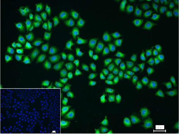
4% Paraformaldehyde-fixed Hela (H) cell; Triton X-100 at r.t. for 20 min; Antibody incubation with (NFKB p65) polyclonal Antibody, unconjugated (bs-0465R) 1:100, 90 min at 37°C; followed by conjugated Goat Anti-Rabbit IgG antibody (green, bs-40295G-FITC) at 37°C for 90 min, DAPI (blue, C02-04002) was used to stain the cell nuclei. PBS instead of the primary antibody was used as the blank control.
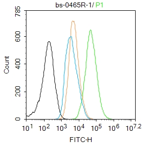
Blank control:A431.
Primary Antibody (green line): Rabbit Anti-NFKB p65 antibody (bs-3485R)
Dilution: 1μg /10^6 cells;
Isotype Control Antibody (orange line): Rabbit IgG .
Secondary Antibody : Goat anti-rabbit IgG-FITC
Dilution: 1μg /test.
Protocol
The cells were fixed with 4% PFA (10min at room temperature)and then permeabilized with 90% ice-cold methanol for 20 min at-20℃. The cells were then incubated in 5%BSA to block non-specific protein-protein interactions for 30 min at room temperature .Cells stained with Primary Antibody for 30 min at room temperature. The secondary antibody used for 40 min at room temperature. Acquisition of 20,000 events was performed.
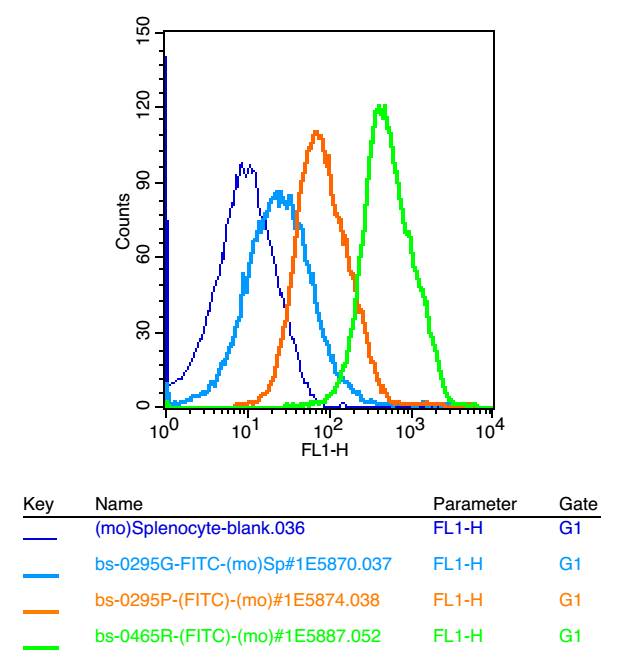
Blank control: mouse splenocytes(blue)
Isotype Control Antibody: Rabbit IgG(orange) ;
Secondary Antibody: Goat anti-rabbit IgG-FITC(white blue),
Dilution: 1:100 in 1 X PBS containing 0.5% BSA ;
Primary Antibody Dilution: 1μl in 100 μL1X PBS containing 0.5% BSA(green).
|
| 1、抗体溶解方法 | |
| 2、抗体修复方式 | |
| 3、常用试剂的配制 | |
| 4、免疫组化操作步骤 | |
| 5、免疫组化问题解答 | |
| 6、Western Blotting 操作步骤 | |
| 7、Western Blotting 问题解答 | |
| 8、关于肽链的设计 | |
| 9、多肽的溶解与保存 | |
| 10、酶标抗体效价测定程序 | |


