

| 产品编号 | bs-0231R |
| 英文名称 | PDGFRA Rabbit pAb |
| 中文名称 | 血小板源性生长因子受体A/PDGFRα抗体 |
| 别 名 | CD140A; PDGFR-2; PDGFR2; APDGFR; PDGFACE; PGFRA_HUMAN; PDGFRA; PDGF-R-alpha; PDGFR-alpha; Alpha platelet-derived growth factor receptor; Alpha-type platelet-derived growth factor receptor; CD140 antigen-like family member A; CD140a antigen; Platelet-deriv |
 |
Specific References (6) | bs-0231R has been referenced in 6 publications.
[IF=14.808] Xue Gong. et al. Circular RNA circEsyt2 regulates vascular smooth muscle cell remodeling via splicing regulation. J Clin Invest. 2021 Dec 15;131(24):e147031 IF ; Mouse.
[IF=6.684] Yang Shuo. et al. The Protective Effects of γ-Tocotrienol on Muscle Stem Cells Through Inhibiting Reactive Oxidative Stress Production. Front Cell Dev Biol. 2022 Mar;0:588 IHC ; Mouse.
[IF=6.208] Casandra Walker. et al. Impact of Fetal Exposure to Endocrine Disrupting Chemical Mixtures on FOXA3 Gene and Protein Expression in Adult Rat Testes. INT J MOL SCI. 2023 Jan;24(2):1211 IF ; Rat.
[IF=4.927] Wenjing Song. et al. Sigma-1 Receptor Activation Improves Oligodendrogenesis and Promotes White-Matter Integrity after Stroke in Mice with Diabetic Mellitus. MOLECULES. 2023 Jan;28(1):390 IHC ; Mouse.
[IF=4.411] Hao Ruan. et al. Fedratinib Attenuates Bleomycin-Induced Pulmonary Fibrosis via the JAK2/STAT3 and TGF-β1 Signaling Pathway. Molecules. 2021 Jan;26(15):4491 IF ; Mouse.
[IF=1.89] Hu, Jianguo, Biao Zeng, and Xingwei Jiang. "The expression of marker for endometrial stem cell and fibrosis was increased in intrauterine adhesious."International journal of clinical and experimental pathology 8.2 (2015): 1525. IHC-P ; Mouse.
|
| 研究领域 | 心血管 信号转导 生长因子和激素 |
| 抗体来源 | Rabbit |
| 克隆类型 | Polyclonal |
| 克 隆 号 | |
| 交叉反应 | Human,Mouse,Rat (predicted: Pig,Cow,Chicken,Dog,Horse) |
| 产品应用 | IHC-P=1:100-500,IHC-F=1:100-500,IF=1:100-500,Flow-Cyt=1μg/Test
not yet tested in other applications. optimal dilutions/concentrations should be determined by the end user. |
| 理论分子量 | 117 kDa |
| 细胞定位 | 细胞膜 |
| 性 状 | Liquid |
| 浓 度 | 1mg/ml |
| 免 疫 原 | KLH conjugated synthetic peptide derived from human PDGF-R-A: 1021-1089/1089 <Cytoplasmic> |
| 亚 型 | IgG |
| 纯化方法 | affinity purified by Protein A |
| 缓 冲 液 | 0.01M TBS (pH7.4) with 1% BSA, 0.02% Proclin300 and 50% Glycerol. |
| 保存条件 | Shipped at 4℃. Store at -20℃ for one year. Avoid repeated freeze/thaw cycles. |
| 注意事项 | This product as supplied is intended for research use only, not for use in human, therapeutic or diagnostic applications. |
| PubMed | PubMed |
| 产品介绍 |
This gene encodes a cell surface tyrosine kinase receptor for members of the platelet-derived growth factor family. These growth factors are mitogens for cells of mesenchymal origin. The identity of the growth factor bound to a receptor monomer determines whether the functional receptor is a homodimer or a heterodimer, composed of both platelet-derived growth factor receptor alpha and beta polypeptides. Studies suggest that this gene plays a role in organ development, wound healing, and tumor progression. Mutations in this gene have been associated with idiopathic hypereosinophilic syndrome, somatic and familial gastrointestinal stromal tumors, and a variety of other cancers. [provided by RefSeq, Mar 2012]. Function: Tyrosine-protein kinase that acts as a cell-surface receptor for PDGFA, PDGFB and PDGFC and plays an essential role in the regulation of embryonic development, cell proliferation, survival and chemotaxis. Depending on the context, promotes or inhibits cell proliferation and cell migration. Plays an important role in the differentiation of bone marrow-derived mesenchymal stem cells. Required for normal skeleton development and cephalic closure during embryonic development. Required for normal development of the mucosa lining the gastrointestinal tract, and for recruitment of mesenchymal cells and normal development of intestinal villi. Plays a role in cell migration and chemotaxis in wound healing. Plays a role in platelet activation, secretion of agonists from platelet granules, and in thrombin-induced platelet aggregation. Binding of its cognate ligands - homodimeric PDGFA, homodimeric PDGFB, heterodimers formed by PDGFA and PDGFB or homodimeric PDGFC -leads to the activation of several signaling cascades; the response depends on the nature of the bound ligand and is modulated by the formation of heterodimers between PDGFRA and PDGFRB. Phosphorylates PIK3R1, PLCG1, and PTPN11. Activation of PLCG1 leads to the production of the cellular signaling molecules diacylglycerol and inositol 1,4,5-trisphosphate, mobilization of cytosolic Ca(2+) and the activation of protein kinase C. Phosphorylates PIK3R1, the regulatory subunit of phosphatidylinositol 3-kinase, and thereby mediates activation of the AKT1 signaling pathway. Mediates activation of HRAS and of the MAP kinases MAPK1/ERK2 and/or MAPK3/ERK1. Promotes activation of STAT family members STAT1, STAT3 and STAT5A and/or STAT5B. Receptor signaling is down-regulated by protein phosphatases that dephosphorylate the receptor and its down-stream effectors, and by rapid internalization of the activated receptor. Subunit: Interacts with homodimeric PDGFA, PDGFB and PDGFC, and with heterodimers formed by PDGFA and PDGFB. Monomer in the absence of bound ligand. Interaction with dimeric PDGFA, PDGFB and/or PDGFC leads to receptor dimerization, where both PDGFRA homodimers and heterodimers with PDGFRB are observed. Interacts (tyrosine phosphorylated) with SHB (via SH2 domain). Interacts (tyrosine phosphorylated) with SHF (via SH2 domain). Interacts (tyrosine phosphorylated) with SRC (via SH2 domain). Interacts (tyrosine phosphorylated) with PIK3R1. Interacts (tyrosine phosphorylated) with PLCG1 (via SH2 domain). Interacts (tyrosine phosphorylated) with CRK, GRB2 and GRB7. Interacts with human cytomegalovirus/HHV-5 envelop glycoprotein B/gB. Subcellular Location: Cell membrane; Single-pass type I membrane protein. Note=The activated receptor is rapidly internalized and degraded. Tissue Specificity: Detected in platelets (at protein level). Widely expressed. Detected in brain, fibroblasts, smooth muscle, heart, and embryo. Expressed in primary and metastatic colon tumors and in normal colon tissue. Post-translational modifications: N-glycosylated. Ubiquitinated, leading to its degradation (Probable). Autophosphorylated on tyrosine residues upon ligand binding. Autophosphorylation occurs in trans, i.e. one subunit of the dimeric receptor phosphorylates tyrosine residues on the other subunit. Phosphorylation at Tyr-731 and Tyr-742 is important for interaction with PIK3R1. Phosphorylation at Tyr-720 and Tyr-754 is important for interaction with PTPN11. Phosphorylation at Tyr-762 is important for interaction with CRK. Phosphorylation at Tyr-572 and Tyr-574 is important for interaction with SRC and SRC family members. Phosphorylation at Tyr-988 and Tyr-1018 is important for interaction with PLCG1. DISEASE: Note=A chromosomal aberration involving PDGFRA is found in some cases of hypereosinophilic syndrome. Interstitial chromosomal deletion del(4)(q12q12) causes the fusion of FIP1L1 and PDGFRA (FIP1L1-PDGFRA). Mutations that cause overexpression and/or constitutive activation of PDGFRA may be a cause of hypereosinophilic syndrome. Defects in PDGFRA are a cause of gastrointestinal stromal tumor (GIST) [MIM:606764]. Note=Mutations that cause constitutive activation of PDGFRA may be a cause of gastrointestinal stromal tumor (GIST). Similarity: Belongs to the protein kinase superfamily. Tyr protein kinase family. CSF-1/PDGF receptor subfamily. Contains 5 Ig-like C2-type (immunoglobulin-like) domains. Contains 1 protein kinase domain. SWISS: P16234 Gene ID: 5156 Database links: Entrez Gene: 5156 Human Entrez Gene: 18595 Mouse Omim: 173490 Human SwissProt: P16234 Human SwissProt: P26618 Mouse Unigene: 74615 Human Unigene: 221403 Mouse Unigene: 55127 Rat 细胞膜受体(Membrane Receptors) PDGFR-α是膜受体,具有酪氨酸酶的活性,与其配体PDGF结合后激活与细胞增殖有关的酶及基因。 PDGFR亦表达于上皮、内皮细胞,前列腺、皮肤、肾小球等上皮细胞均有PDGFR表达. 亦有学者报道血小板源性生长因子受体α抗体在细胞胞浆胞膜、胞核都有不同的表达 还有人认为:PDGF及其受体一般表达于浸润病变组织的炎症细胞附近,组织缺血损伤、肾小球高压、免疫因素作用、炎症细胞浸润、实质细胞活化等导致PDGFR表达增强 |
| 产品图片 |
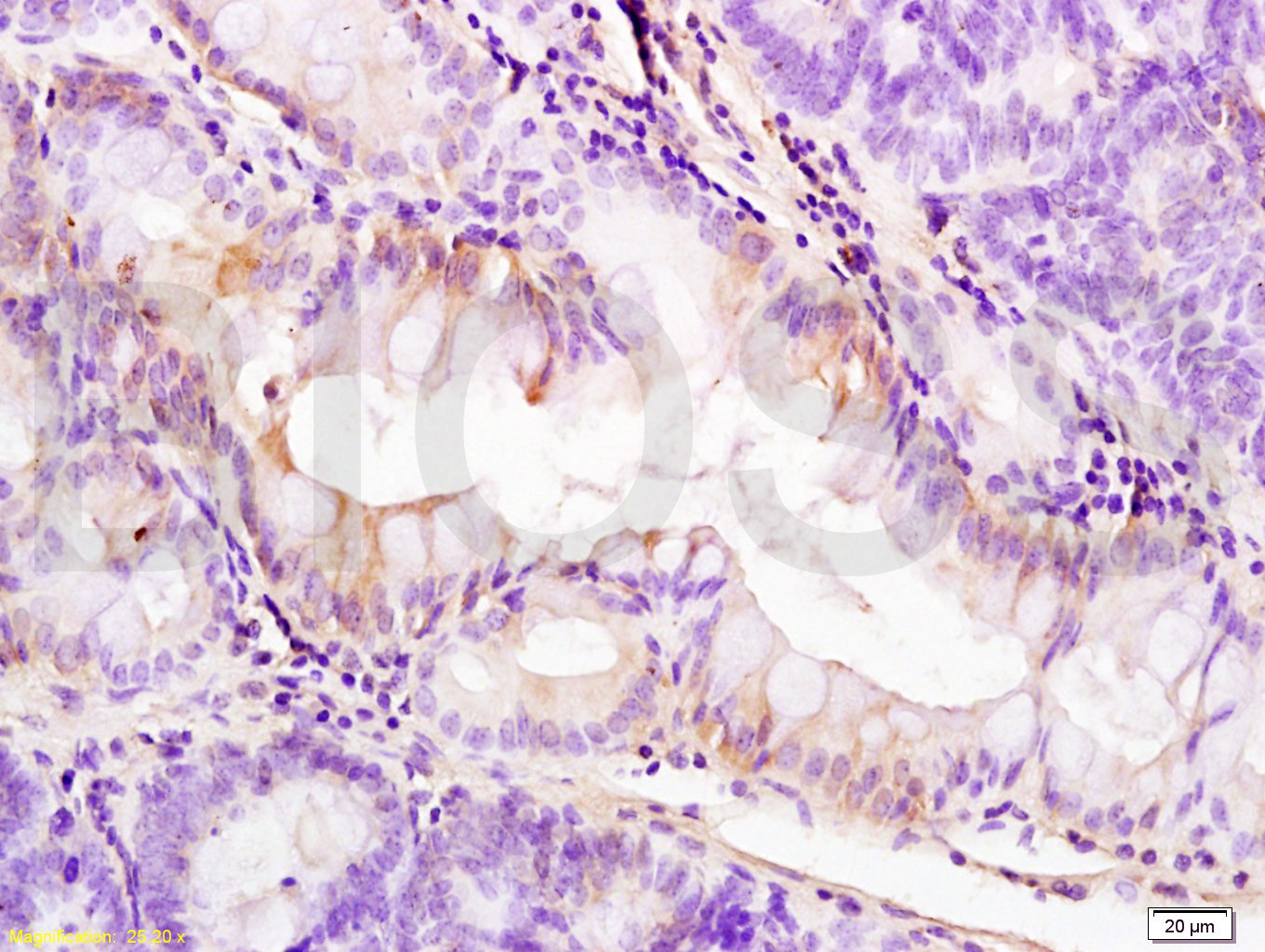
Tissue/cell: rat colitis tissue; 4% Paraformaldehyde-fixed and paraffin-embedded;
Antigen retrieval: citrate buffer ( 0.01M, pH 6.0 ), Boiling bathing for 15min; Block endogenous peroxidase by 3% Hydrogen peroxide for 30min; Blocking buffer (normal goat serum,C-0005) at 37℃ for 20 min;
Incubation: Anti-PDGFRA Polyclonal Antibody, Unconjugated(bs-0231R) 1:200, overnight at 4°C, followed by conjugation to the secondary antibody(SP-0023) and DAB(C-0010) staining
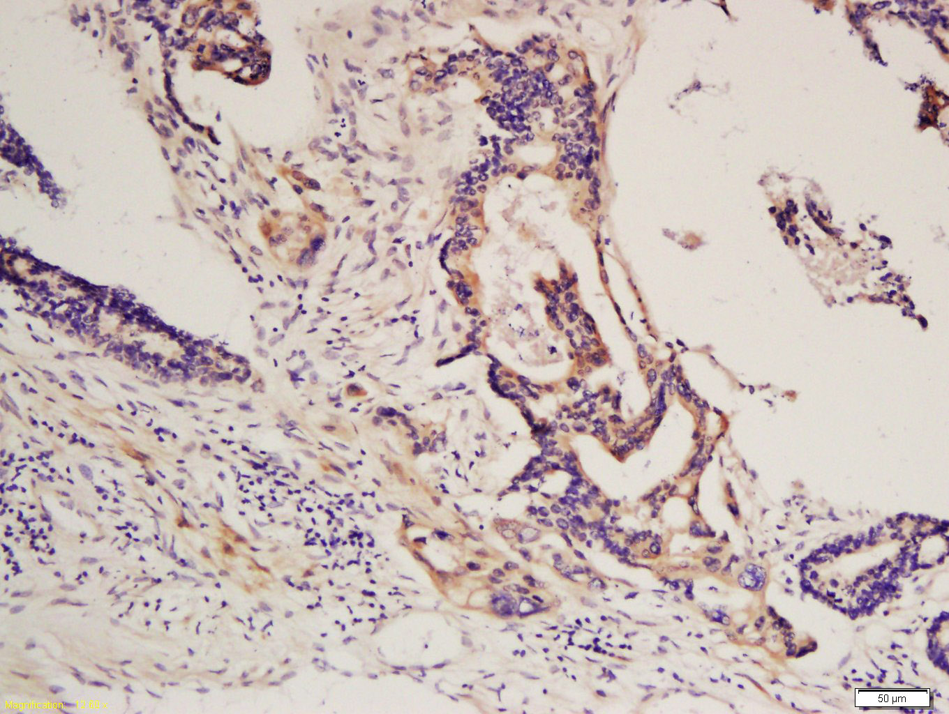
Tissue/cell: human colon carcinoma; 4% Paraformaldehyde-fixed and paraffin-embedded;
Antigen retrieval: citrate buffer ( 0.01M, pH 6.0 ), Boiling bathing for 15min; Block endogenous peroxidase by 3% Hydrogen peroxide for 30min; Blocking buffer (normal goat serum,C-0005) at 37℃ for 20 min;
Incubation: Anti-PDGFRA Polyclonal Antibody, Unconjugated(bs-0231R) 1:200, overnight at 4°C, followed by conjugation to the secondary antibody(SP-0023) and DAB(C-0010) staining
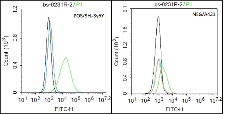
Black line : Positive blank control (SH-Sy5Y); Negative blank control (A431)
Green line : Primary Antibody (Rabbit Anti-PDGFRA antibody (bs-0231R) )
Orange line:Isotype Control Antibody (Rabbit IgG) .
Blue line : Secondary Antibody (Goat anti-rabbit IgG-AF488)
SH-Sy5Y(Positive)and A431(Negative control)cells (black) were fixed with 4% PFA for 10min at room temperature,permeabilized with 90% ice-cold methanol for 20 min at -20℃, and incubated in 5% BSA blocking buffer for 30 min at room temperature. Cells were then stained with PDGFRA Antibody(bs-0231R)at 1:50 dilution in blocking buffer and incubated for 30 min at room temperature, washed twice with 2% BSA in PBS, followed by secondary antibody(blue) incubation for 40 min at room temperature. Acquisitions of 20,000 events were performed. Cells stained with primary antibody (green), and isotype control (orange).
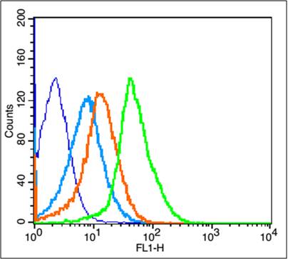
Blank control: Mouse Kidney (blue). Primary Antibody:Rabbit Anti-PDGFRA antibody (bs-0231R,Green); Dilution: 1μg in 100 μL 1X PBS containing 0.5% BSA; Isotype Control Antibody: Rabbit IgG(orange) ,used under the same conditions; Secondary Antibody: Goat anti-rabbit IgG-FITC(white blue), Dilution: 1:200 in 1 X PBS containing 0.5% BSA.
Protocol
The cells were fixed with 2% paraformaldehyde for 10 min at 37℃. Primary antibody (bs-0231R, 1μg /1x10^6 cells) were incubated for 30 min at room temperature, followed by 1 X PBS containing 0.5% BSA + 1 0% goat serum (15 min) to block non-specific protein-protein interactions. Then the Goat Anti-rabbit IgG/FITC antibody was added into the blocking buffer mentioned above to react with the primary antibody at 1/200 dilution for 40 min on ice. Acquisition of 20,000 events was performed.
|
| 1、抗体溶解方法 | |
| 2、抗体修复方式 | |
| 3、常用试剂的配制 | |
| 4、免疫组化操作步骤 | |
| 5、免疫组化问题解答 | |
| 6、Western Blotting 操作步骤 | |
| 7、Western Blotting 问题解答 | |
| 8、关于肽链的设计 | |
| 9、多肽的溶解与保存 | |
| 10、酶标抗体效价测定程序 | |


