

| 产品编号 | bs-0636R |
| 英文名称 | phospho-P38 MAPK (Thr180 + Tyr182) Rabbit pAb |
| 中文名称 | 磷酸化-丝裂原活化蛋白激酶p38抗体(P-p38 MAPK) |
| 别 名 | MAPK14 | p38 MAPK (phospho-T180/Y182); p-p38 MAPK; phospho-p38 MAPK; p-MAPK14; MAPK14 | p38 MAPK (phospho-Thr180/Tyr182); CSBP; CSBP1; CSBP2; CSPB1; EXIP; Mxi2; PRKM14; PRKM15; RK; SAPK2A; p38; p38ALPHA; Crk1; p38-alpha; p38MAPK; p38a; Hog; p38Hog; MK14_C |
 |
Specific References (41) | bs-0636R has been referenced in 41 publications.
|
| 产品类型 | 磷酸化抗体 |
| 研究领域 | 肿瘤 细胞生物 免疫学 信号转导 细胞凋亡 转录调节因子 激酶和磷酸酶 |
| 抗体来源 | Rabbit |
| 克隆类型 | Polyclonal |
| 克 隆 号 | |
| 交叉反应 | Human,Mouse,Rat (predicted: Rabbit,Dog) |
| 产品应用 | IHC-P=1:100-500,IHC-F=1:100-500,IF=1:100-500
not yet tested in other applications. optimal dilutions/concentrations should be determined by the end user. |
| 理论分子量 | 42 kDa |
| 细胞定位 | 细胞核 细胞浆 |
| 性 状 | Liquid |
| 浓 度 | 1mg/ml |
| 免 疫 原 | KLH conjugated Synthesised phosphopeptide derived from human p38 MAPK around the phosphorylation site of Thr180/Tyr182: M(p-T)G(p-Y)VA |
| 亚 型 | IgG |
| 纯化方法 | affinity purified by Protein A |
| 缓 冲 液 | 0.01M TBS (pH7.4) with 1% BSA, 0.02% Proclin300 and 50% Glycerol. |
| 保存条件 | Shipped at 4℃. Store at -20℃ for one year. Avoid repeated freeze/thaw cycles. |
| 注意事项 | This product as supplied is intended for research use only, not for use in human, therapeutic or diagnostic applications. |
| PubMed | PubMed |
| 产品介绍 |
The protein encoded by this gene is a member of the MAP kinase family. MAP kinases act as an integration point for multiple biochemical signals, and are involved in a wide variety of cellular processes such as proliferation, differentiation, transcription regulation and development. This kinase is activated by various environmental stresses and proinflammatory cytokines. The activation requires its phosphorylation by MAP kinase kinases(MKKs), or its autophosphorylation triggered by the interaction of MAP3K7IP1/TAB1 protein with this kinase. The substrates of this kinase include transcription regulator ATF2, MEF2C, and MAX, cell cycle regulator CDC25B, and tumor suppressor p53, which suggest the roles of this kinase in stress related transcription and cell cycle regulation, as well as in genotoxic stress response. Four alternatively spliced transcript variants of this gene encoding distinct isoforms have been reported. Function: Serine/threonine kinase which acts as an essential component of the MAP kinase signal transduction pathway. MAPK14 is one of the four p38 MAPKs which play an important role in the cascades of cellular responses evoked by extracellular stimuli such as proinflammatory cytokines or physical stress leading to direct activation of transcription factors. Accordingly, p38 MAPKs phosphorylate a broad range of proteins and it has been estimated that they may have approximately 200 to 300 substrates each. Some of the targets are downstream kinases which are activated through phosphorylation and further phosphorylate additional targets. RPS6KA5/MSK1 and RPS6KA4/MSK2 can directly phosphorylate and activate transcription factors such as CREB1, ATF1, the NF-kappa-B isoform RELA/NFKB3, STAT1 and STAT3, but can also phosphorylate histone H3 and the nucleosomal protein HMGN1. RPS6KA5/MSK1 and RPS6KA4/MSK2 play important roles in the rapid induction of immediate-early genes in response to stress or mitogenic stimuli, either by inducing chromatin remodeling or by recruiting the transcription machinery. On the other hand, two other kinase targets, MAPKAPK2/MK2 and MAPKAPK3/MK3, participate in the control of gene expression mostly at the post-transcriptional level, by phosphorylating ZFP36 (tristetraprolin) and ELAVL1, and by regulating EEF2K, which is important for the elongation of mRNA during translation. MKNK1/MNK1 and MKNK2/MNK2, two other kinases activated by p38 MAPKs, regulate protein synthesis by phosphorylating the initiation factor EIF4E2. MAPK14 interacts also with casein kinase II, leading to its activation through autophosphorylation and further phosphorylation of TP53/p53. In the cytoplasm, the p38 MAPK pathway is an important regulator of protein turnover. For example, CFLAR is an inhibitor of TNF-induced apoptosis whose proteasome-mediated degradation is regulated by p38 MAPK phosphorylation. In a similar way, MAPK14 phosphorylates the ubiquitin ligase SIAH2, regulating its activity towards EGLN3. MAPK14 may also inhibit the lysosomal degradation pathway of autophagy by interfering with the intracellular trafficking of the transmembrane protein ATG9. Another function of MAPK14 is to regulate the endocytosis of membrane receptors by different mechanisms that impinge on the small GTPase RAB5A. In addition, clathrin-mediated EGFR internalization induced by inflammatory cytokines and UV irradiation depends on MAPK14-mediated phosphorylation of EGFR itself as well as of RAB5A effectors. Ectodomain shedding of transmembrane proteins is regulated by p38 MAPKs as well. In response to inflammatory stimuli, p38 MAPKs phosphorylate the membrane-associated metalloprotease ADAM17. Such phosphorylation is required for ADAM17-mediated ectodomain shedding of TGF-alpha family ligands, which results in the activation of EGFR signaling and cell proliferation. Another p38 MAPK substrate is FGFR1. FGFR1 can be translocated from the extracellular space into the cytosol and nucleus of target cells, and regulates processes such as rRNA synthesis and cell growth. FGFR1 translocation requires p38 MAPK activation. In the nucleus, many transcription factors are phosphorylated and activated by p38 MAPKs in response to different stimuli. Classical examples include ATF1, ATF2, ATF6, ELK1, PTPRH, DDIT3, TP53/p53 and MEF2C and MEF2A. The p38 MAPKs are emerging as important modulators of gene expression by regulating chromatin modifiers and remodelers. The promoters of several genes involved in the inflammatory response, such as IL6, IL8 and IL12B, display a p38 MAPK-dependent enrichment of histone H3 phosphorylation on 'Ser-10' (H3S10ph) in LPS-stimulated myeloid cells. This phosphorylation enhances the accessibility of the cryptic NF-kappa-B-binding sites marking promoters for increased NF-kappa-B recruitment. Phosphorylates CDC25B and CDC25C which is required for binding to 14-3-3 proteins and leads to initiation of a G2 delay after ultraviolet radiation. Phosphorylates TIAR following DNA damage, releasing TIAR from GADD45A mRNA and preventing mRNA degradation. The p38 MAPKs may also have kinase-independent roles, which are thought to be due to the binding to targets in the absence of phosphorylation. Protein O-Glc-N-acylation catalyzed by the OGT is regulated by MAPK14, and, although OGT does not seem to be phosphorylated by MAPK14, their interaction increases upon MAPK14 activation induced by glucose deprivation. This interaction may regulate OGT activity by recruiting it to specific targets such as neurofilament H, stimulating its O-Glc-N-acylation. Required in mid-fetal development for the growth of embryo-derived blood vessels in the labyrinth layer of the placenta. Also plays an essential role in developmental and stress-induced erythropoiesis, through regulation of EPO gene expression. Isoform MXI2 activation is stimulated by mitogens and oxidative stress and only poorly phosphorylates ELK1 and ATF2. Isoform EXIP may play a role in the early onset of apoptosis. Phosphorylates S100A9 at 'Thr-113'. Subunit: Binds to a kinase interaction motif within the protein tyrosine phosphatase, PTPRR (By similarity). This interaction retains MAPK14 in the cytoplasm and prevents nuclear accumulation. Interacts with SPAG9 and GADD45A. Interacts with CDC25B, CDC25C, DUSP1, DUSP10, DUSP16, NP60, FAM48A and TAB1. Interacts with casein kinase II subunits CSNK2A1 and CSNK2B. Subcellular Location: Cytoplasm. Nucleus. Tissue Specificity: Brain, heart, placenta, pancreas and skeletal muscle. Expressed to a lesser extent in lung, liver and kidney. Post-translational modifications: Dually phosphorylated on Thr-180 and Tyr-182 by the MAP2Ks MAP2K3/MKK3, MAP2K4/MKK4 and MAP2K6/MKK6 in response to inflammatory citokines, environmental stress or growth factors, which a ctivates the enzyme. Dual phosphorylation can also be mediated by TAB1-mediated autophosphorylation. TCR engagement in T-cells also leads to Tyr-323 phosphorylation by ZAP70. Dephosphorylated and inactivated by DUPS1, DUSP10 and DUSP16. Acetylated at Lys-53 and Lys-152 by KAT2B and EP300. Acetylation at Lys-53 increases the affinity for ATP and enhances kinase activity. Lys-53 and Lys-152 are deacetylated by HDAC3. Ubiquitinated. Ubiquitination leads to degradation by the proteasome pathway. Similarity: Belongs to the protein kinase superfamily. CMGC Ser/Thr protein kinase family. MAP kinase subfamily. Contains 1 protein kinase domain. SWISS: Q16539 Gene ID: 1432 Database links: Entrez Gene: 1432 Human Entrez Gene: 26416 Mouse GenBank: NM_001315 Human GenBank: NM_139012 Human GenBank: NM_011951 Mouse Omim: 600289 Human SwissProt: Q16539 Human SwissProt: P47811 Mouse Unigene: 485233 Human Unigene: 311337 Mouse Unigene: 88085 Rat 激酶和磷酸酶(Kinases and Phosphatases) 丝裂原活化蛋白激酶p38(p38 MAPK、磷酸化pERK)参与细胞生长、增殖、分化、死亡及细胞间的功能同步等多种生理过程. P-p38MAPK是丝裂原活化蛋白激酶家族中的成员之一,大量研究显示p38在能量代谢中具有广泛的作用。p38参与脂肪组织、骨骼肌、胰岛细胞和肝脏等组织、器官的能量代谢.分子量:38KDa p38 MAPK:作为细胞信号传递系统的交汇点,细胞内普遍存在的一条信号转导通路。细胞外的物理应激因子,如高渗透压、热休克、紫外线以及细胞因子、内毒素脂多糖(LPS)等都能激活该途径,诱导细胞内蛋白质合成与分泌、细胞分化及凋亡等生物效应。p38 MAPK还能与细胞内其他信号通路之间相互作用,是细胞内信号传递系统的交汇点或共同通路。p38 MAPK一旦被激活后,可以使一些转录因子如CREB、转录活化因子-1(activating factor-1, ATF-1)、ATF-2及活化蛋白-1(AP-1)等的丝氨酸和苏氨酸位点磷酸化,活化这些转录因子,从而调节目的基因的表达。 p38(丝氨酸位点)磷酸化后可以直接激活转录因子,参与机体的应激反应。 |
| 产品图片 |
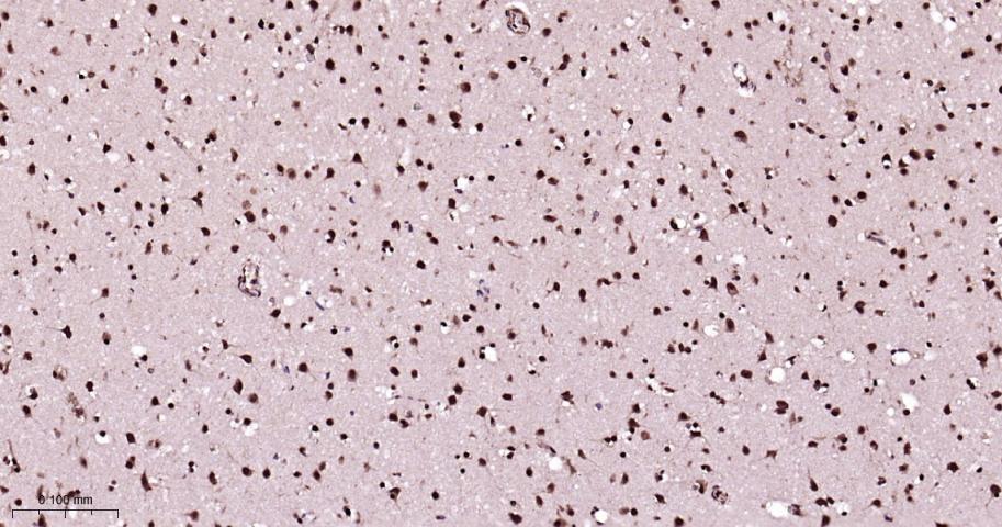
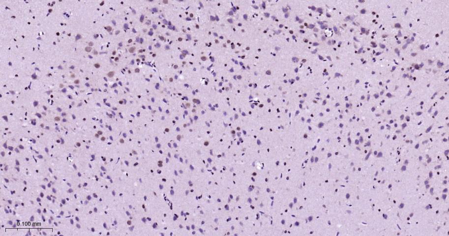
Paraformaldehyde-fixed, paraffin embedded Human Brain; Antigen retrieval by boiling in sodium citrate buffer (pH6.0) for 15 min; Antibody incubation with Phospho-P38 MAPK (Thr180 + Tyr182) Polyclonal Antibody, Unconjugated(bs-0636R) at 1:200 overnight at 4°C, followed by conjugation to the bs-0295G-HRP and DAB (C-0010) staining.
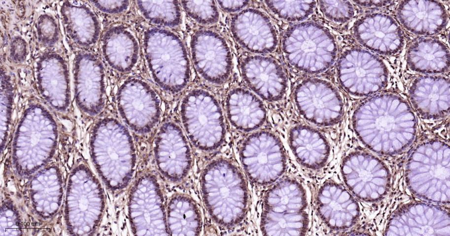
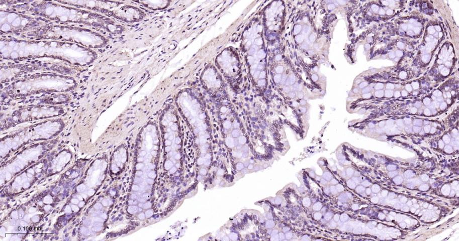
Paraformaldehyde-fixed, paraffin embedded Human colon; Antigen retrieval by boiling in sodium citrate buffer (pH6.0) for 15 min; Antibody incubation with Phospho-P38 MAPK (Thr180 + Tyr182) Polyclonal Antibody, Unconjugated(bs-0636R) at 1:200 overnight at 4°C, followed by conjugation to the bs-0295G-HRP and DAB (C-0010) staining.
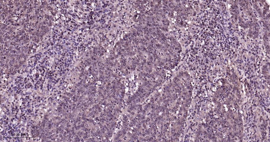
Paraformaldehyde-fixed, paraffin embedded Human colon cancer; Antigen retrieval by boiling in sodium citrate buffer (pH6.0) for 15 min; Antibody incubation with Phospho-P38 MAPK (Thr180 + Tyr182) Polyclonal Antibody, Unconjugated(bs-0636R) at 1:200 overnight at 4°C, followed by conjugation to the bs-0295G-HRP and DAB (C-0010) staining.
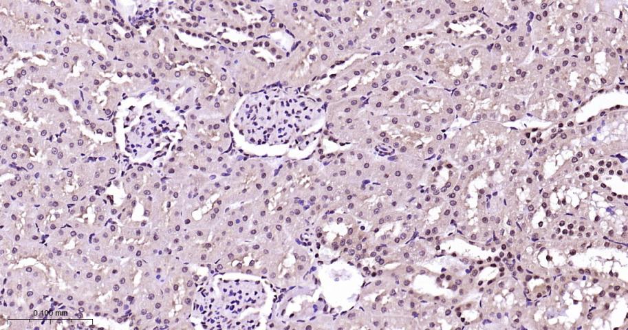
Paraformaldehyde-fixed, paraffin embedded Rat kidney; Antigen retrieval by boiling in sodium citrate buffer (pH6.0) for 15 min; Antibody incubation with Phospho-P38 MAPK (Thr180 + Tyr182) Polyclonal Antibody, Unconjugated(bs-0636R) at 1:200 overnight at 4°C, followed by conjugation to the bs-0295G-HRP and DAB (C-0010) staining.
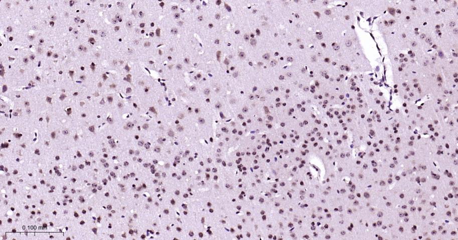
Paraformaldehyde-fixed, paraffin embedded Rat brain; Antigen retrieval by boiling in sodium citrate buffer (pH6.0) for 15 min; Antibody incubation with Phospho-P38 MAPK (Thr180 + Tyr182) Polyclonal Antibody, Unconjugated(bs-0636R) at 1:200 overnight at 4°C, followed by conjugation to the bs-0295G-HRP and DAB (C-0010) staining.
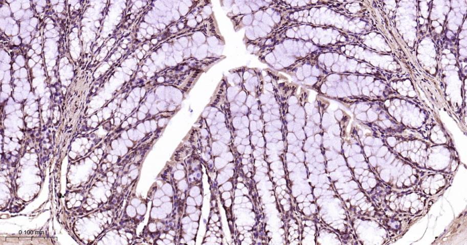
Paraformaldehyde-fixed, paraffin embedded Rat colon; Antigen retrieval by boiling in sodium citrate buffer (pH6.0) for 15 min; Antibody incubation with Phospho-P38 MAPK (Thr180 + Tyr182) Polyclonal Antibody, Unconjugated(bs-0636R) at 1:200 overnight at 4°C, followed by conjugation to the bs-0295G-HRP and DAB (C-0010) staining.
Paraformaldehyde-fixed, paraffin embedded Mouse brain; Antigen retrieval by boiling in sodium citrate buffer (pH6.0) for 15 min; Antibody incubation with Phospho-P38 MAPK (Thr180 + Tyr182) Polyclonal Antibody, Unconjugated(bs-0636R) at 1:200 overnight at 4°C, followed by conjugation to the bs-0295G-HRP and DAB (C-0010) staining.
Paraformaldehyde-fixed, paraffin embedded Mouse colon; Antigen retrieval by boiling in sodium citrate buffer (pH6.0) for 15 min; Antibody incubation with Phospho-P38 MAPK (Thr180 + Tyr182) Polyclonal Antibody, Unconjugated(bs-0636R) at 1:200 overnight at 4°C, followed by conjugation to the bs-0295G-HRP and DAB (C-0010) staining.
|
| 1、抗体溶解方法 | |
| 2、抗体修复方式 | |
| 3、常用试剂的配制 | |
| 4、免疫组化操作步骤 | |
| 5、免疫组化问题解答 | |
| 6、Western Blotting 操作步骤 | |
| 7、Western Blotting 问题解答 | |
| 8、关于肽链的设计 | |
| 9、多肽的溶解与保存 | |
| 10、酶标抗体效价测定程序 | |


