

| 产品编号 | bs-2066R |
| 英文名称 | phospho-GSK-3 Beta (Ser9) Rabbit pAb |
| 中文名称 | 磷酸化糖原合酶激酶-3β抗体 |
| 别 名 | GSK3B | GSK3 beta (phospho-S9); p-GSK3 beta; phospho-GSK3 beta; p-GSK3B; GSK3B | GSK3 beta (phospho-Ser9); 7330414F15Rik; 8430431H08Rik; GSK-3; GSK-3beta; GSK3; GSK3B_HUMAN; GSK3B; GSK-3 beta; Serine/threonine-protein kinase GSK3B; 2.7.11.26; GSK3B_MOUSE; |
 |
Specific References (18) | bs-2066R has been referenced in 18 publications.
|
| 产品类型 | 磷酸化抗体 |
| 研究领域 | 细胞生物 神经生物学 信号转导 细胞凋亡 激酶和磷酸酶 |
| 抗体来源 | Rabbit |
| 克隆类型 | Polyclonal |
| 克 隆 号 | |
| 交叉反应 | Human,Mouse,Rat |
| 产品应用 | WB=1:500-2000,IHC-P=1:100-500,IHC-F=1:100-500,IF=1:100-500,Flow-Cyt=1ug/Test,ICC/IF=1:100-500
not yet tested in other applications. optimal dilutions/concentrations should be determined by the end user. |
| 理论分子量 | 47 kDa |
| 检测分子量 | 47 |
| 细胞定位 | 细胞核 细胞浆 细胞膜 |
| 性 状 | Liquid |
| 浓 度 | 1mg/ml |
| 免 疫 原 | KLH conjugated Synthesised phosphopeptide derived from human GSK-3 Beta around the phosphorylation site of Ser9: TT(p-S)FA |
| 亚 型 | IgG |
| 纯化方法 | affinity purified by Protein A |
| 缓 冲 液 | 0.01M TBS (pH7.4) with 1% BSA, 0.02% Proclin300 and 50% Glycerol. |
| 保存条件 | Shipped at 4℃. Store at -20℃ for one year. Avoid repeated freeze/thaw cycles. |
| 注意事项 | This product as supplied is intended for research use only, not for use in human, therapeutic or diagnostic applications. |
| PubMed | PubMed |
| 产品介绍 |
The protein encoded by this gene is a serine-threonine kinase, belonging to the glycogen synthase kinase subfamily. It is involved in energy metabolism, neuronal cell development, and body pattern formation. Polymorphisms in this gene have been implicated in modifying risk of Parkinson disease, and studies in mice show that overexpression of this gene may be relevant to the pathogenesis of Alzheimer disease. Alternatively spliced transcript variants encoding different isoforms have been found for this gene.[provided by RefSeq, Sep 2009] Function: Constitutively active protein kinase that acts as a negative regulator in the hormonal control of glucose homeostasis, Wnt signaling and regulation of transcription factors and microtubules, by phosphorylating and inactivating glycogen synthase (GYS1 or GYS2), EIF2B, CTNNB1/beta-catenin, APC, AXIN1, JUN, NFATC1/NFATC, MAPT/TAU and MACF1. Requires primed phosphorylation of the majority of its substrates. In skeletal muscle, contributes to insulin regulation of glycogen synthesis by phosphorylating and inhibiting GYS1 activity and hence glycogen synthesis. May also mediate the development of insulin resistance by regulating activation of transcription factors. Regulates protein synthesis by controlling the activity of initiation factor 2B (EIF2BE/EIF2B5) in the same manner as glycogen synthase. In Wnt signaling, GSK3B forms a multimeric complex with APC, AXIN1 and CTNNB1/beta-catenin and phosphorylates the N-terminus of CTNNB1 leading to its degradation mediated by ubiquitin/proteasomes. Phosphorylates JUN at sites proximal to its DNA-binding domain, thereby reducing its affinity for DNA. Phosphorylates NFATC1/NFATC on conserved serine residues promoting NFATC1/NFATC nuclear export, shutting off NFATC1/NFATC gene regulation, and thereby opposing the action of calcineurin. Phosphorylates MAPT/TAU on 'Thr-548', decreasing significantly MAPT/TAU ability to bind and stabilize microtubules. MAPT/TAU is the principal component of neurofibrillary tangles in Alzheimer disease. Plays an important role in ERBB2-dependent stabilization of microtubules at the cell cortex. Phosphorylates MACF1, inhibiting its binding to microtubules which is critical for its role in bulge stem cell migration and skin wound repair. Probably regulates NF-kappa-B (NFKB1) at the transcriptional level and is required for the NF-kappa-B-mediated anti-apoptotic response to TNF-alpha (TNF/TNFA). Negatively regulates replication in pancreatic beta-cells, resulting in apoptosis, loss of beta-cells and diabetes. Phosphorylates MUC1 in breast cancer cells, decreasing the interaction of MUC1 with CTNNB1/beta-catenin. Is necessary for the establishment of neuronal polarity and axon outgrowth. Phosphorylates MARK2, leading to inhibit its activity. Phosphorylates SIK1 at 'Thr-182', leading to sustain its activity. Subunit: Monomer. Interacts with ARRB2 and DISC1. Interacts with CABYR, MMP2, MUC1, NIN and PRUNE Interacts with AXIN1; the interaction mediates hyperphosphorylation of CTNNB1 leading to its ubiquitination and destruction. Interacts with and phosphorylates SNAI1. Interacts with DNM1L (via a C-terminal domain). Found in a complex composed of MACF1, APC, AXIN1, CTNNB1 and GSK3B. Subcellular Location: Cytoplasm. Nucleus. Cell membrane. Note=The phosphorylated form shows localization to cytoplasm and cell membrane. The MEMO1-RHOA-DIAPH1 signaling pathway controls localization of the phosophorylated form to the cell membrane. Tissue Specificity: Expressed in testis, thymus, prostate and ovary and weakly expressed in lung, brain and kidney. Post-translational modifications: Phosphorylated by AKT1 and ILK1. Upon insulin-mediated signaling, the activated PKB/AKT1 protein kinase phosphorylates and desactivates GSK3B, resulting in the dephosphorylation and activation of GYS1. Activated by phosphorylation at Tyr-216. Similarity: Belongs to the protein kinase superfamily. CMGC Ser/Thr protein kinase family. GSK-3 subfamily. Contains 1 protein kinase domain. SWISS: P49841 Gene ID: 2932 Database links: Entrez Gene: 2932 Human Entrez Gene: 56637 Mouse Omim: 605004 Human SwissProt: P49841 Human SwissProt: Q9WV60 Mouse Unigene: 445733 Human Unigene: 394930 Mouse |
| 产品图片 |
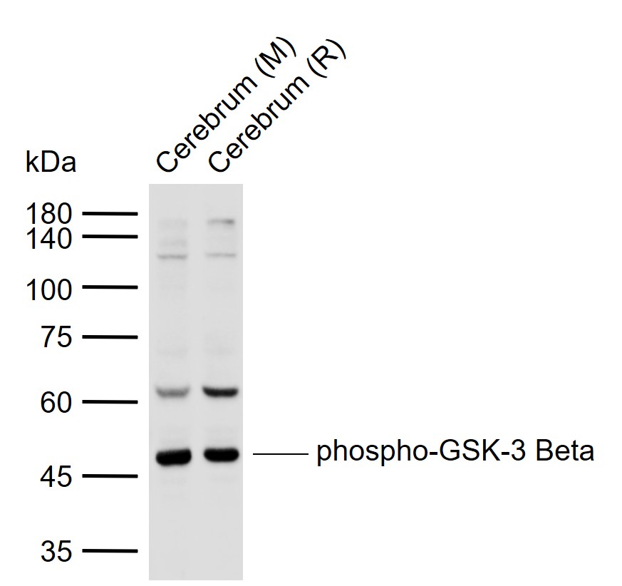
Sample:
Lane 1: Mouse Cerebrum tissue lysates
Lane 2: Rat Cerebrum tissue lysates
Primary: Anti-phospho-GSK-3 Beta (Ser9) (bs-2066R) at 1/1000 dilution
Secondary: IRDye800CW Goat Anti-Rabbit IgG at 1/20000 dilution
Predicted band size: 47 kDa
Observed band size: 47 kDa
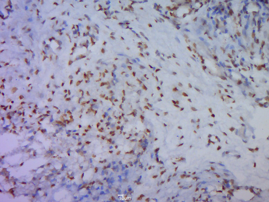
Paraformaldehyde-fixed, paraffin embedded (Mouse placenta); Antigen retrieval by boiling in sodium citrate buffer (pH6.0) for 15min; Block endogenous peroxidase by 3% hydrogen peroxide for 20 minutes; Blocking buffer (normal goat serum) at 37°C for 30min; Antibody incubation with (Beta(Ser9)) Polyclonal Antibody, Unconjugated (bs-2066R p-GSK-3) at 1:500 overnight at 4°C, followed by a conjugated secondary (sp-0023) for 20 minutes and DAB staining.
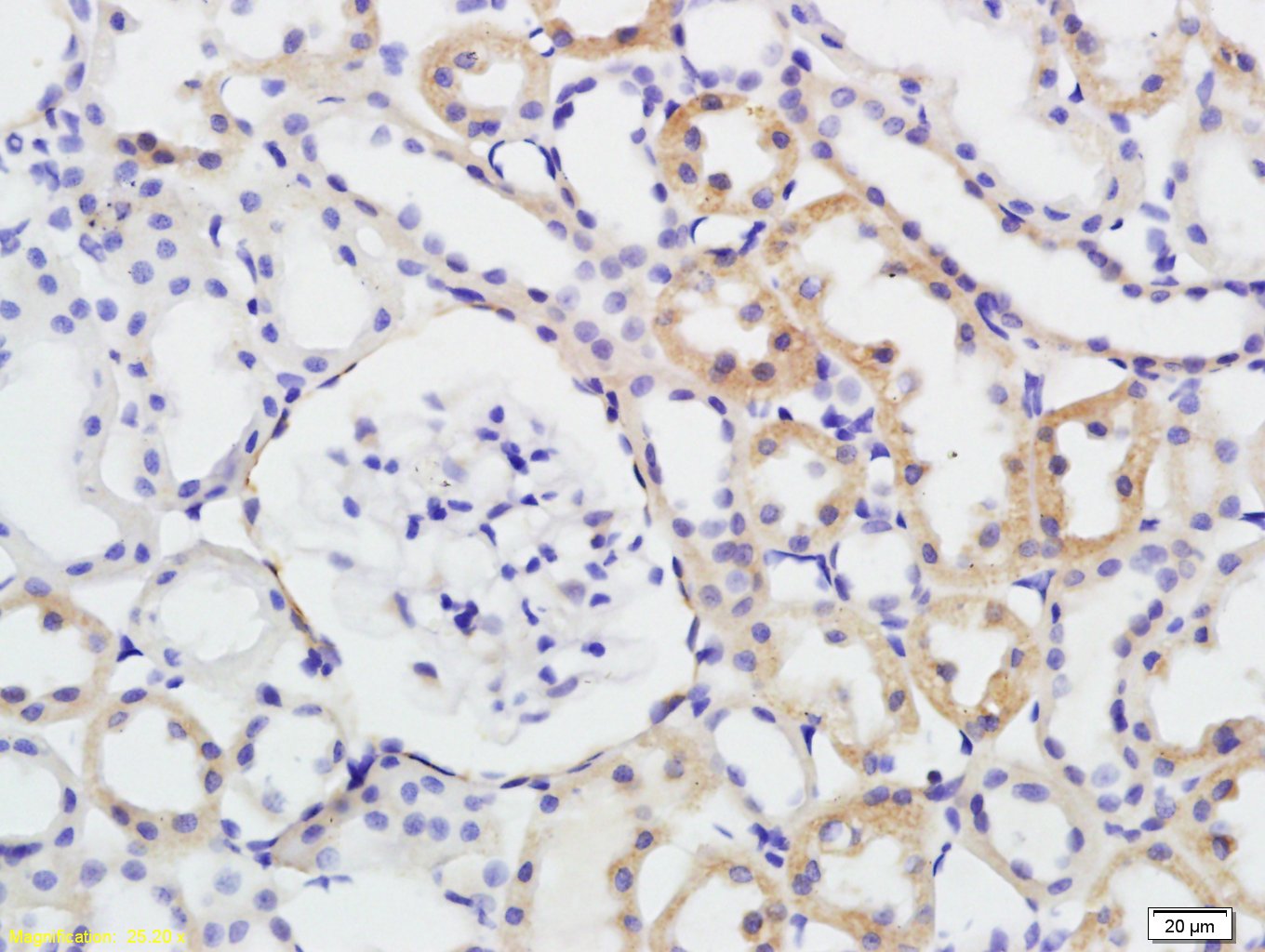
Tissue/cell: rat kidney tissue; 4% Paraformaldehyde-fixed and paraffin-embedded;
Antigen retrieval: citrate buffer ( 0.01M, pH 6.0 ), Boiling bathing for 15min; Block endogenous peroxidase by 3% Hydrogen peroxide for 30min; Blocking buffer (normal goat serum,C-0005) at 37℃ for 20 min;
Incubation: Anti-phospho-GSK-3 Beta(Ser9) Polyclonal Antibody, Unconjugated(bs-2066R) 1:200, overnight at 4°C, followed by conjugation to the secondary antibody(SP-0023) and DAB(C-0010) staining
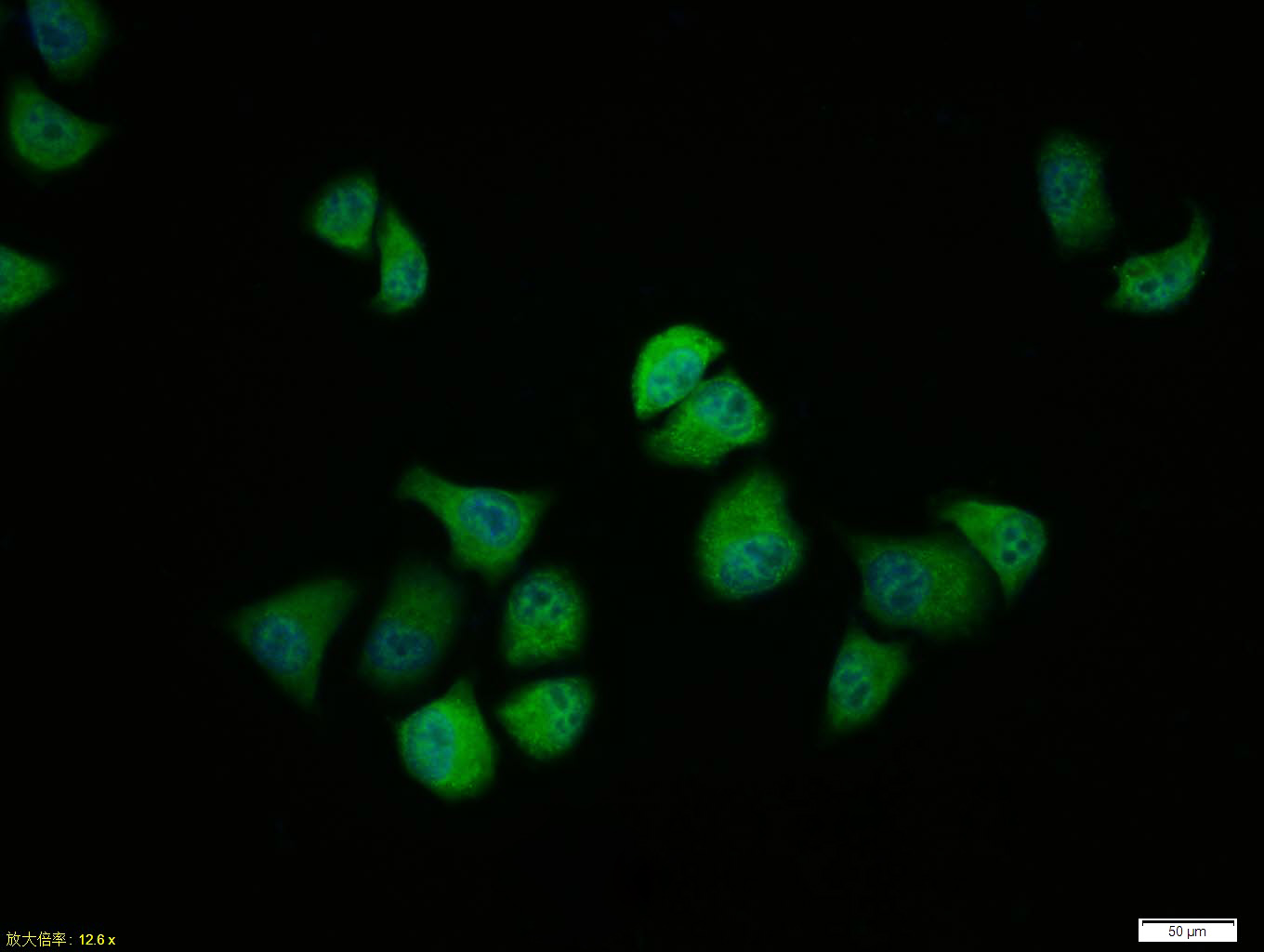
Tissue/cell:Hela cell; 4% Paraformaldehyde-fixed; Triton X-100 at room temperature for 20 min; Blocking buffer (normal goat serum, C-0005) at 37°C for 20 min; Antibody incubation with (phospho-GSK-3 Beta (Ser9)) polyclonal Antibody, Unconjugated (bs-2066R) 1:100, 90 minutes at 37°C; followed by a FITC conjugated Goat Anti-Rabbit IgG antibody at 37°C for 90 minutes, DAPI (blue, C02-04002) was used to stain the cell nuclei.
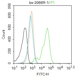
Blank control: A431.
Primary Antibody (green line): Rabbit Anti-phospho-GSK-3 Beta (Ser9) antibody (bs-2066R)
Dilution: 1μg /10^6 cells;
Isotype Control Antibody (orange line): Rabbit IgG .
Secondary Antibody : Goat anti-rabbit IgG-AF488
Dilution: 1μg /test.
Protocol
The cells were fixed with 4% PFA (10min at room temperature)and then permeabilized with 90% ice-cold methanol for 20 min at-20℃. The cells were then incubated in 5%BSA to block non-specific protein-protein interactions for 30 min at room temperature .Cells stained with Primary Antibody for 30 min at room temperature. The secondary antibody used for 40 min at room temperature. Acquisition of 20,000 events was performed.
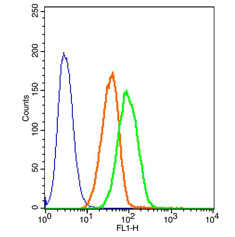
The blue histogram is unstained cells(A549 cells).
The Orange histogram is cells stained with Rabbit IgG/FITC (bs-0295P-FITC).
The green histogram is cells stained with Rabbit Anti-phospho-GSK-3 Beta(Ser9)/FITC Conjugated antibody (bs-2066R-FITC).
Isotype control: Cell lines treated with Rabbit IgG/FITC(bs-0295P-FITC) instead of the primary antibody to confirm that primary antibody binding is specific. Concentration: 5μL in 100 μL 1 X PBS containing 0.5% BSA.
|
| 1、抗体溶解方法 | |
| 2、抗体修复方式 | |
| 3、常用试剂的配制 | |
| 4、免疫组化操作步骤 | |
| 5、免疫组化问题解答 | |
| 6、Western Blotting 操作步骤 | |
| 7、Western Blotting 问题解答 | |
| 8、关于肽链的设计 | |
| 9、多肽的溶解与保存 | |
| 10、酶标抗体效价测定程序 | |


