

| 产品编号 | bs-1287R |
| 英文名称 | IKB alpha Rabbit pAb |
| 中文名称 | 核因子κB抑制蛋白α抗体 |
| 别 名 | EDAID2; IKBA; MAD-3; NFKBI; IKBA_HUMAN; NFKBIA; I-kappa-B-alpha (IkB-alpha | IkappaBalpha); Major histocompatibility complex enhancer-binding protein MAD3; MAD3; |
 |
Specific References (76) | bs-1287R has been referenced in 76 publications.
|
| 研究领域 | 肿瘤 免疫学 信号转导 转录调节因子 激酶和磷酸酶 |
| 抗体来源 | Rabbit |
| 克隆类型 | Polyclonal |
| 克 隆 号 | |
| 交叉反应 | Human,Mouse,Rat |
| 产品应用 | WB=1:500-2000,IHC-P=1:100-500,IHC-F=1:100-500,IF=1:100-500,Flow-Cyt=1μg/Test,ICC/IF=1:100-500
not yet tested in other applications. optimal dilutions/concentrations should be determined by the end user. |
| 理论分子量 | 36 kDa |
| 检测分子量 | 39 |
| 细胞定位 | 细胞核 细胞浆 |
| 性 状 | Liquid |
| 浓 度 | 1mg/ml |
| 免 疫 原 | KLH conjugated synthetic peptide derived from human NFKBIA: 1-120/314 |
| 亚 型 | IgG |
| 纯化方法 | affinity purified by Protein A |
| 缓 冲 液 | 0.01M TBS (pH7.4) with 1% BSA, 0.02% Proclin300 and 50% Glycerol. |
| 保存条件 | Shipped at 4℃. Store at -20℃ for one year. Avoid repeated freeze/thaw cycles. |
| 注意事项 | This product as supplied is intended for research use only, not for use in human, therapeutic or diagnostic applications. |
| PubMed | PubMed |
| 产品介绍 |
This gene encodes a member of the NF-kappa-B inhibitor family, which contain multiple ankrin repeat domains. The encoded protein interacts with REL dimers to inhibit NF-kappa-B/REL complexes which are involved in inflammatory responses. The encoded protein moves between the cytoplasm and the nucleus via a nuclear localization signal and CRM1-mediated nuclear export. Mutations in this gene have been found in ectodermal dysplasia anhidrotic with T-cell immunodeficiency autosomal dominant disease. [provided by RefSeq, Aug 2011] Function: Inhibits the activity of dimeric NF-kappa-B/REL complexes by trapping REL dimers in the cytoplasm through masking of their nuclear localization signals. On cellular stimulation by immune and proinflammatory responses, becomes phosphorylated promoting ubiquitination and degradation, enabling the dimeric RELA to translocate to the nucleus and activate transcription. Subunit: Interacts with RELA; the interaction requires the nuclear import signal. Interacts with NKIRAS1 and NKIRAS2. Part of a 70-90 kDa complex at least consisting of CHUK, IKBKB, NFKBIA, RELA, IKBKAP and MAP3K14. Interacts with HBV protein X. Interacts with RWDD3; the interaction enhances sumoylation. Interacts (when phosphorylated at the 2 serine residues in the destruction motif D-S-G-X(2,3,4)-S) with BTRC. Associates with the SCF(BTRC) complex, composed of SKP1, CUL1 and BTRC; the association is mediated via interaction with BTRC. Part of a SCF(BTRC)-like complex lacking CUL1, which is associated with RELA; RELA interacts directly with NFKBIA. Interacts with PRMT2. Interacts with PRKACA in platelets; this interaction is disrupted by thrombin and collagen. Interacts with HIF1AN. Subcellular Location: Cytoplasm. Nucleus. Tissue Specificity: Highly expressed in lymph node, thymus followed by liver, brain, muscle, kidney, gastrointestinal and reproductive tract. Post-translational modifications: Phosphorylated; disables inhibition of NF-kappa-B DNA-binding activity. Phosphorylation at positions 32 and 36 is prerequisite to recognition by UBE2D3 leading to polyubiquitination and subsequent degradation. Sumoylated; sumoylation requires the presence of the nuclear import signal. Monoubiquitinated at Lys-21 and/or Lys-22 by UBE2D3. Ubiquitin chain elongation is then performed by CDC34 in cooperation with the SCF(FBXW11) E3 ligase complex, building ubiquitin chains from the UBE2D3-primed NFKBIA-linked ubiquitin. The resulting polyubiquitination leads to protein degradation. Also ubiquitinated by SCF(BTRC) following stimulus-dependent phosphorylation at Ser-32 and Ser-36. Deubiquitinated by porcine reproductive and respiratory syndrome virus Nsp2 protein, which thereby interferes with NFKBIA degradation and impairs subsequent NF-kappa-B activation. DISEASE: Defects in NFKBIA are the cause of ectodermal dysplasia anhidrotic with T-cell immunodeficiency autosomal dominant (ADEDAID) [MIM:612132]. Ectodermal dysplasia defines a heterogeneous group of disorders due to abnormal development of two or more ectodermal structures. ADEDAID is an ectodermal dysplasia associated with decreased production of pro-inflammatory cytokines and certain interferons, rendering patients susceptible to infection. Similarity: Belongs to the NF-kappa-B inhibitor family. Contains 5 ANK repeats. SWISS: P25963 Gene ID: 4792 Database links: Entrez Gene: 4792 Human Entrez Gene: 18035 Mouse SwissProt: P25963 Human SwissProt: Q9Z1E3 Mouse 转录调节因子(Transcriptin Regulators) |
| 产品图片 |
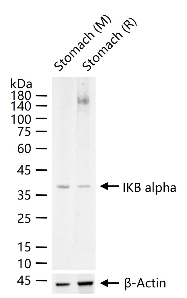
25 ug total protein per lane of various lysates (see on figure) probed with IKB alpha polyclonal antibody, unconjugated (bs-1287R) at 1:1000 dilution and 4°C overnight incubation. Followed by conjugated secondary antibody incubation at r.t. for 60 min.

Paraformaldehyde-fixed, paraffin embedded (Mouse brain); Antigen retrieval by boiling in sodium citrate buffer (pH6.0) for 15min; Block endogenous peroxidase by 3% hydrogen peroxide for 20 minutes; Blocking buffer (normal goat serum) at 37°C for 30min; Antibody incubation with (IKB alpha) Polyclonal Antibody, Unconjugated (bs-1287R) at 1:400 overnight at 4°C, followed by operating according to SP Kit(Rabbit) (sp-0023) instructionsand DAB staining.
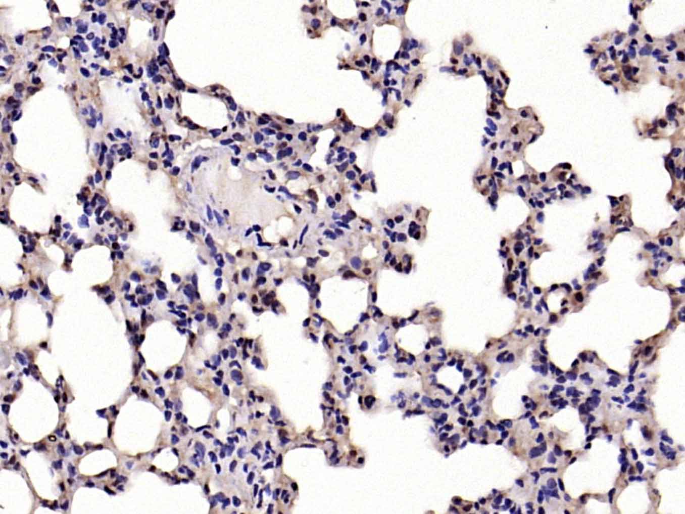
Paraformaldehyde-fixed, paraffin embedded (mouse lung); Antigen retrieval by boiling in sodium citrate buffer (pH6.0) for 15min; Block endogenous peroxidase by 3% hydrogen peroxide for 20 minutes; Blocking buffer (normal goat serum) at 37°C for 30min; Antibody incubation with (IKB alpha) Polyclonal Antibody, Unconjugated (bs-1287R) at 1:200 overnight at 4°C, followed by operating according to SP Kit(Rabbit) (sp-0023) instructionsand DAB staining.
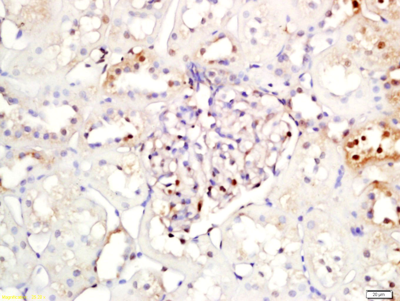
Tissue/cell: rat kidney tissue; 4% Paraformaldehyde-fixed and paraffin-embedded;
Antigen retrieval: citrate buffer ( 0.01M, pH 6.0 ), Boiling bathing for 15min; Block endogenous peroxidase by 3% Hydrogen peroxide for 30min; Blocking buffer (normal goat serum,C-0005) at 37℃ for 20 min;
Incubation: Anti-IKB alpha Polyclonal Antibody, Unconjugated(bs-1287R) 1:200, overnight at 4°C, followed by conjugation to the secondary antibody(SP-0023) and DAB(C-0010) staining
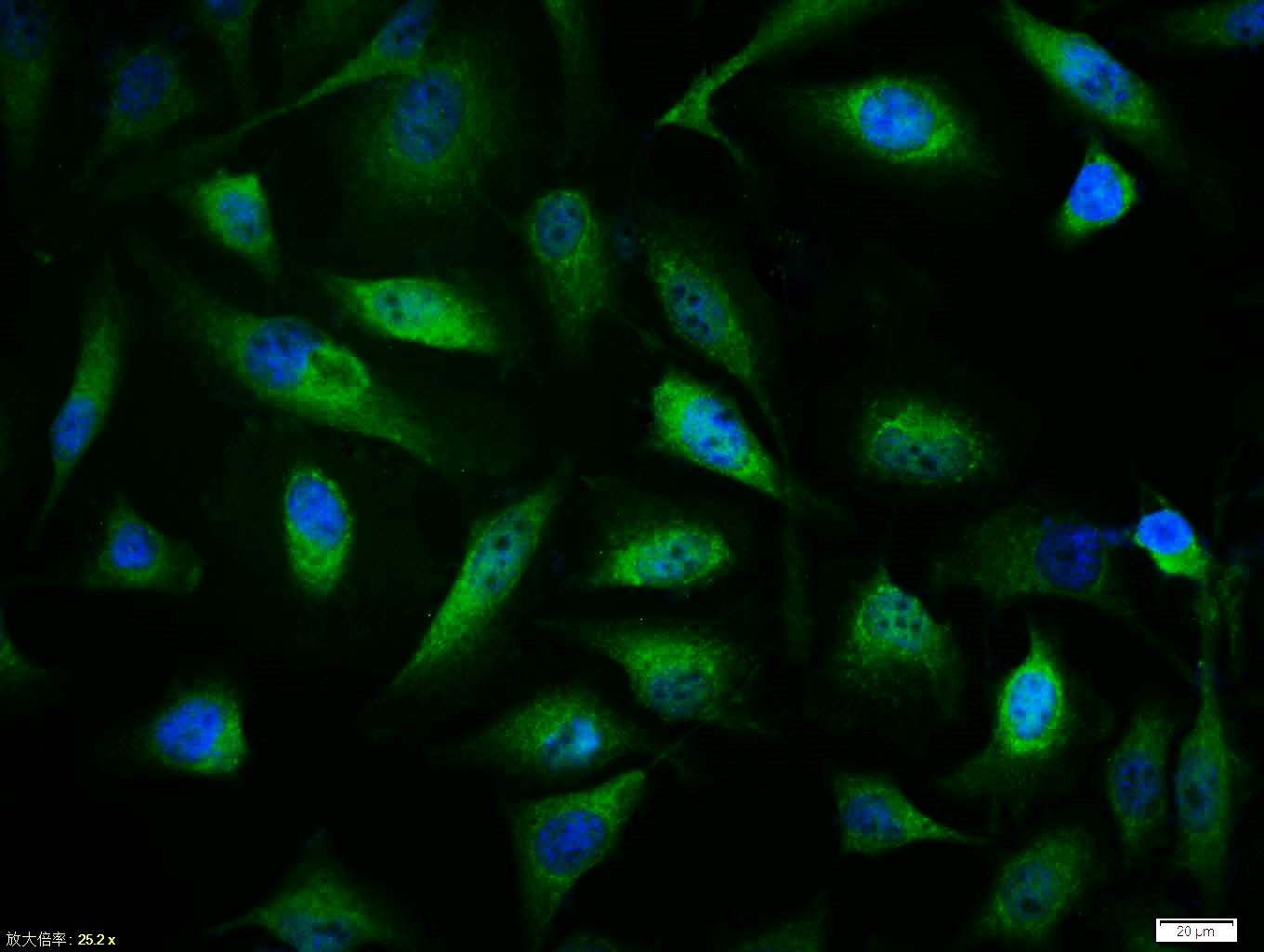
Tissue/cell:Hela cell; 4% Paraformaldehyde-fixed; Triton X-100 at room temperature for 20 min; Blocking buffer (normal goat serum,C-0005) at 37°C for 20 min; Antibody incubation with (IKB alpha) polyclonal Antibody, Unconjugated (bs-1287R) 1:100, 90 minutes at 37°C; followed by a FITC conjugated Goat Anti-Rabbit IgG antibody at 37°C for 90 minutes, DAPI (blue, C02-04002) was used to stain the cell nuclei.
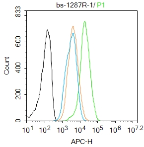
Blank control: Hela.
Primary Antibody (green line): Rabbit Anti-IKB alpha antibody (bs-1287R)
Dilution: 1μg /10^6 cells;
Isotype Control Antibody (orange line): Rabbit IgG .
Secondary Antibody : Goat anti-rabbit IgG-AF647
Dilution: 1μg /test.
Protocol
The cells were fixed with 4% PFA (10min at room temperature)and then permeabilized with 90% ice-cold methanol for 20 min at -20℃. The cells were then incubated in 5%BSA to block non-specific protein-protein interactions for 30 min at room temperature .Cells stained with Primary Antibody for 30 min at room temperature. The secondary antibody used for 40 min at room temperature. Acquisition of 20,000 events was performed.
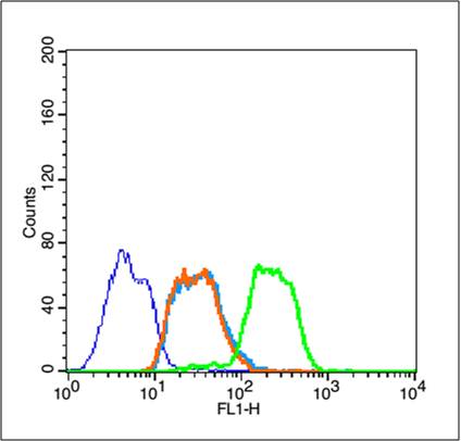
Blank control (blue line): Jurkat (blue).
Primary Antibody (green line): Rabbit Anti-IKB alpha antibody (bs-1287R)
Dilution: 1μg /10^6 cells;
Isotype Control Antibody (orange line): Rabbit IgG .
Secondary Antibody (white blue line): Goat anti-rabbit IgG-FITC
Dilution: 1μg /test.
Protocol
The cells were fixed with 2% paraformaldehyde (10 min) , then permeabilized with 90% ice-cold methanol for 30 min on ice. Cells stained with Primary Antibody for 30 min at room temperature. The cells were then incubated in 1 X PBS/2%BSA/10% goat serum to block non-specific protein-protein interactions followed by the antibody for 15 min at room temperature. The secondary antibody used for 40 min at room temperature. Acquisition of 20,000 events was performed.
|
| 1、抗体溶解方法 | |
| 2、抗体修复方式 | |
| 3、常用试剂的配制 | |
| 4、免疫组化操作步骤 | |
| 5、免疫组化问题解答 | |
| 6、Western Blotting 操作步骤 | |
| 7、Western Blotting 问题解答 | |
| 8、关于肽链的设计 | |
| 9、多肽的溶解与保存 | |
| 10、酶标抗体效价测定程序 | |


