

| 产品编号 | bs-0805R |
| 英文名称 | CD56 Rabbit pAb |
| 中文名称 | 神经细胞粘附分子1抗体 |
| 别 名 | CD56; MSK39; NCAM; E-NCAM; NCAM-1; N-CAM; N-CAM-1; NCAM-C; NCAMC; NCAM1_HUMAN; NCAM1; NCAM1_MOUSE; NCAM1_RAT; neural cell adhesion molecule 1 |
 |
Specific References (3) | bs-0805R has been referenced in 3 publications.
[IF=15.304] Yao Lei. et al. Phytochemical natural killer cells reprogram tumor microenvironment for potent immunotherapy of solid tumors. BIOMATERIALS. 2022 Jun;:121635 WB, IF, FC ; Mouse.
[IF=2.6] Wu TH et al. The Combination of Astragalus membranaceus and Angelica sinensis Inhibits Lung Cancer and Cachexia through Its Immunomodulatory Function. J Oncol. 2019 Nov 3;2019:9206951. FCM ; Mouse.
[IF=0.976] Sahiner et al. Impact of intravesical hyaluronic acid treatment on bladder inflammation in interstitial cystitis rat model. (2018) Int.Braz.J.Urol. 44:1014-1022 IHC ; Rat.
|
| 研究领域 | 肿瘤 神经生物学 干细胞 细胞粘附分子 细胞类型标志物 |
| 抗体来源 | Rabbit |
| 克隆类型 | Polyclonal |
| 交叉反应 | Human,Mouse,Rat (predicted: Rabbit,Pig,Cow,Chicken,Dog,GuineaPig,Horse) |
| 产品应用 | WB=1:500-2000,IHC-P=1:100-500,IHC-F=1:100-500,IF=1:100-500,Flow-Cyt=1μg/Test,ICC/IF=1:100-500
not yet tested in other applications. optimal dilutions/concentrations should be determined by the end user. |
| 理论分子量 | 92kDa |
| 检测分子量 | 95 |
| 细胞定位 | 细胞膜 分泌型蛋白 |
| 性 状 | Liquid |
| 浓 度 | 1mg/ml |
| 免 疫 原 | KLH conjugated synthetic peptide derived from human CD56: 621-720/858 <Extracellular> |
| 亚 型 | IgG |
| 纯化方法 | affinity purified by Protein A |
| 缓 冲 液 | 0.01M TBS (pH7.4) with 1% BSA, 0.02% Proclin300 and 50% Glycerol. |
| 保存条件 | Shipped at 4℃. Store at -20℃ for one year. Avoid repeated freeze/thaw cycles. |
| 注意事项 | This product as supplied is intended for research use only, not for use in human, therapeutic or diagnostic applications. |
| PubMed | PubMed |
| 产品介绍 |
This gene encodes a cell adhesion protein which is a member of the immunoglobulin superfamily. The encoded protein is involved in cell-to-cell interactions as well as cell-matrix interactions during development and differentiation. The encoded protein has been shown to be involved in development of the nervous system, and for cells involved in the expansion of T cells and dendritic cells which play an important role in immune surveillance. Alternative splicing results in multiple transcript variants. [provided by RefSeq, Jun 2011] Function: This protein is a cell adhesion molecule involved in neuron-neuron adhesion, neurite fasciculation, outgrowth of neurites, etc. Subcellular Location: Isoform 1, 2,: Cell membrane; Single-pass type I membrane protein. Isoform 3, 4: Cell membrane; Lipid-anchor, GPI-anchor. Isoform 5, 6: Secreted. Similarity: Contains 2 fibronectin type-III domains. Contains 5 Ig-like C2-type (immunoglobulin-like) domains. SWISS: P13591 Gene ID: 4684 Database links: Entrez Gene: 4684 Human Entrez Gene: 17967 Mouse Omim: 116930 Human SwissProt: P13591 Human SwissProt: P13595 Mouse Unigene: 503878 Human Unigene: 711235 Human Unigene: 733031 Human Unigene: 439182 Mouse Unigene: 4974 Mouse Unigene: 11283 Rat 细胞粘附蛋白(Call Adhesion Protein) 神经元标志物 NCAM-1为神经细胞粘附分子,主要分布于神经组织,神经—肌肉接头,神经—内分泌腺和某些内分泌腺以及大多数神经外胚层来源的细胞、组织和肿瘤中。 NCAM 1主要用于视网膜母细胞瘤、髓母细胞瘤、星形细胞瘤、神经母细胞瘤等肿瘤方面的研究。 |
| 产品图片 |
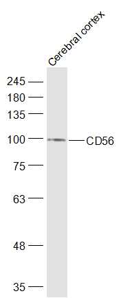
Sample:
Cerebral cortex (Mouse) Lysate at 40 ug
Primary: Anti-CD56 (bs-0805R) at 1/1000 dilution
Secondary: IRDye800CW Goat Anti-Rabbit IgG at 1/20000 dilution
Predicted band size: 92 kD
Observed band size: 95 kD

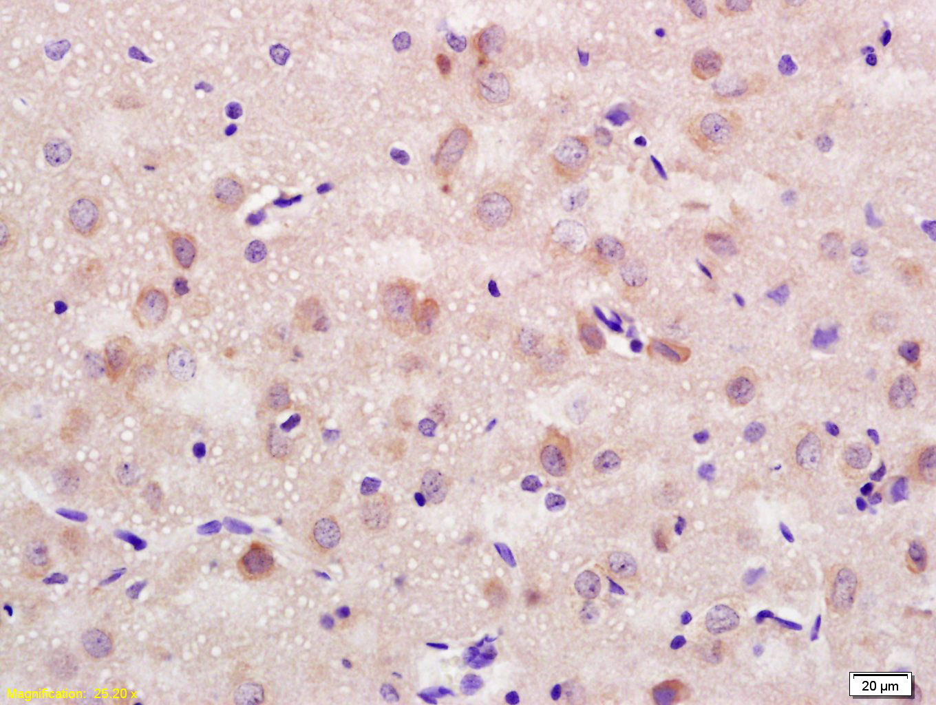
Paraformaldehyde-fixed, paraffin embedded (Mouse brain); Antigen retrieval by boiling in sodium citrate buffer (pH6.0) for 15min; Block endogenous peroxidase by 3% hydrogen peroxide for 20 minutes; Blocking buffer (normal goat serum) at 37°C for 30min; Antibody incubation with (CD56) Polyclonal Antibody, Unconjugated (bs-0805R) at 1:500 overnight at 4°C, followed by a conjugated secondary (sp-0023) for 20 minutes and DAB staining.
Tissue/cell: rat brain tissue; 4% Paraformaldehyde-fixed and paraffin-embedded;
Antigen retrieval: citrate buffer ( 0.01M, pH 6.0 ), Boiling bathing for 15min; Block endogenous peroxidase by 3% Hydrogen peroxide for 30min; Blocking buffer (normal goat serum,C-0005) at 37℃ for 20 min;
Incubation: Anti-CD56/NCAM1 Polyclonal Antibody, Unconjugated(bs-0805R) 1:200, overnight at 4°C, followed by conjugation to the secondary antibody(SP-0023) and DAB(C-0010) staining
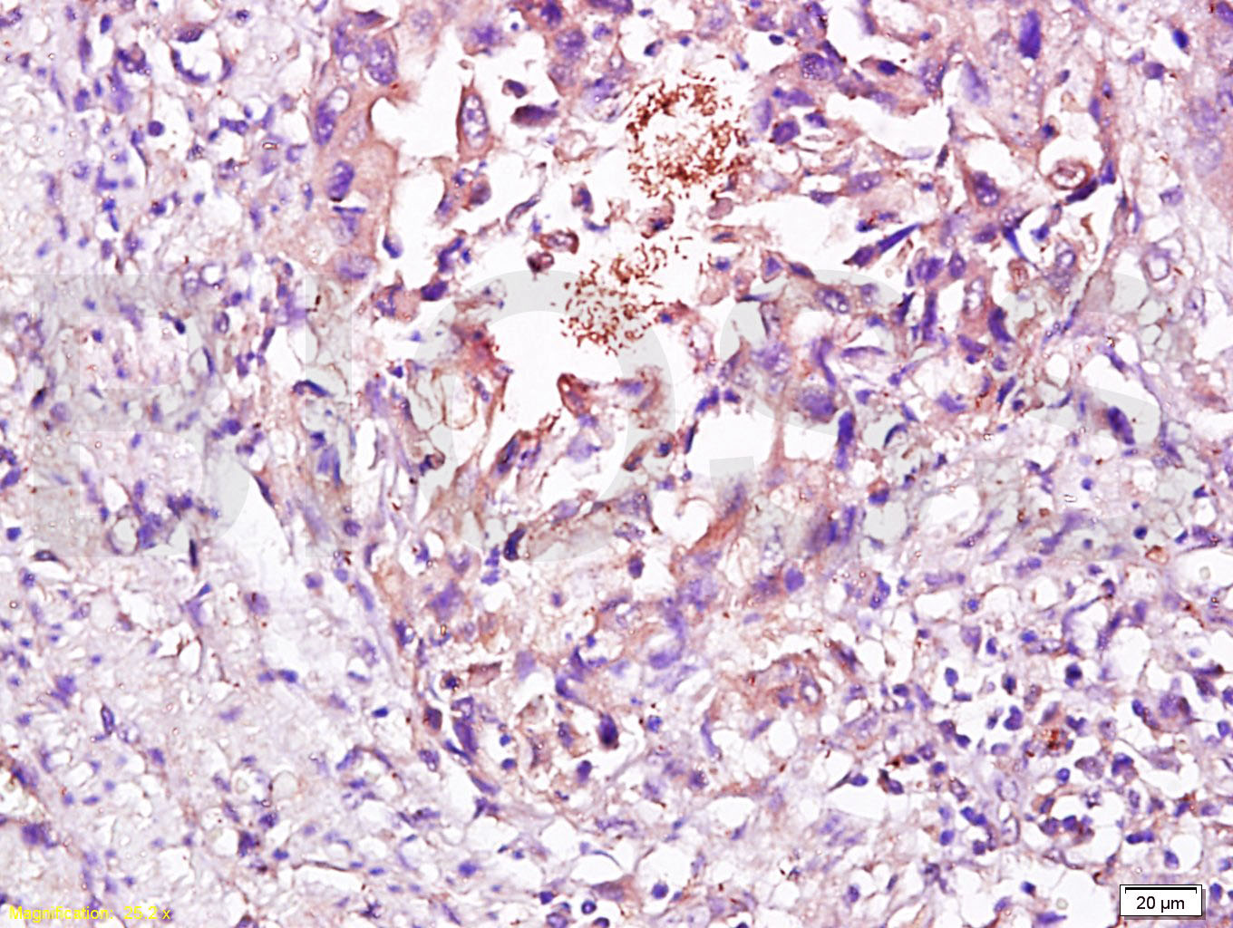
Tissue/cell: Human esophageal carcinoma; 4% Paraformaldehyde-fixed and paraffin-embedded;
Antigen retrieval: citrate buffer ( 0.01M, pH 6.0 ), Boiling bathing for 15min; Block endogenous peroxidase by 3% Hydrogen peroxide for 30min; Blocking buffer (normal goat serum,C-0005) at 37℃ for 20 min;
Incubation: Anti-CD56/NCAM1 Polyclonal Antibody, Unconjugated(bs-0805R) 1:200, overnight at 4°C, followed by conjugation to the secondary antibody(SP-0023) and DAB(C-0010) staining
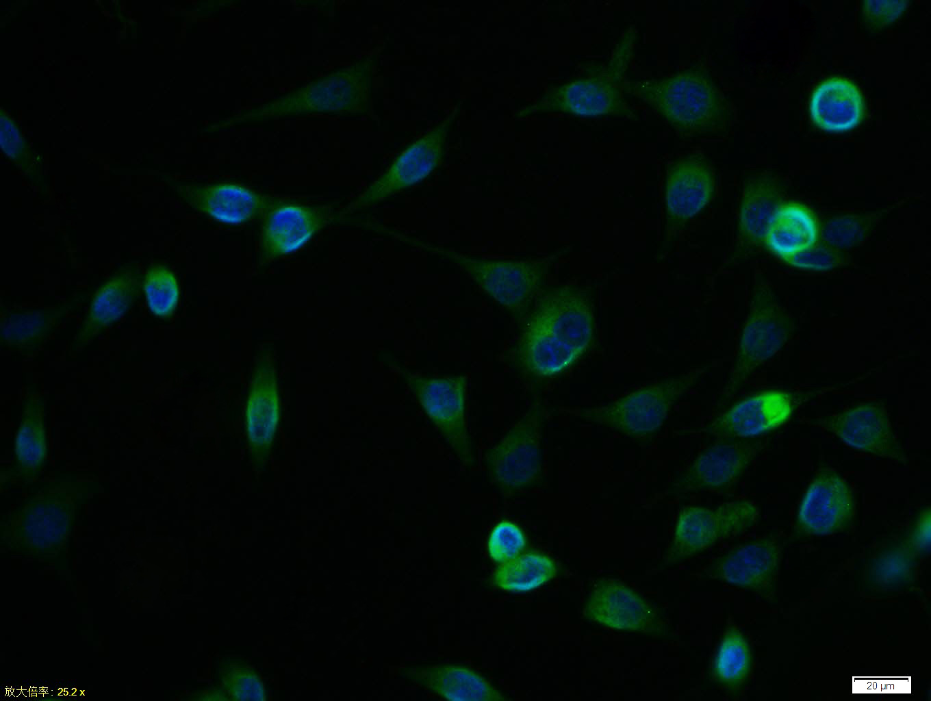
Tissue/cell:SH-SY5Y cell; 4% Paraformaldehyde-fixed; Triton X-100 at room temperature for 20 min; Blocking buffer (normal goat serum,C-0005) at 37°C for 20 min; Antibody incubation with (CD56) polyclonal Antibody, Unconjugated (bs-0805R) 1:100, 90 minutes at 37°C; followed by a FITC conjugated Goat Anti-Rabbit IgG antibody at 37°C for 90 minutes, DAPI (blue, C02-04002) was used to stain the cell nuclei.
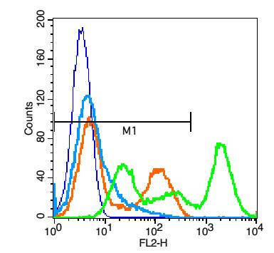
Blank control: Jurkat cells(blue). Primary Antibody:Rabbit Anti-CD56 antibody(bs-0805R), Dilution: 1μg in 100 μL 1X PBS containing 0.5% BSA; Isotype Control Antibody: Rabbit IgG(orange) ,used under the same conditions ); Secondary Antibody: Goat anti-rabbit IgG-PE(white blue), Dilution: 1:200 in 1 X PBS containing 0.5% BSA.
Protocol
The cells were fixed with 2% paraformaldehyde (10 min) . Primary antibody (bs-0805R, 1μg /1x10^6 cells) were incubated for 30 min on the ice, followed by 1 X PBS containing 0.5% BSA + 1 0% goat serum (15 min) to block non-specific protein-protein interactions. Then the Goat Anti-rabbit IgG/PE antibody was added into the blocking buffer mentioned above to react with the primary antibody at 1/200 dilution for 30 min on ice. Acquisition of 20,000 events was performed.
|
| 1、抗体溶解方法 | |
| 2、抗体修复方式 | |
| 3、常用试剂的配制 | |
| 4、免疫组化操作步骤 | |
| 5、免疫组化问题解答 | |
| 6、Western Blotting 操作步骤 | |
| 7、Western Blotting 问题解答 | |
| 8、关于肽链的设计 | |
| 9、多肽的溶解与保存 | |
| 10、酶标抗体效价测定程序 | |


