

| 产品编号 | bs-2138R |
| 英文名称 | PARP1 Rabbit pAb |
| 中文名称 | 多腺苷二磷酸多聚酶抗体(N端) |
| 别 名 | ADPRT; ADPRT 1; ADPRT1; ARTD1; PARP; PARP-1; PARS; PPOL; Poly-PARP; pADPRT-1; 5830444G22Rik; Adprp; msPARP; sPARP-1; PARP1_HUMAN; PARP1; ADP-ribosyltransferase diphtheria toxin-like 1 (ARTD1); DNA ADP-ribosyltransferase PARP1; NAD(+) ADP-ribosyltransferas |
 |
Specific References (19) | bs-2138R has been referenced in 19 publications.
|
| 研究领域 | 染色质和核信号 细胞凋亡 |
| 抗体来源 | Rabbit |
| 克隆类型 | Polyclonal |
| 克 隆 号 | |
| 交叉反应 | Human |
| 产品应用 | WB=1:500-2000,Flow-Cyt=0.2μg/Test
not yet tested in other applications. optimal dilutions/concentrations should be determined by the end user. |
| 理论分子量 | 111 kDa |
| 检测分子量 | 115 |
| 细胞定位 | 细胞核 |
| 性 状 | Liquid |
| 浓 度 | 1mg/ml |
| 免 疫 原 | KLH conjugated synthetic peptide derived from human PARP: 201-300/1014 |
| 亚 型 | IgG |
| 纯化方法 | affinity purified by Protein A |
| 缓 冲 液 | 0.01M TBS (pH7.4) with 1% BSA, 0.02% Proclin300 and 50% Glycerol. |
| 保存条件 | Shipped at 4℃. Store at -20℃ for one year. Avoid repeated freeze/thaw cycles. |
| 注意事项 | This product as supplied is intended for research use only, not for use in human, therapeutic or diagnostic applications. |
| PubMed | PubMed |
| 产品介绍 |
This gene encodes a chromatin-associated enzyme, poly(ADP-ribosyl)transferase, which modifies various nuclear proteins by poly(ADP-ribosyl)ation. The modification is dependent on DNA and is involved in the regulation of various important cellular processes such as differentiation, proliferation, and tumor transformation and also in the regulation of the molecular events involved in the recovery of cell from DNA damage. In addition, this enzyme may be the site of mutation in Fanconi anemia, and may participate in the pathophysiology of type I diabetes. [provided by RefSeq, Jul 2008]. Function: Involved in the base excision repair (BER) pathway, by catalyzing the poly(ADP-ribosyl)ation of a limited number of acceptor proteins involved in chromatin architecture and in DNA metabolism. This modification follows DNA damages and appears as an obligatory step in a detection/signaling pathway leading to the reparation of DNA strand breaks. Mediates the poly(ADP-ribosyl)ation of APLF and CHFR. Positively regulates the transcription of MTUS1 and negatively regulates the transcription of MTUS2/TIP150. With EEF1A1 and TXK, forms a complex that acts as a T-helper 1 (Th1) cell-specific transcription factor and binds the promoter of IFN-gamma to directly regulate its transcription, and is thus involved importantly in Th1 cytokine production. Subunit: Component of a base excision repair (BER) complex, containing at least XRCC1, PARP2, POLB and LRIG3. Homo- and heterodimer with PARP2. Interacts with PARP3, APTX and SRY. The SWAP complex consists of NPM1, NCL, PARP1 and SWAP70. Interacts with TIAM2 and ZNF423 (By similarity). Interacts (when poly-ADP-ribosylated) with CHD1L. Interacts with the DNA polymerase alpha catalytic subunit POLA1; this interaction functions as part of the control of replication fork progression. Interacts with EEF1A1, RNF4 and TXK. Subcellular Location: Mitochondrion outer membrane; Single-pass membrane protein. Nucleus membrane; Single-pass membrane protein. Endoplasmic reticulum membrane; Single-pass membrane protein. Nucleus. Post-translational modifications: Phosphorylated by PRKDC and TXK. Phosphorylated upon DNA damage, probably by ATM or ATR. Poly-ADP-ribosylated by PARP2. Poly-ADP-ribosylation mediates the recruitment of CHD1L to DNA damage sites. S-nitrosylated, leading to inhibit transcription regulation activity. Similarity: Contains 1 BRCT domain. Contains 1 PARP alpha-helical domain. Contains 1 PARP catalytic domain. Contains 2 PARP-type zinc fingers. SWISS: P09874 Gene ID: 142 Database links: Entrez Gene: 142 Human Entrez Gene: 11545 Mouse Omim: 173870 Human SwissProt: P09874 Human SwissProt: P11103 Mouse Unigene: 177766 Human Unigene: 277779 Mouse Unigene: 11327 Rat PARP(poly ADP-ribose polymerase)是DNA修复酶。 PARP是细胞凋亡核心成员半胱胺酸蛋白酶(caspase)的切割底物。因此,它在DNA损伤修复与细胞凋亡中发挥着重要作用。Anti-PARP p85 是特意的PARPp85片段的特异抗体,由caspase剪切116kDa完整分子而得到的。 PARP是存在于多数真核细胞中的一个多功能蛋白质翻译后修饰酶。它通过识别结构损伤的DNA片段而被激活,对聚腺苷二磷酸核糖聚合酶PARP被认为是DNA损伤的感受器。它还能对许多核蛋白进行聚腺苷二磷酸核糖基化。因此,在DNA损伤修复与细胞凋亡中发挥着重要作用,端锚聚合酶在癌细胞端粒结构的调控机制中有重要作用。 |
| 产品图片 |
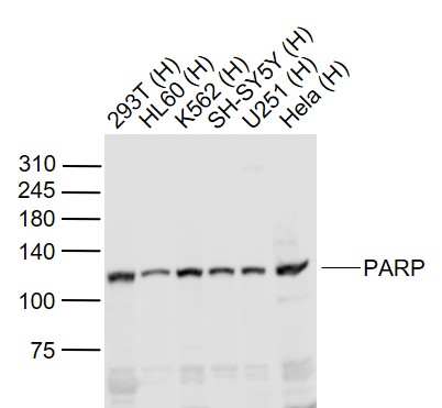
Sample:
Lane 1: 293T (Human) Cell Lysate at 30 ug
Lane 2: HL60 (Human) Cell Lysate at 30 ug
Lane 3: K562 (Human) Cell Lysate at 30 ug
Lane 4: SH-SY5Y (Human) Cell Lysate at 30 ug
Lane 5: U251 (Human) Cell Lysate at 30 ug
Lane 6: Hela (Human) Cell Lysate at 30 ug
Primary: Anti-PARP (bs-2138R) at 1/1000 dilution
Secondary: IRDye800CW Goat Anti-Rabbit IgG at 1/20000 dilution
Predicted band size: 115 kD
Observed band size: 115 kD
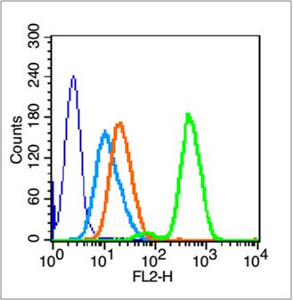
Blank control (blue line): HL60 cells (blue).
Primary Antibody (green line): Rabbit Anti- PARP antibody (bs-2138R)
Dilution: 0.2μg /10^6 cells;
Isotype Control Antibody (orange line): Rabbit IgG .
Secondary Antibody (white blue line): Goat anti-rabbit IgG-PE
Dilution: 1μg /test.
Protocol
The cells were fixed with 70% methanol (Overnight at 4℃) and then permeabilized with 90% ice-cold methanol for 20 min at -20℃. Cells stained with Primary Antibody for 30 min at room temperature. The cells were then incubated in 1 X PBS/2%BSA/10% goat serum to block non-specific protein-protein interactions followed by the antibody for 15 min at room temperature. The secondary antibody used for 40 min at room temperature. Acquisition of 20,000 events was performed.
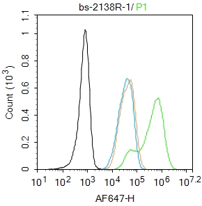
Blank control:293T.
Primary Antibody (green line): Rabbit Anti-PARP1 antibody (bs-2138R)
Dilution: 1μg /10^6 cells;
Isotype Control Antibody (orange line): Rabbit IgG .
Secondary Antibody : Goat anti-rabbit IgG-AF647
Dilution: 1μg /test.
Protocol
The cells were fixed with 4% PFA (10min at room temperature)and then permeabilized with 90% ice-cold methanol for 20 min at -20℃.The cells were then incubated in 5%BSA to block non-specific protein-protein interactions for 30 min at room temperature .Cells stained with Primary Antibody for 30 min at room temperature. The secondary antibody used for 40 min at room temperature. Acquisition of 20,000 events was performed.
|
| 1、抗体溶解方法 | |
| 2、抗体修复方式 | |
| 3、常用试剂的配制 | |
| 4、免疫组化操作步骤 | |
| 5、免疫组化问题解答 | |
| 6、Western Blotting 操作步骤 | |
| 7、Western Blotting 问题解答 | |
| 8、关于肽链的设计 | |
| 9、多肽的溶解与保存 | |
| 10、酶标抗体效价测定程序 | |


