

| 产品编号 | bs-1330R |
| 英文名称 | PLEKHO1 Rabbit pAb |
| 中文名称 | 酪蛋白激酶2相互作用蛋白1抗体 |
| 别 名 | CKIP-1; CKIP1; JBP; OC120; 2810052M02Rik; JZA-20; Jza2; RGD1311487; PKHO1_HUMAN; PLEKHO1; PH domain-containing family O member 1; C-Jun-binding protein (JBP); Casein kinase 2-interacting protein 1 (CK2-interacting protein 1 | CKIP-1); Osteoclast maturatio |
| 研究领域 | 肿瘤 细胞凋亡 |
| 抗体来源 | Rabbit |
| 克隆类型 | Polyclonal |
| 克 隆 号 | |
| 交叉反应 | Human,Mouse,Rat |
| 产品应用 | WB=1:500-2000,IHC-P=1:100-500,IHC-F=1:100-500,IF=1:100-500
not yet tested in other applications. optimal dilutions/concentrations should be determined by the end user. |
| 理论分子量 | 45 kDa |
| 检测分子量 | 45 |
| 细胞定位 | 细胞核 细胞浆 细胞膜 |
| 性 状 | Liquid |
| 浓 度 | 1mg/ml |
| 免 疫 原 | KLH conjugated synthetic peptide derived from human PLEKHO1: 51-170/409 |
| 亚 型 | IgG |
| 纯化方法 | affinity purified by Protein A |
| 缓 冲 液 | 0.01M TBS (pH7.4) with 1% BSA, 0.02% Proclin300 and 50% Glycerol. |
| 保存条件 | Shipped at 4℃. Store at -20℃ for one year. Avoid repeated freeze/thaw cycles. |
| 注意事项 | This product as supplied is intended for research use only, not for use in human, therapeutic or diagnostic applications. |
| PubMed | PubMed |
| 产品介绍 |
Plays a role in the regulation of the actin cytoskeleton through its interactions with actin capping protein (CP). May function to target CK2 to the plasma membrane thereby serving as an adapter to facilitate the phosphorylation of CP by protein kinase 2(CK2). Appears to target ATM to the plasma membrane. Appears to also inhibit tumor cell growth by inhibiting AKT-mediated cell-survival. Also implicated in PI3K-regulated muscle differentiation, the regulation of AP-1 activity (plasma membrane bound AP-1 regulator that translocates to the nucleus) and the promotion of apoptosis induced by tumor necrosis factor TNF. When bound to PKB, it inhibits it probably by decreasing PKB level of phosphorylation. Function: Plays a role in the regulation of the actin cytoskeleton through its interactions with actin capping protein (CP). May function to target CK2 to the plasma membrane thereby serving as an adapter to facilitate the phosphorylation of CP by protein kinase 2 (CK2). Appears to target ATM to the plasma membrane. Appears to also inhibit tumor cell growth by inhibiting AKT-mediated cell-survival. Also implicated in PI3K-regulated muscle differentiation, the regulation of AP-1 activity (plasma membrane bound AP-1 regulator that translocates to the nucleus) and the promotion of apoptosis induced by tumor necrosis factor TNF. When bound to PKB, it inhibits it probably by decreasing PKB level of phosphorylation. Subunit: Heterodimer or homodimer. Subcellular Location: Cell membrane. Nucleus. Cytoplasm. Predominantly localized to the plasma membrane. In C2C12 cells, with the absence of growth factor, it is found in the nucleus. It rapidly translocates to the plasma membrane after insulin stimulation. In response to TNF, it translocates from the plasma membrane to the cytoplasm and then to the nucleus accompanied by cleavage by caspase-3. However, the subcellular location is highly dependent of the cell type, and this explains why it is found exclusively at the plasma membrane, in some type of cells. Tissue Specificity: Abundantly expressed in skeletal muscle and heart, moderately in kidney, liver, brain and placenta and sparingly in the pancreas and lung. Easily detectable in cancer cell lines such as MOLT-4, HEK293 and Jurkat cells. Post-translational modifications: C-terminal fragments could be released during apoptosis via caspase-3-dependent cleavage. Similarity: Contains 1 PH domain. SWISS: Q53GL0 Gene ID: 51177 Database links: Entrez Gene: 51177 Human Entrez Gene: 67220 Mouse Omim: 608335 Human SwissProt: Q53GL0 Human SwissProt: Q9JIY0 Mouse Unigene: 438824 Human Unigene: 458147 Mouse Unigene: 21037 Rat 酪蛋白激酶2相互作用蛋白1是近年来发现的一种重要分子,它通过与其他分子的相互作用在许多细胞行为中都发挥着重要的作用。 CKIP-1还具有促进细胞凋亡的作用。CKIP-1可以与caspase-3形成正反馈环,增强肿瘤细胞的凋亡。 |
| 产品图片 |
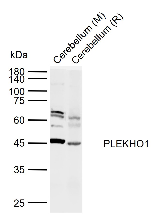
Sample:
Lane 1: Mouse Cerebellum tissue lysates
Lane 2: Rat Cerebellum tissue lysates
Primary: Anti-PLEKHO1 (bs-1330R) at 1/1000 dilution
Secondary: IRDye800CW Goat Anti-Rabbit IgG at 1/20000 dilution
Predicted band size: 45 kDa
Observed band size: 45 kDa
Paraformaldehyde-fixed, paraffin embedded (rat cerebellum); Antigen retrieval by boiling in sodium citrate buffer (pH6.0) for 15min; Block endogenous peroxidase by 3% hydrogen peroxide for 20 minutes; Blocking buffer (normal goat serum) at 37°C for 30min; Antibody incubation with (PLEKHO1) Polyclonal Antibody, Unconjugated (bs-1330R) at 1:200 overnight at 4°C, followed by operating according to SP Kit(Rabbit) (sp-0023) instructionsand DAB staining.
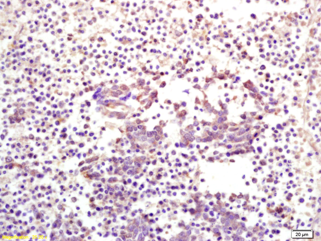
Tissue/cell: human lung carcinoma; 4% Paraformaldehyde-fixed and paraffin-embedded;
Antigen retrieval: citrate buffer ( 0.01M, pH 6.0 ), Boiling bathing for 15min; Block endogenous peroxidase by 3% Hydrogen peroxide for 30min; Blocking buffer (normal goat serum,C-0005) at 37℃ for 20 min;
Incubation: Anti-CKIP-1 Polyclonal Antibody, Unconjugated(bs-1330R) 1:200, overnight at 4°C, followed by conjugation to the secondary antibody(SP-0023) and DAB(C-0010) staining
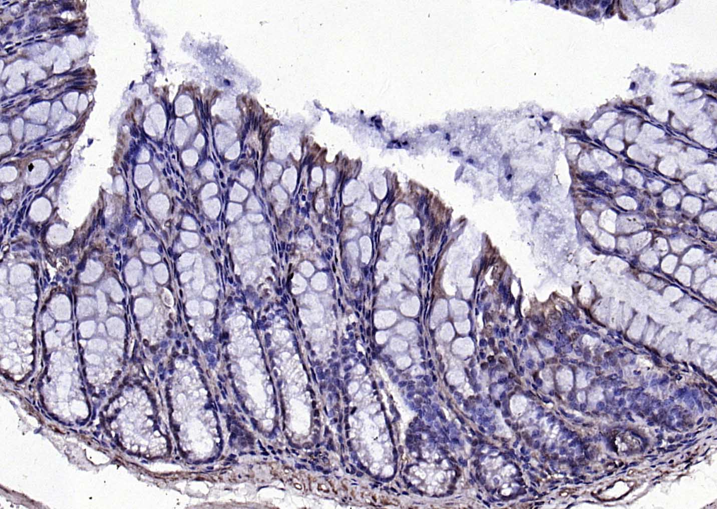
Paraformaldehyde-fixed, paraffin embedded (mouse colon); Antigen retrieval by boiling in sodium citrate buffer (pH6.0) for 15min; Block endogenous peroxidase by 3% hydrogen peroxide for 20 minutes; Blocking buffer (normal goat serum) at 37°C for 30min; Antibody incubation with (PLEKHO1) Polyclonal Antibody, Unconjugated (bs-1330R) at 1:200 overnight at 4°C, followed by operating according to SP Kit(Rabbit) (sp-0023) instructionsand DAB staining.
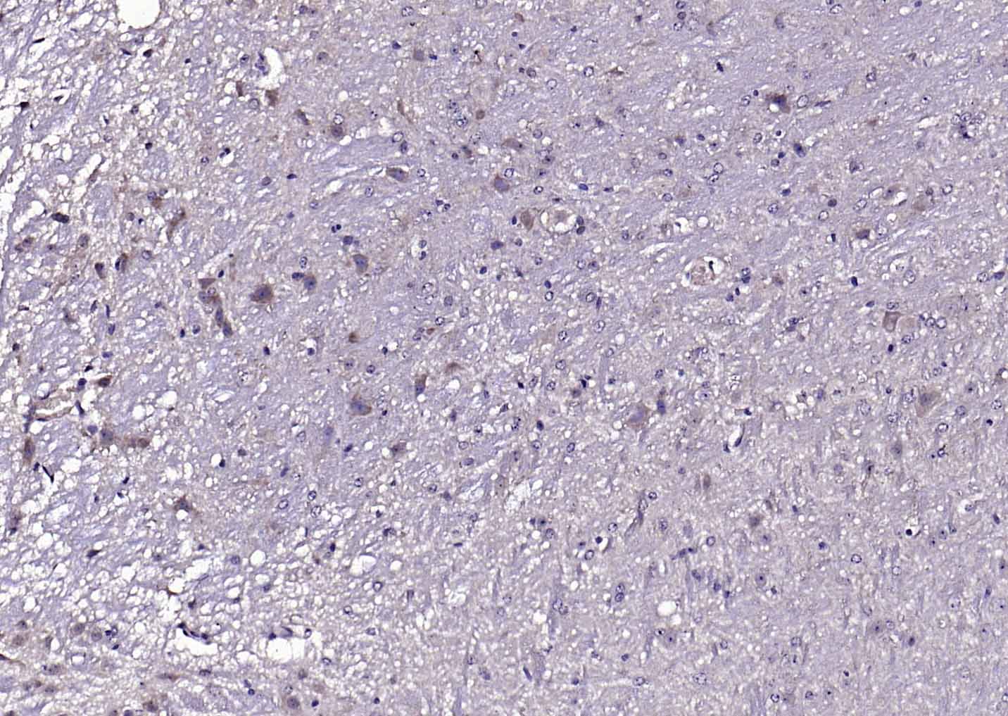
Paraformaldehyde-fixed, paraffin embedded (mouse cerebellum); Antigen retrieval by boiling in sodium citrate buffer (pH6.0) for 15min; Block endogenous peroxidase by 3% hydrogen peroxide for 20 minutes; Blocking buffer (normal goat serum) at 37°C for 30min; Antibody incubation with (PLEKHO1) Polyclonal Antibody, Unconjugated (bs-1330R) at 1:200 overnight at 4°C, followed by operating according to SP Kit(Rabbit) (sp-0023) instructionsand DAB staining.
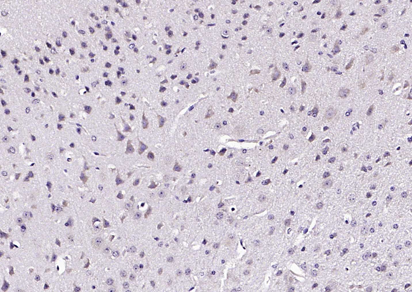
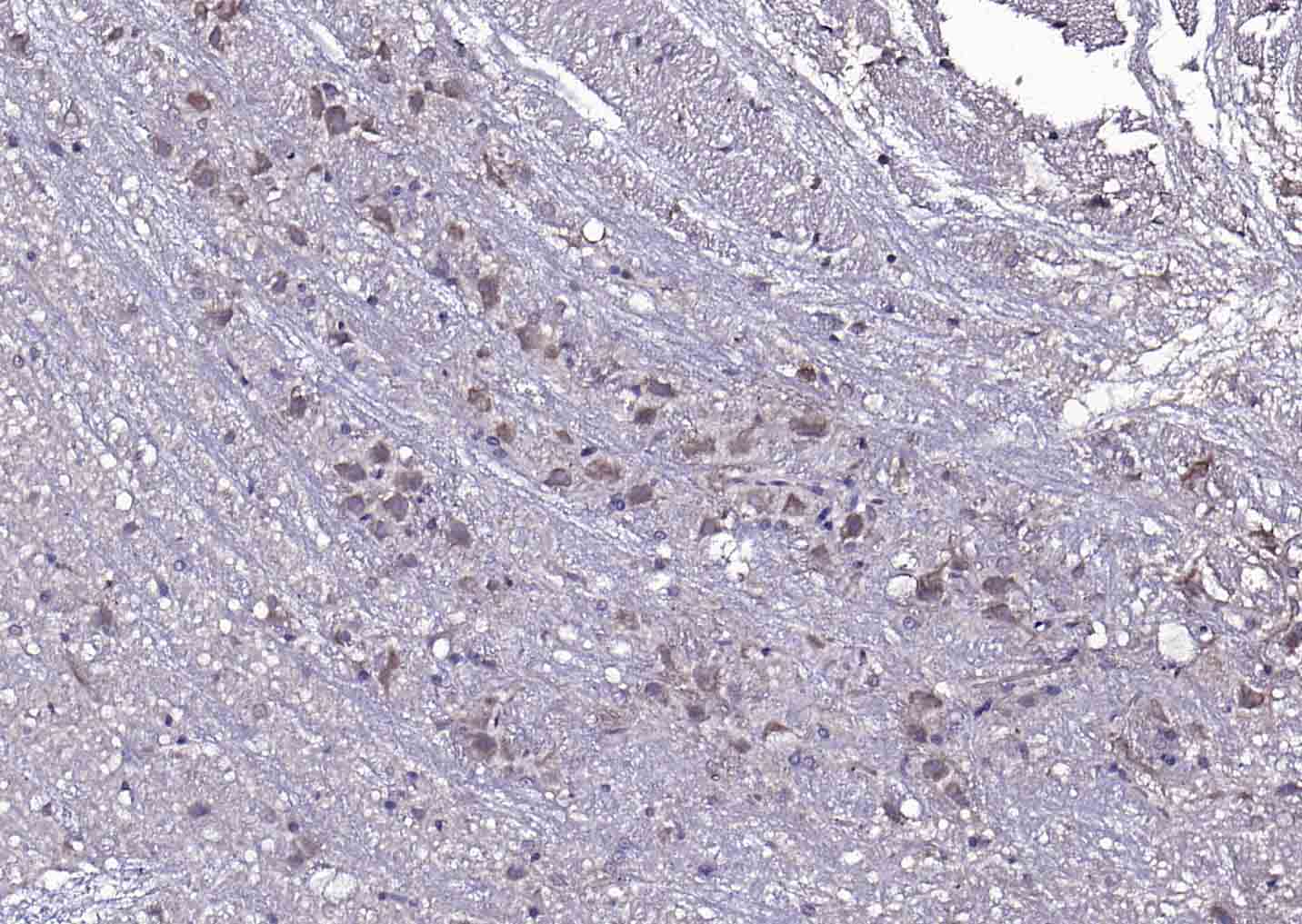
Paraformaldehyde-fixed, paraffin embedded (mouse brain); Antigen retrieval by boiling in sodium citrate buffer (pH6.0) for 15min; Block endogenous peroxidase by 3% hydrogen peroxide for 20 minutes; Blocking buffer (normal goat serum) at 37°C for 30min; Antibody incubation with (PLEKHO1) Polyclonal Antibody, Unconjugated (bs-1330R) at 1:200 overnight at 4°C, followed by operating according to SP Kit(Rabbit) (sp-0023) instructionsand DAB staining.
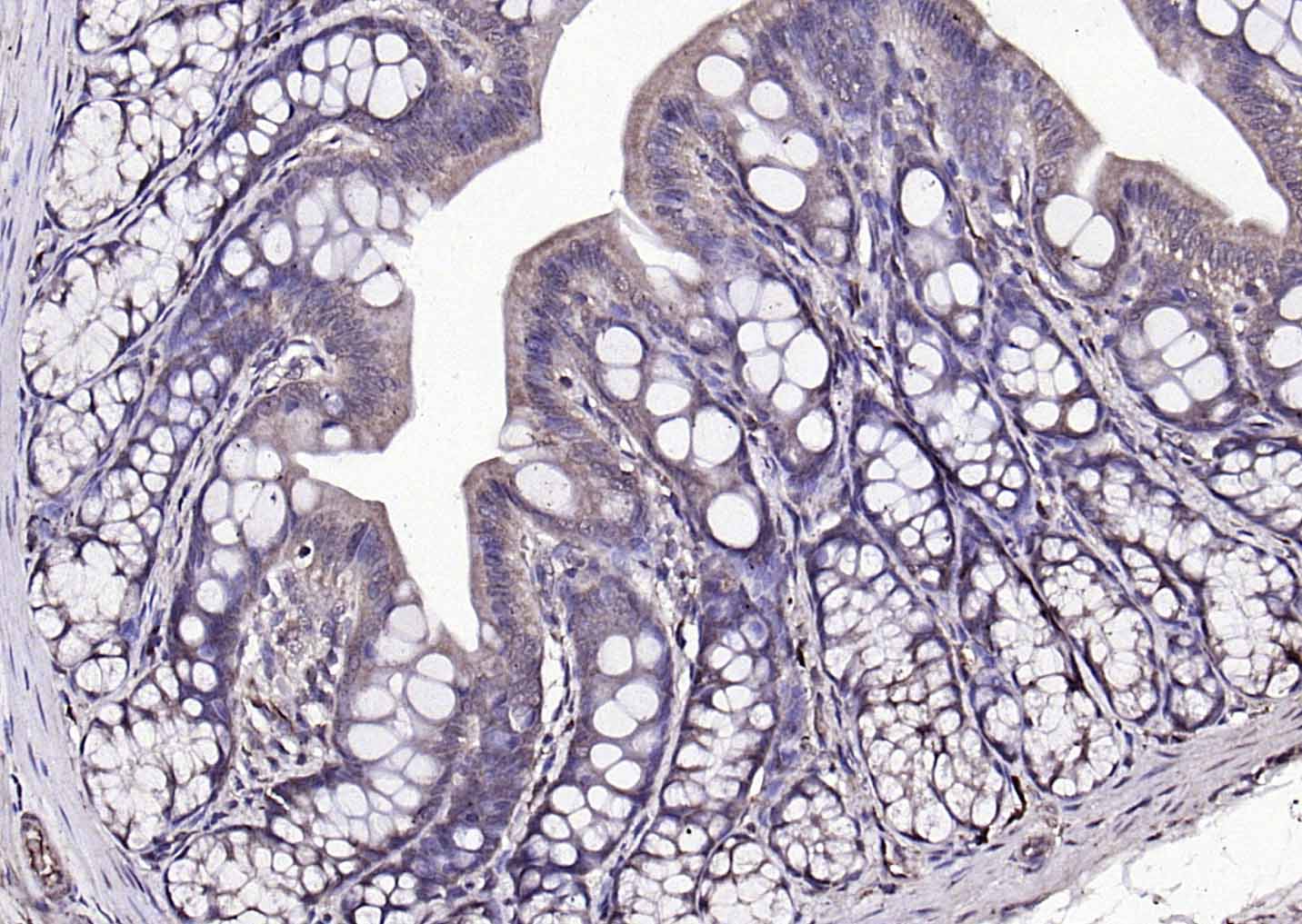
Paraformaldehyde-fixed, paraffin embedded (rat colon); Antigen retrieval by boiling in sodium citrate buffer (pH6.0) for 15min; Block endogenous peroxidase by 3% hydrogen peroxide for 20 minutes; Blocking buffer (normal goat serum) at 37°C for 30min; Antibody incubation with (PLEKHO1) Polyclonal Antibody, Unconjugated (bs-1330R) at 1:200 overnight at 4°C, followed by operating according to SP Kit(Rabbit) (sp-0023) instructionsand DAB staining.
|
| 1、抗体溶解方法 | |
| 2、抗体修复方式 | |
| 3、常用试剂的配制 | |
| 4、免疫组化操作步骤 | |
| 5、免疫组化问题解答 | |
| 6、Western Blotting 操作步骤 | |
| 7、Western Blotting 问题解答 | |
| 8、关于肽链的设计 | |
| 9、多肽的溶解与保存 | |
| 10、酶标抗体效价测定程序 | |


