

| 产品编号 | bs-0486R |
| 英文名称 | Integrin beta 1 Rabbit pAb |
| 中文名称 | 整合素β1(CD29)抗体 |
| 别 名 | CD29; FNRB; GPIIA; MDF2; MSK12; VLA-BETA; VLAB; 4633401G24Rik; Gm9863; ITB1_HUMAN; ITGB1; Fibronectin receptor subunit beta; Glycoprotein IIa (GPIIA); VLA-4 subunit beta; ITB1_MOUSE; ITB1_RAT; Beta oligodendroglia (Beta OL); |
 |
Specific References (29) | bs-0486R has been referenced in 29 publications.
|
| 研究领域 | 细胞生物 免疫学 细胞粘附分子 |
| 抗体来源 | Rabbit |
| 克隆类型 | Polyclonal |
| 克 隆 号 | |
| 交叉反应 | Human,Mouse,Rat (predicted: Rabbit,Pig,Sheep,Cow,Chicken,Dog,Horse) |
| 产品应用 | WB=1:500-2000,IHC-P=1:100-500,IHC-F=1:100-500,IF=1:100-500,Flow-Cyt=1μg/Test
not yet tested in other applications. optimal dilutions/concentrations should be determined by the end user. |
| 理论分子量 | 88 kDa |
| 检测分子量 | 130 |
| 细胞定位 | 细胞浆 细胞膜 |
| 性 状 | Liquid |
| 浓 度 | 1mg/ml |
| 免 疫 原 | KLH conjugated synthetic peptide derived from human Integrin beta 1: 25-100/798 <Extracellular> |
| 亚 型 | IgG |
| 纯化方法 | affinity purified by Protein A |
| 缓 冲 液 | 0.01M TBS (pH7.4) with 1% BSA, 0.02% Proclin300 and 50% Glycerol. |
| 保存条件 | Shipped at 4℃. Store at -20℃ for one year. Avoid repeated freeze/thaw cycles. |
| 注意事项 | This product as supplied is intended for research use only, not for use in human, therapeutic or diagnostic applications. |
| PubMed | PubMed |
| 产品介绍 |
Integrins alpha-1/beta-1, alpha-2/beta-1, alpha-10/beta-1 and alpha-11/beta-1 are receptors for collagen. Integrins alpha-1/beta-1 and alpha-2/beta-2 recognize the proline-hydroxylated sequence G-F-P-G-E-R in collagen. Integrins alpha-2/beta-1, alpha-3/beta-1, alpha-4/beta-1, alpha-5/beta-1, alpha-8/beta-1, alpha-10/beta-1, alpha-11/beta-1 and alpha-V/beta-1 are receptors for fibronectin. Alpha-4/beta-1 recognizes one or more domains within the alternatively spliced CS-1 and CS-5 regions of fibronectin. Integrin alpha-5/beta-1 is a receptor for fibrinogen. Integrin alpha-1/beta-1, alpha-2/beta-1, alpha-6/beta-1 and alpha-7/beta-1 are receptors for lamimin. Integrin alpha-4/beta-1 is a receptor for VCAM1. It recognizes the sequence Q-I-D-S in VCAM1. Integrin alpha-9/beta-1 is a receptor for VCAM1, cytotactin and osteopontin. It recognizes the sequence A-E-I-D-G-I-E-L in cytotactin. Integrin alpha-3/beta-1 is a receptor for epiligrin, thrombospondin and CSPG4. Alpha-3/beta-1 may mediate with LGALS3 the stimulation by CSPG4 of endothelial cells migration. Integrin alpha-V/beta-1 is a receptor for vitronectin. Beta-1 integrins recognize the sequence R-G-D in a wide array of ligands. Isoform beta-1B interferes with isoform beta-1A resulting in a dominant negative effect on cell adhesion and migration (in vitro). In case of HIV-1 infection, the interaction with extracellular viral Tat protein seems to enhance angiogenesis in Kaposi's sarcoma lesions. Function: Integrins alpha-1/beta-1, alpha-2/beta-1, alpha-10/beta-1 and alpha-11/beta-1 are receptors for collagen. Integrins alpha-1/beta-1 and alpha-2/beta-2 recognize the proline-hydroxylated sequence G-F-P-G-E-R in collagen. Integrins alpha-2/beta-1, alpha-3/beta-1, alpha-4/beta-1, alpha-5/beta-1, alpha-8/beta-1, alpha-10/beta-1, alpha-11/beta-1 and alpha-V/beta-1 are receptors for fibronectin. Alpha-4/beta-1 recognizes one or more domains within the alternatively spliced CS-1 and CS-5 regions of fibronectin. Integrin alpha-5/beta-1 is a receptor for fibrinogen. Integrin alpha-1/beta-1, alpha-2/beta-1, alpha-6/beta-1 and alpha-7/beta-1 are receptors for lamimin. Integrin alpha-4/beta-1 is a receptor for VCAM1. It recognizes the sequence Q-I-D-S in VCAM1. Integrin alpha-9/beta-1 is a receptor for VCAM1, cytotactin and osteopontin. It recognizes the sequence A-E-I-D-G-I-E-L in cytotactin. Integrin alpha-3/beta-1 is a receptor for epiligrin, thrombospondin and CSPG4. Alpha-3/beta-1 may mediate with LGALS3 the stimulation by CSPG4 of endothelial cells migration. Integrin alpha-V/beta-1 is a receptor for vitronectin. Beta-1 integrins recognize the sequence R-G-D in a wide array of ligands. Isoform beta-1B interferes with isoform beta-1A resulting in a dominant negative effect on cell adhesion and migration (in vitro). In case of HIV-1 infection, the interaction with extracellular viral Tat protein seems to enhance angiogenesis in Kaposi's sarcoma lesions. Subunit: Heterodimer of an alpha and a beta subunit. Beta-1 associates with either alpha-1, alpha-2, alpha-3, alpha-4, alpha-5, alpha-6, alpha-7, alpha-8, alpha-9, alpha-10, alpha-11 or alpha-V. Binds LGALS3BP and ITGB1BP3, when associated with alpha-7, but not with alpha-5. Interacts with FLNA, FLNB and RANBP9. Isoform Beta-1D interacts with ACE2. Isoform Beta-1A interacts with the C-terminal region of FLNC. Interacts with KRT1 in the presence of GNB2L1 and SRC. Interacts with HIV-1 Tat. Binds to human echoviruses 1 and 8 capsid proteins and acts as a receptor for these viruses. Interacts with RAB21. Interacts (via the cytoplasmic region) with RAB25 (via the hypervariable C-terminal region). Interacts with FGR and HCK (By similarity). Interacts with MYO10. Subcellular Location: Cell membrane; Single-pass type I membrane protein. Melanosome. Cleavage furrow. Note=Isoform beta-1B does not localize to focal adhesions. Highly enriched in stage I melanosomes. Located on plasma membrane of neuroblastoma NMB7 cells. In a lung cancer cell line, in prometaphase and metaphase, localizes diffusely at the membrane and in afew intracellular vesicles. In early telophase, detected mainly on the matrix-facing side of the cells. By mid-telophase, concentrated to the ingressing cleavage furrow, mainly to the basal side of the furrow. In late telophase, concentrated to the extending protrusions formed at the opposite ends of the spreading daughter cells, in vesicles at the base of the lamellipodia formed by the separating daughter cells. Tissue Specificity: Isoform beta-1A is widely expressed, other isoforms are generally coexpressed with a more restricted distribution. Isoform beta-1B is expressed in skin, liver, skeletal muscle, cardiac muscle, placenta, umbilical vein endothelial cells, neuroblastoma cells, lymphoma cells, hepatoma cells and astrocytoma cells. Isoform beta-1C and isoform beta-1C-2 are expressed in muscle, kidney, liver, placenta, cervical epithelium, umbilical vein endothelial cells, fibroblast cells, embryonal kidney cells, platelets and several blood cell lines. Isoform beta-C-2, rather than isoform beta-1C, is selectively expressed in peripheral T-cells. Isoform beta-1C is expressed in non-proliferating and differentiated prostate gland epithelial cells and in platelets, on the surface of erythroleukemia cells and in various hematopoietic cell lines. Isoform beta-1D is expressed specifically in striated muscle (skeletal and cardiac muscle). Post-translational modifications: The cysteine residues are involved in intrachain disulfide bonds. Similarity: Belongs to the integrin beta chain family. Contains 1 VWFA domain. SWISS: P05556 Gene ID: 3688 Database links: Entrez Gene: 3688 Human Entrez Gene: 16412 Mouse Omim: 135630 Human SwissProt: P05556 Human SwissProt: P09055 Mouse Unigene: 643813 Human Unigene: 263396 Mouse Unigene: 25733 Rat |
| 产品图片 |
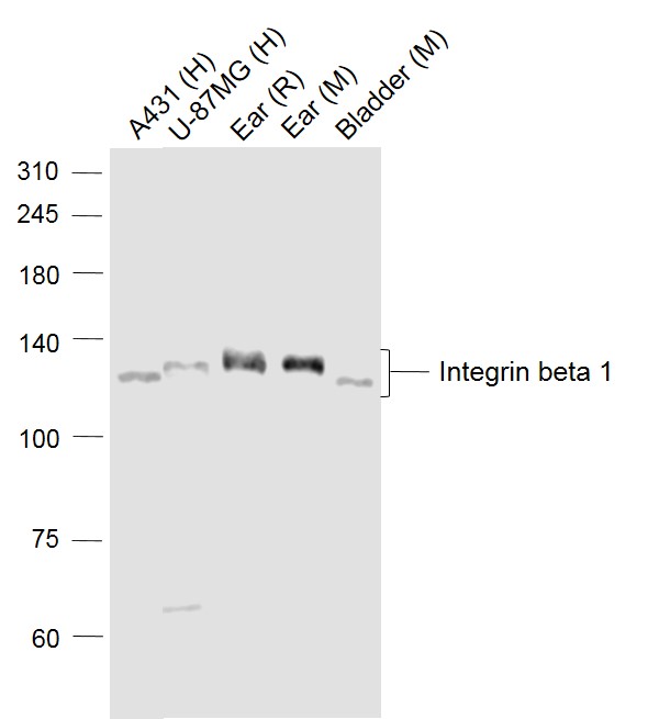
Sample:
Lane 1: A431 (Human) Cell Lysate at 30 ug
Lane 2: U-87MG (Human) Cell Lysate at 30 ug
Lane 3: Ear (Rat) Lysate at 40 ug
Lane 4: Ear (Mouse) Lysate at 40 ug
Lane 5: Bladder (Mouse) Lysate at 40 ug
Primary: Anti-Integrin beta 1 (bs-0486R) at 1/1000 dilution
Secondary: IRDye800CW Goat Anti-Rabbit IgG at 1/20000 dilution
Predicted band size: 130 kD
Observed band size: 130 kD
Paraformaldehyde-fixed, paraffin embedded (Mouse brain); Antigen retrieval by boiling in sodium citrate buffer (pH6.0) for 15min; Block endogenous peroxidase by 3% hydrogen peroxide for 20 minutes; Blocking buffer (normal goat serum) at 37°C for 30min; Antibody incubation with (Integrin beta 1) Polyclonal Antibody, Unconjugated (bs-0486R) at 1:400 overnight at 4°C, followed by operating according to SP Kit(Rabbit) (sp-0023) instructions and DAB staining.
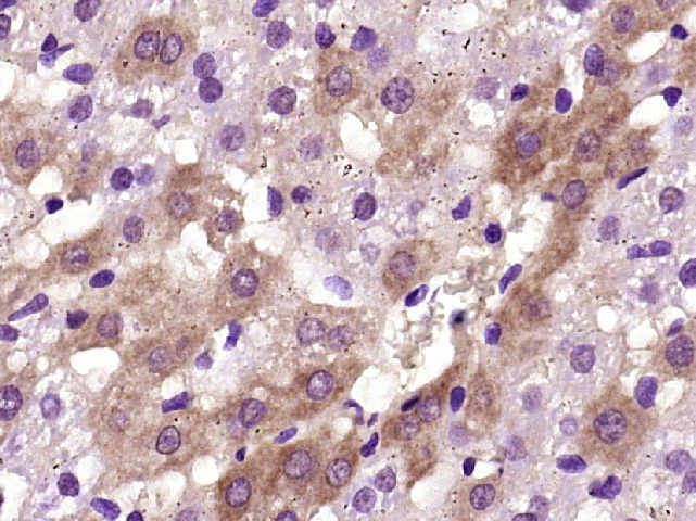
Paraformaldehyde-fixed, paraffin embedded (Rat liver); Antigen retrieval by boiling in sodium citrate buffer (pH6.0) for 15min; Block endogenous peroxidase by 3% hydrogen peroxide for 20 minutes; Blocking buffer (normal goat serum) at 37°C for 30min; Antibody incubation with (Integrin beta 1) Polyclonal Antibody, Unconjugated (bs-0486R) at 1:400 overnight at 4°C, followed by operating according to SP Kit(Rabbit) (sp-0023) instructionsand DAB staining.
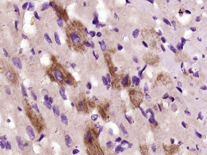
Paraformaldehyde-fixed, paraffin embedded (Rat heart); Antigen retrieval by boiling in sodium citrate buffer (pH6.0) for 15min; Block endogenous peroxidase by 3% hydrogen peroxide for 20 minutes; Blocking buffer (normal goat serum) at 37°C for 30min; Antibody incubation with (Integrin beta 1) Polyclonal Antibody, Unconjugated (bs-0486R) at 1:400 overnight at 4°C, followed by operating according to SP Kit(Rabbit) (sp-0023) instructionsand DAB staining.

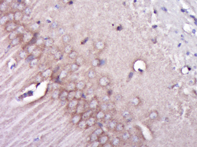
Paraformaldehyde-fixed, paraffin embedded (Rat brain); Antigen retrieval by boiling in sodium citrate buffer (pH6.0) for 15min; Block endogenous peroxidase by 3% hydrogen peroxide for 20 minutes; Blocking buffer (normal goat serum) at 37°C for 30min; Antibody incubation with (Integrin beta 1) Polyclonal Antibody, Unconjugated (bs-0486R) at 1:500 overnight at 4°C, followed by a conjugated secondary (sp-0023) for 20 minutes and DAB staining.
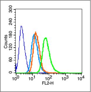
Blank control (blue line): Hela (blue).
Primary Antibody (green line): Rabbit Anti-Integrin beta 1 antibody (bs-0486R)
Dilution: 1μg /10^6 cells;
Isotype Control Antibody (orange line): Rabbit IgG .
Secondary Antibody (white blue line): Goat anti-rabbit IgG-PE
Dilution: 1μg /test.
Protocol
The cells were fixed with 70% methanol (Overnight at 4℃) and then permeabilized with 90% ice-cold methanol for 20 min at -20℃. Cells stained with Primary Antibody for 30 min at room temperature. The cells were then incubated in 1 X PBS/2%BSA/10% goat serum to block non-specific protein-protein interactions followed by the antibody for 15 min at room temperature. The secondary antibody used for 40 min at room temperature. Acquisition of 20,000 events was performed.
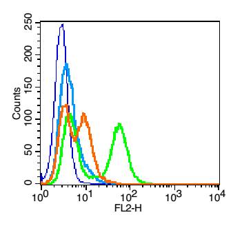
Blank control: HUVEC cells(blue). Primary Antibody:Rabbit Anti-CD29 antibody(bs-0486R), Dilution: 1μg in 100 μL 1X PBS containing 0.5% BSA; Isotype Control Antibody: Rabbit IgG(orange) ,used under the same conditions ); Secondary Antibody: Goat anti-rabbit IgG-PE(white blue), Dilution: 1:200 in 1 X PBS containing 0.5% BSA.
Protocol
The cells were fixed with 2% paraformaldehyde (10 min) .Primary antibody (bs-0486R, 1μg /1x10^6 cells) were incubated for 30 min on the ice, followed by 1 X PBS containing 0.5% BSA + 1 0% goat serum (15 min) to block non-specific protein-protein interactions. Then the Goat Anti-rabbit IgG/PE antibody was added into the blocking buffer mentioned above to react with the primary antibody at 1/200 dilution for 30 min on ice. Acquisition of 20,000 events was performed.
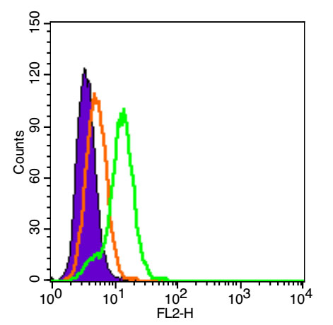
Blank control (Black line):HUVEC(Black).
Primary Antibody (green line): Rabbit Anti-Integrin beta 1 antibody (bs-0486R)
Dilution: 3μg /10^6 cells;
Isotype Control Antibody (orange line): Rabbit IgG-PE.
Protocol
The cells were fixed with 4% PFA (10min at room temperature)and then were incubated in 5%BSA to block non-specific protein-protein interactions for 30 min at room temperature .Cells stained with Primary Antibody for 30 min at room temperature. Acquisition of 20,000 events was performed.
|
| 1、抗体溶解方法 | |
| 2、抗体修复方式 | |
| 3、常用试剂的配制 | |
| 4、免疫组化操作步骤 | |
| 5、免疫组化问题解答 | |
| 6、Western Blotting 操作步骤 | |
| 7、Western Blotting 问题解答 | |
| 8、关于肽链的设计 | |
| 9、多肽的溶解与保存 | |
| 10、酶标抗体效价测定程序 | |


