

| 产品编号 | bs-1347R |
| 英文名称 | phospho-Rb (Ser780) Rabbit pAb |
| 中文名称 | 磷酸化成视网膜细胞瘤抑癌蛋白抗体 |
| 别 名 | RB1 | Rb (phospho-S780); p-Rb; phospho-Rb; p-RB1; RB1 | Rb (phospho-Ser780); OSRC; PPP1R130; RB; p105-Rb; p110-RB1; pRb; pp110; Rb-1; pp105; RB_HUMAN; RB1; pRb (Rb); RB_MOUSE; RB_RAT; |
 |
Specific References (4) | bs-1347R has been referenced in 4 publications.
[IF=8.755] Sijie Wang. et al. PFKFB4 facilitates palbociclib resistance in oestrogen receptor-positive breast cancer by enhancing stemness. CELL PROLIFERAT. 2022 Sep;:e13337 WB ; Human.
[IF=5.652] Haijun Sun. et al. WD Repeat Domain 43 promotes malignant progression of non-small cell lung cancer by regulating CDK2. INT J BIOCHEM CELL B. 2022 Aug;:106293 WB ; Human.
[IF=4.302] Ju B et al. miR-193a/b-3p relieves hepatic fibrosis and restrains proliferation and activation of hepaticstellate cells. J Cell Mol Med. 2019 Apr 3. WB ; Human.
[IF=3.332] Muhammad T et al. Aloperine in combination with therapeutic adenoviral vector synergistically suppressed the growth of non-small cell lung cancer. J Cancer Res Clin Oncol. 2020 Feb 22. WB ; Human.
|
| 产品类型 | 磷酸化抗体 |
| 研究领域 | 肿瘤 细胞生物 免疫学 染色质和核信号 表观遗传学 |
| 抗体来源 | Rabbit |
| 克隆类型 | Polyclonal |
| 克 隆 号 | |
| 交叉反应 | Human,Mouse,Rat (predicted: Cow,Chicken,Dog) |
| 产品应用 | WB=1:500-2000,IHC-P=1:100-500,IHC-F=1:100-500,IF=1:100-500,Flow-Cyt=1ug/test,ICC/IF=1:100-500
not yet tested in other applications. optimal dilutions/concentrations should be determined by the end user. |
| 理论分子量 | 106 kDa |
| 检测分子量 | 125 |
| 细胞定位 | 细胞核 |
| 性 状 | Liquid |
| 浓 度 | 1mg/ml |
| 免 疫 原 | KLH conjugated Synthesised phosphopeptide derived from human Rb around the phosphorylation site of Ser780: L(p-S)PI |
| 亚 型 | IgG |
| 纯化方法 | affinity purified by Protein A |
| 缓 冲 液 | 0.01M TBS (pH7.4) with 1% BSA, 0.02% Proclin300 and 50% Glycerol. |
| 保存条件 | Shipped at 4℃. Store at -20℃ for one year. Avoid repeated freeze/thaw cycles. |
| 注意事项 | This product as supplied is intended for research use only, not for use in human, therapeutic or diagnostic applications. |
| PubMed | PubMed |
| 产品介绍 |
The protein encoded by this gene is a negative regulator of the cell cycle and was the first tumor suppressor gene found. The encoded protein also stabilizes constitutive heterochromatin to maintain the overall chromatin structure. The active, hypophosphorylated form of the protein binds transcription factor E2F1. Defects in this gene are a cause of childhood cancer retinoblastoma (RB), bladder cancer, and osteogenic sarcoma. Function: Key regulator of entry into cell division that acts as a tumor suppressor. Promotes G0-G1 transition when phosphorylated by CDK3/cyclin-C. Acts as a transcription repressor of E2F1 target genes. The underphosphorylated, active form of RB1 interacts with E2F1 and represses its transcription activity, leading to cell cycle arrest. Directly involved in heterochromatin formation by maintaining overall chromatin structure and, in particular, that of constitutive heterochromatin by stabilizing histone methylation. ecruits and targets histone methyltransferases SUV39H1, SUV420H1 and SUV420H2, leading to epigenetic transcriptional repression. Controls histone H4 'Lys-20' trimethylation. Inhibits the intrinsic kinase activity of TAF1. Mediates transcriptional repression by SMARCA4/BRG1 by recruiting a histone deacetylase (HDAC) complex to the c-FOS promoter. In resting neurons, transcription of the c-FOS promoter is inhibited by BRG1-dependent recruitment of a phospho-RB1-HDAC1 repressor complex. Upon calcium influx, RB1 is dephosphorylated by calcineurin, which leads to release of the epressor complex (By similarity). In case of viral infections, interactions with SV40 large T antigen, HPV E7 protein or adenovirus E1A protein induce the disassembly of RB1-E2F1 complex thereby disrupting RB1's activity. Subunit: Interacts with ATAD5. Interacts with PRMT2, CDK1 and CDK2. The hypophosphorylated form interacts with and sequesters the E2F1 transcription factor. Interacts with heterodimeric E2F/DP transcription factor complexes containing TFDP1 and either E2F1, E2F3, E2F4 or E2F5, or TFDP2 and E2F4. The unphosphorylated form interacts with EID1, ARID3B, KDM5A, SUV39H1, MJD2A/JHDM3A and THOC1. Interacts with the N-terminal domain of TAF1. Interacts with AATF, DNMT1, LIN9, LMNA, SUV420H1, SUV420H2, PELP1 and TMPO-alpha. May interact with NDC80. Interacts with GRIP1 and UBR4. Interacts with ARID4A and KDM5B. Interacts with E4F1 and LIMD1. Interacts with SMARCA4/BRG1 AND HDAC1. Interacts with adenovirus E1A protein, HPV E7 protein and SV40 large T antigen. Interacts with PSMA3 and USP4. Interacts (when methylated at Lys-860) with L3MBTL1. Subcellular Location: Nucleus. Tissue Specificity: Expressed in the retina. Post-translational modifications: Phosphorylated by CDK6 and CDK4, and subsequently by CDK2 at Ser-567 in G1, thereby releasing E2F1 which is then able to activate cell growth. Dephosphorylated at the late M phase. SV40 large T antigen, HPV E7 and adenovirus E1A bind to the underphosphorylated, active form of pRb. Phosphorylation at Thr-821 and Thr-826 promotes interaction between the C-terminal domain C and the Pocket domain, and thereby inhibits interactions with heterodimeric E2F/DP transcription factor complexes. Dephosphorylated at Ser-795 by calcineruin upon calcium stimulation. CDK3/cyclin-C-mediated phosphorylation at Ser-807 and Ser-811 is required for G0-G1 transition. Phosphorylated by CDK1 and CDK2 upon TGFB1-mediated apoptosis. N-terminus is methylated by METTL11A/NTM1. Monomethylated at Lys-860 by SMYD2, promoting interaction with L3MBTL1. DISEASE: Defects in RB1 are the cause of childhood cancer retinoblastoma (RB) [MIM:180200]. RB is a congenital malignant tumor that arises from the nuclear layers of the retina. It occurs in about 1:20'000 live births and represents about 2% of childhood malignancies. It is bilateral in about 30% of cases. Although most RB appear sporadically, about 20% are transmitted as an autosomal dominant trait with incomplete penetrance. The diagnosis is usually made before the age of 2 years when strabismus or a gray to yellow reflex from pupil ('cat eye') is investigated. Defects in RB1 are a cause of susceptibility to bladder cancer (BLC) [MIM:109800]. A malignancy originating in tissues of the urinary bladder. It often presents with multiple tumors appearing at different times and at different sites in the bladder. Most bladder cancers are transitional cell carcinomas. They begin in cells that normally make up the inner lining of the bladder. Other types of bladder cancer include squamous cell carcinoma (cancer that begins in thin, flat cells) and adenocarcinoma (cancer that begins in cells that make and release mucus and other fluids). Bladder cancer is a complex disorder with both genetic and environmental influences. Defects in RB1 are a cause of osteogenic sarcoma (OSRC) [MIM:259500]. Similarity: Belongs to the retinoblastoma protein (RB) family. SWISS: P06400 Gene ID: 5925 Database links: Entrez Gene: 5925 Human Entrez Gene: 19645 Mouse Omim: 614041 Human SwissProt: P06400 Human SwissProt: P13405 Mouse Unigene: 408528 Human Unigene: 273862 Mouse Unigene: 55115 Rat 核标志物 |
| 产品图片 |
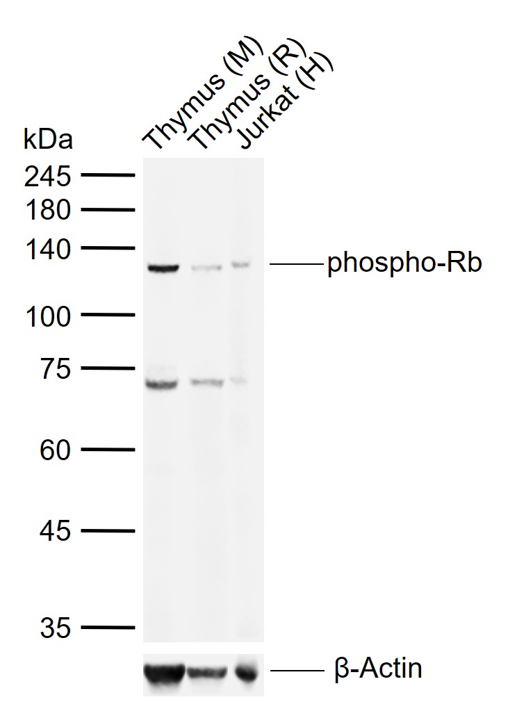
Sample:
Lane 1: Mouse Thymus tissue lysates
Lane 2: Rat Thymus tissue lysates
Lane 3: Human Jurkat cell lysates
Primary: Anti-phospho-Rb (Ser780) (bs-1347R) at 1/1000 dilution
Secondary: IRDye800CW Goat Anti-Rabbit IgG at 1/20000 dilution
Predicted band size: 106 kDa
Observed band size: 125 kDa
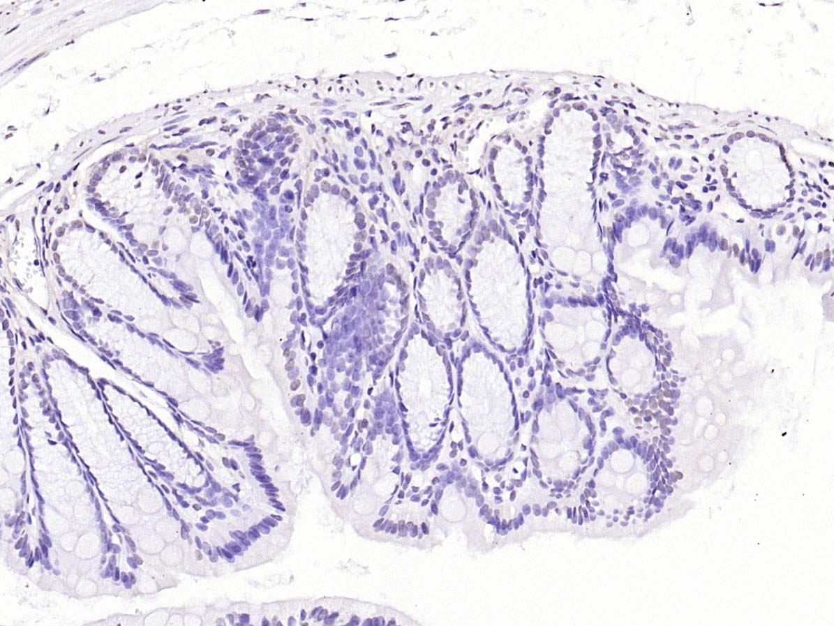
Paraformaldehyde-fixed, paraffin embedded (mouse colon); Antigen retrieval by boiling in sodium citrate buffer (pH6.0) for 15min; Block endogenous peroxidase by 3% hydrogen peroxide for 20 minutes; Blocking buffer (normal goat serum) at 37°C for 30min; Antibody incubation with (phospho-Rb (Ser780)) Polyclonal Antibody, Unconjugated (bs-1347R) at 1:200 overnight at 4°C, followed by operating according to SP Kit(Rabbit) (sp-0023) instructionsand DAB staining.
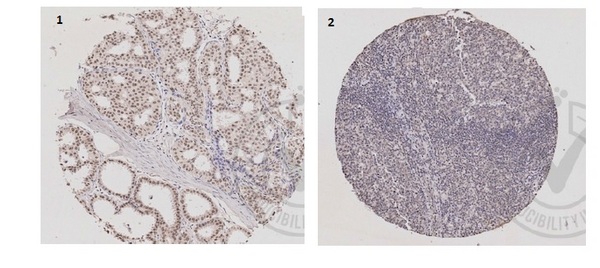
Independently Validated Antibody, image provided by Science Direct, badge number 029648:Formalin-fixed and paraffin embedded human breast tissue labeled with Anti-phospho-Rb/p105-Rb(Ser780) Polyclonal Antibody, Unconjugated (bs-1347R) at 1:200 followed by conjugation to the secondary antibody and DAB staining. Negative control, Human lymph node tissue, also stained postive
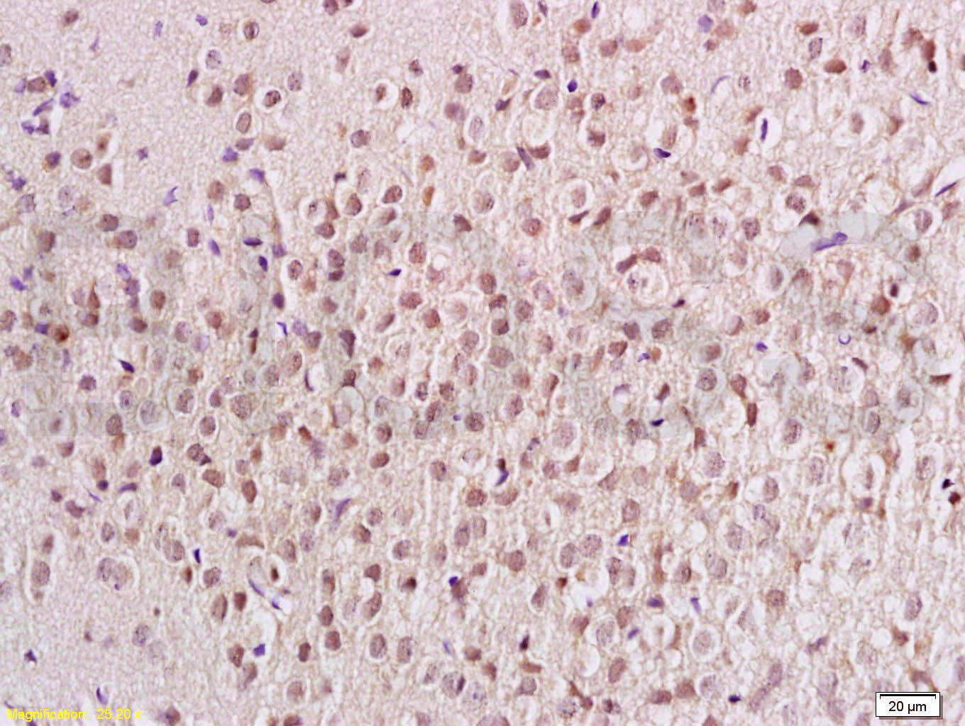
Tissue/cell: rat brain tissue; 4% Paraformaldehyde-fixed and paraffin-embedded;
Antigen retrieval: citrate buffer ( 0.01M, pH 6.0 ), Boiling bathing for 15min; Block endogenous peroxidase by 3% Hydrogen peroxide for 30min; Blocking buffer (normal goat serum,C-0005) at 37℃ for 20 min;
Incubation: Anti-phospho-Rb/p105-Rb(Ser780) Polyclonal Antibody, Unconjugated(bs-1347R) 1:200, overnight at 4°C, followed by conjugation to the secondary antibody(SP-0023) and DAB(C-0010) staining
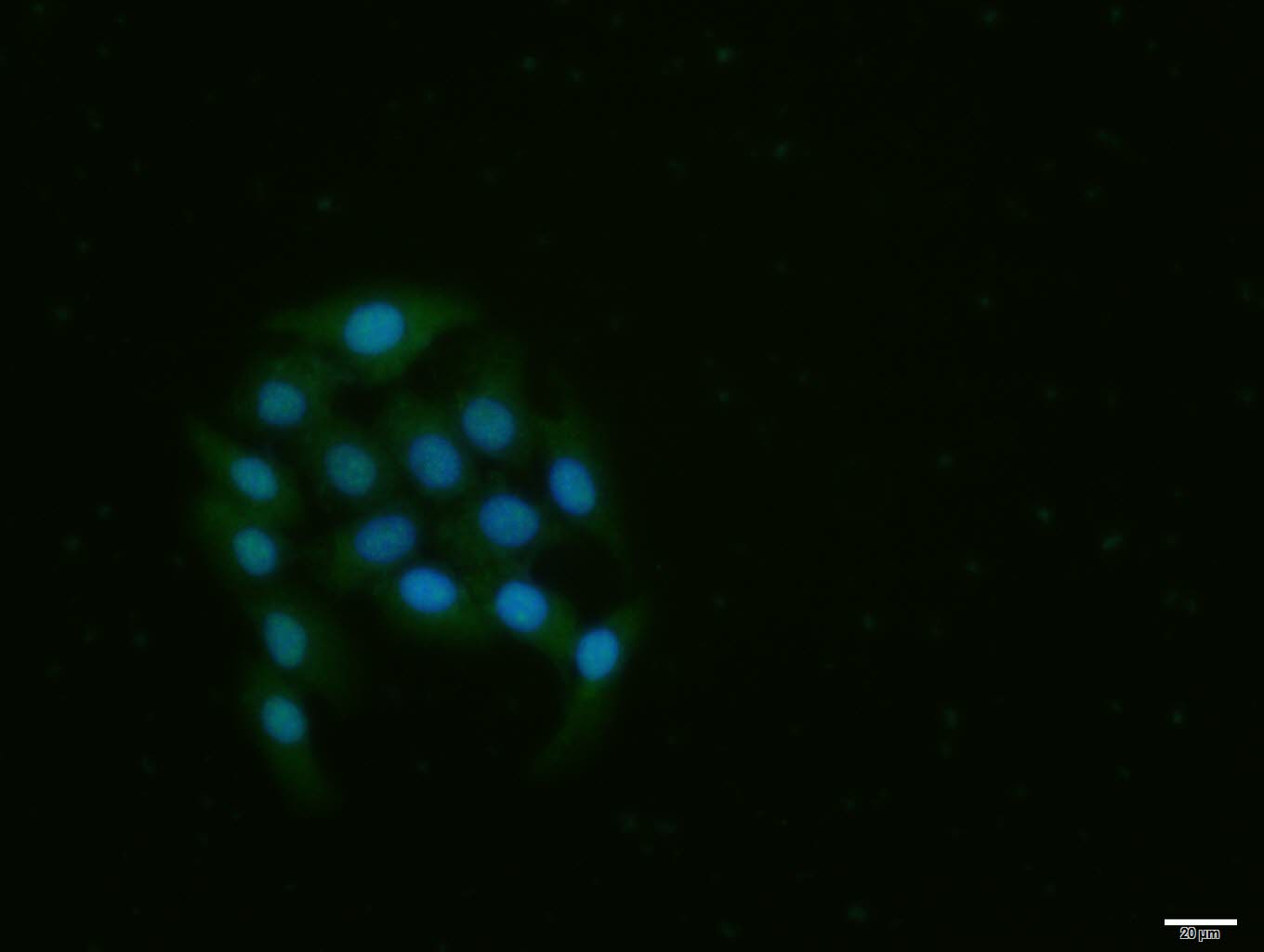
MCF-7 cell; 4% Paraformaldehyde-fixed; Triton X-100 at room temperature for 20 min; Blocking buffer (normal goat serum, C-0005) at 37°C for 20 min; Antibody incubation with (phospho-Rb (Ser780)) polyclonal Antibody, Unconjugated (bs-1347R) 1:100, 90 minutes at 37°C; followed by a conjugated Goat Anti-Rabbit IgG antibody at 37°C for 90 minutes, DAPI (blue, C02-04002) was used to stain the cell nuclei.
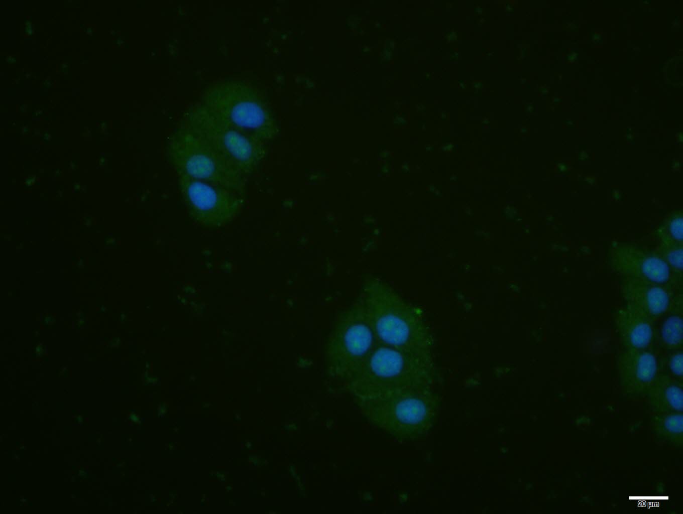
MCF-7 cell; 4% Paraformaldehyde-fixed; Triton X-100 at room temperature for 20 min; Blocking buffer (normal goat serum, C-0005) at 37°C for 20 min; Antibody incubation with (phospho-Rb (Ser780)) polyclonal Antibody, Unconjugated (bs-1347R) 1:100, 90 minutes at 37°C; followed by a conjugated Goat Anti-Rabbit IgG antibody at 37°C for 90 minutes, DAPI (blue, C02-04002) was used to stain the cell nuclei.
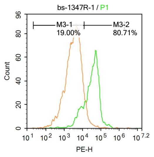
Molt-4 cells were fixed with 4% PFA for 10min at room temperature ,permeabilized with 90% ice-cold methanol for 20 min at -20℃, and incubated in 5% BSA blocking buffer for 30 min at room temperature. Cells were then stained with phospho-Rb (Ser780) Antibody(bs-1347R)at 1:500 dilution in blocking buffer and incubated for 30 min at room temperature, washed twice with 2%BSA in PBS, followed by secondary antibody incubation for 40 min at room temperature. Acquisitions of 20,000 events were performed. Cells stained with primary antibody (green), and isotype control (orange).
|
| 1、抗体溶解方法 | |
| 2、抗体修复方式 | |
| 3、常用试剂的配制 | |
| 4、免疫组化操作步骤 | |
| 5、免疫组化问题解答 | |
| 6、Western Blotting 操作步骤 | |
| 7、Western Blotting 问题解答 | |
| 8、关于肽链的设计 | |
| 9、多肽的溶解与保存 | |
| 10、酶标抗体效价测定程序 | |


