

| 产品编号 | bs-1508R |
| 英文名称 | Cathepsin L Rabbit pAb |
| 中文名称 | 组织蛋白酶L抗体 |
| 别 名 | CATL; CTSL1; MEP; 1190035F06Rik; fs; nkt; CATHL; CATL1_HUMAN; CTSL; Cathepsin L1; Major excreted protein (MEP); 3.4.22.15; CATL1_MOUSE; p39 cysteine proteinase; CATL1_RAT; Cyclic protein 2 (CP-2); |
 |
Specific References (2) | bs-1508R has been referenced in 2 publications.
[IF=7.56] Hernáez B, Guerra M, Salas ML, Andrés G (2016) African Swine Fever Virus Undergoes Outer Envelope Disruption, Capsid Disassembly and Inner Envelope Fusion before Core Release from Multivesicular Endosomes. PLoS Pathog 12(4): e1005595. other ; Pig.
[IF=4.406] Mathew Suji Eapen. et al. The Pathophysiology of COVID-19 and SARS-CoV-2 Infection: Dysregulation of endocytic machinery and ACE2 in small airways of smokers and COPD patients can augment their susceptibility to SARS-CoV-2 (COVID-19) infections. Am J Physiol-Lung C. 2021 Jan 1; 320(1): L158–L163 IHC ; Human.
|
| 研究领域 | 激酶和磷酸酶 合成与降解 |
| 抗体来源 | Rabbit |
| 克隆类型 | Polyclonal |
| 克 隆 号 | |
| 交叉反应 | Human,Rat (predicted: Mouse) |
| 产品应用 | WB=1:500-2000,IHC-P=1:100-500,IHC-F=1:100-500,IF=1:100-500,Flow-Cyt=1ug/Test
not yet tested in other applications. optimal dilutions/concentrations should be determined by the end user. |
| 理论分子量 | 19/30/37 kDa |
| 检测分子量 | 40 |
| 细胞定位 | 细胞浆 |
| 性 状 | Liquid |
| 浓 度 | 1mg/ml |
| 免 疫 原 | KLH conjugated synthetic peptide derived from human cathepsin L1 proprotein: 71-170/334 |
| 亚 型 | IgG |
| 纯化方法 | affinity purified by Protein A |
| 缓 冲 液 | 0.01M TBS (pH7.4) with 1% BSA, 0.02% Proclin300 and 50% Glycerol. |
| 保存条件 | Shipped at 4℃. Store at -20℃ for one year. Avoid repeated freeze/thaw cycles. |
| 注意事项 | This product as supplied is intended for research use only, not for use in human, therapeutic or diagnostic applications. |
| PubMed | PubMed |
| 产品介绍 |
The protein encoded by this gene is a lysosomal cysteine proteinase that plays a major role in intracellular protein catabolism. Its substrates include collagen and elastin, as well as alpha-1 protease inhibitor, a major controlling element of neutrophil elastase activity. The encoded protein has been implicated in several pathologic processes, including myofibril necrosis in myopathies and in myocardial ischemia, and in the renal tubular response to proteinuria. This protein, which is a member of the peptidase C1 family, is a dimer composed of disulfide-linked heavy and light chains, both produced from a single protein precursor. At least two transcript variants encoding the same protein have been found for this gene. Function: Important for the overall degradation of proteins in lysosomes. Subunit: Dimer of a heavy and a light chain linked by disulfide bonds. Subcellular Location: Lysosome. Similarity: Belongs to the peptidase C1 family. SWISS: P07711 Gene ID: 1514 Database links: Entrez Gene: 1514 Human Entrez Gene: 13039 Mouse Omim: 116880 Human SwissProt: P07711 Human SwissProt: P06797 Mouse Unigene: 726015 Human Unigene: 930 Mouse Unigene: 1294 Rat 合成与降解(Synthesis and Degradation) 组织蛋白酶L(cathepsin L CL)属于木瓜蛋白酶家族中的半胱氨酸蛋白水解酶,以酶原的形式贮存于溶酶体中. 参与许多特殊的生理过程,如激素原的激活、抗原呈递、组织器官的发育等。 在病理状态下,因各种原因所致的细胞损伤(如病原微生物、炎症因子、氧化应激等)可降解细胞成分或细胞间质基质成分,包括层粘素、IV型胶原纤维及纤维连接素等,与人类许多疾病如肿瘤的浸润与转移、关节炎、骨质疏松、阿尔茨海姆病、多发性硬化症及其他慢性炎症性疾病有关. |
| 产品图片 |
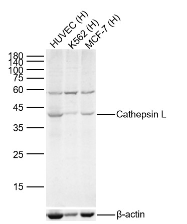
Sample:
Lane 1: Human HUVEC cell Lysates
Lane 2: Human K562 cell Lysates
Lane 3: Human MCF-7 cell Lysates
Primary: Anti-Cathepsin L (bs-1508R) at 1/300 dilution
Secondary: IRDye800CW Goat Anti-Rabbit IgG at 1/20000 dilution
Predicted band size: 19/30/37kDa
Observed band size: 40kDa
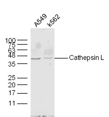
Sample:
A549 Cell (Human) Lysate at 30 ug
K562 Cell (Human) Lysate at 30 ug
Primary: Anti- Cathepsin L (bs-1508R) at 1/300 dilution
Secondary: IRDye800CW Goat Anti-Rabbit IgG at 1/20000 dilution
Predicted band size: 19/30/37 kD
Observed band size: 40 kD

Paraformaldehyde-fixed, paraffin embedded (Human kidney); Antigen retrieval by boiling in sodium citrate buffer (pH6.0) for 15min; Block endogenous peroxidase by 3% hydrogen peroxide for 20 minutes; Blocking buffer (normal goat serum) at 37°C for 30min; Antibody incubation with (Cathepsin L) Polyclonal Antibody, Unconjugated (bs-1508R) at 1:200 overnight at 4°C, followed by operating according to SP Kit(Rabbit) (sp-0023) instructionsand DAB staining.
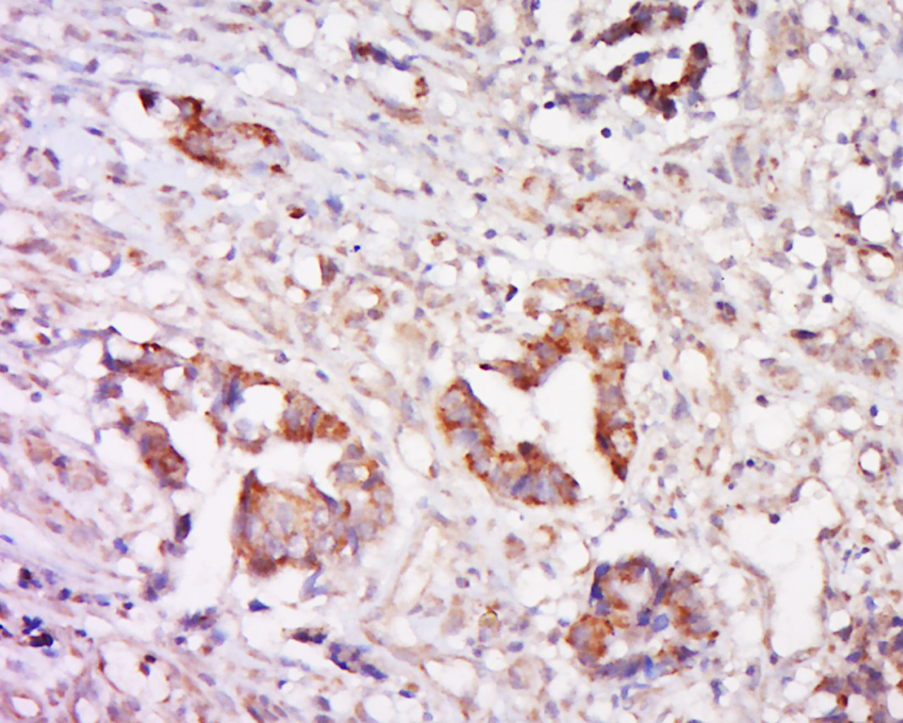
Tissue/cell: human cervical carcinoma; 4% Paraformaldehyde-fixed and paraffin-embedded;
Antigen retrieval: citrate buffer ( 0.01M, pH 6.0 ), Boiling bathing for 15min; Block endogenous peroxidase by 3% Hydrogen peroxide for 30min; Blocking buffer (normal goat serum,C-0005) at 37℃ for 20 min;
Incubation: Anti-Cathepsin Polyclonal Antibody, Unconjugated(bs-1508R) 1:500, overnight at 4°C, followed by conjugation to the secondary antibody(SP-0023) and DAB(C-0010) staining
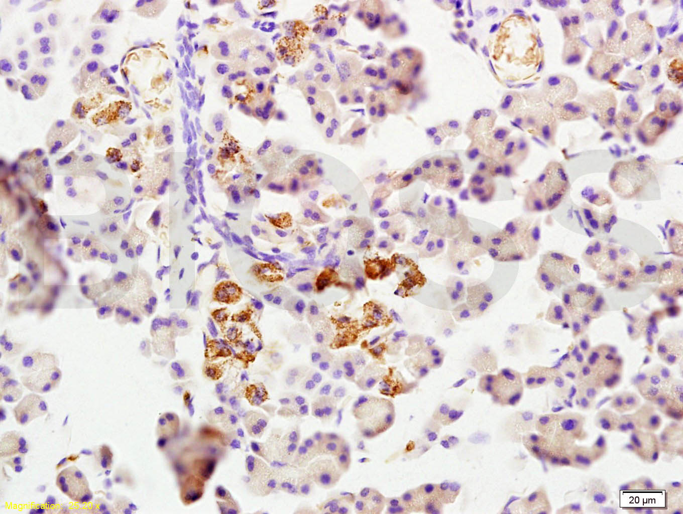
Tissue/cell: rat pancreas tissue; 4% Paraformaldehyde-fixed and paraffin-embedded;
Antigen retrieval: citrate buffer ( 0.01M, pH 6.0 ), Boiling bathing for 15min; Block endogenous peroxidase by 3% Hydrogen peroxide for 30min; Blocking buffer (normal goat serum,C-0005) at 37℃ for 20 min;
Incubation: Anti-Cathepsin L Polyclonal Antibody, Unconjugated(bs-1508R) 1:200, overnight at 4°C, followed by conjugation to the secondary antibody(SP-0023) and DAB(C-0010) staining
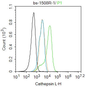
Blank control:A549.
Primary Antibody (green line): Rabbit Anti-Cathepsin L antibody (bs-1508R)
Dilution: 1ug/Test;
Secondary Antibody : Goat anti-rabbit IgG-FITC
Dilution: 0.5ug/Test.
Protocol
The cells were fixed with 4% PFA (10min at room temperature)and then permeabilized with 0.1% PBST for 20 min at room temperature.The cells were then incubated in 5%BSA to block non-specific protein-protein interactions for 30 min at room temperature .Cells stained with Primary Antibody for 30 min at room temperature. The secondary antibody used for 40 min at room temperature. Acquisition of 20,000 events was performed.
|
| 1、抗体溶解方法 | |
| 2、抗体修复方式 | |
| 3、常用试剂的配制 | |
| 4、免疫组化操作步骤 | |
| 5、免疫组化问题解答 | |
| 6、Western Blotting 操作步骤 | |
| 7、Western Blotting 问题解答 | |
| 8、关于肽链的设计 | |
| 9、多肽的溶解与保存 | |
| 10、酶标抗体效价测定程序 | |


