

| 产品编号 | bs-1702R |
| 英文名称 | FUT4 Rabbit pAb |
| 中文名称 | α-(1,3)-岩藻糖基转移酶 4抗体 |
| 别 名 | CD15; ELFT; FCT3A; FUC-TIV; FUTIV; LeX; SSEA-1; FUT4_HUMAN; FUT4; 4-galactosyl-N-acetylglucosaminide 3-alpha-L-fucosyltransferase; ELAM-1 ligand fucosyltransferase; Fucosyltransferase 4; Fucosyltransferase IV (Fuc-TIV | FucT-IV); Galactoside 3-L-fucosyltr |
 |
Specific References (6) | bs-1702R has been referenced in 6 publications.
[IF=7.94] Maolin Zhang. et al. Rapid and efficient generation of cartilage pellets from mouse induced pluripotent stem cells by transcriptional activation of BMP-4 with shaking culture:. J TISSUE ENG. 2022;(): IF ; Mouse.
[IF=6.064] Xinchao Miao. et al. Epiprofin Transcriptional Activation Promotes Ameloblast Induction From Mouse Induced Pluripotent Stem Cells via the BMP-Smad Signaling Axis. FRONT BIOENG BIOTECH. 2022 Jun 21;10:890882 IF ; Mouse.
[IF=2.766] Kim et al. Klotho and S100A8/A9 as Discriminative Markers between Pre-Renal and Intrinsic Acute Kidney Injury. (2016) PLoS.One. 11:e0147255 IF ; Rat.
[IF=1.888] C Geng et al. A simple fabricated microfluidic chip for urine sample-based bladder cancer detection.(2018).J MICROMECH MICROENG. microfluidic chip ; human.
[IF=1.31] Cong, Shan, Guifang Cao, and Dongjun Liu. "Effects of different feeder layers on culture of bovine embryonic stem cell-like cells in vitro." Cytotechnology (2014): 1-11. Bovine.
[IF=0] Zschemisch, Nils-Holger, et al. "Immortalized tumor derived rat fibroblasts as feeder cells facilitate the cultivation of male embryonic stem cells from the rat strain WKY/Ztm." SpringerPlus 3.1 (2014): 588. Rat.
|
| 研究领域 | 肿瘤 细胞生物 免疫学 细胞膜受体 |
| 抗体来源 | Rabbit |
| 克隆类型 | Polyclonal |
| 克 隆 号 | |
| 交叉反应 | Human,Mouse (predicted: Rat) |
| 产品应用 | WB=1:500-2000,IHC-P=1:100-500,IHC-F=1:100-500,IF=1:100-500,Flow-Cyt=1μg/Test
not yet tested in other applications. optimal dilutions/concentrations should be determined by the end user. |
| 理论分子量 | 58 kDa |
| 检测分子量 | 63 |
| 细胞定位 | 细胞浆 细胞膜 |
| 性 状 | Liquid |
| 浓 度 | 1mg/ml |
| 免 疫 原 | KLH conjugated synthetic peptide derived from human FUT4: 251-295/433 |
| 亚 型 | IgG |
| 纯化方法 | affinity purified by Protein A |
| 缓 冲 液 | 0.01M TBS (pH7.4) with 1% BSA, 0.02% Proclin300 and 50% Glycerol. |
| 保存条件 | Shipped at 4℃. Store at -20℃ for one year. Avoid repeated freeze/thaw cycles. |
| 注意事项 | This product as supplied is intended for research use only, not for use in human, therapeutic or diagnostic applications. |
| PubMed | PubMed |
| 产品介绍 |
The Lewis histo-blood group system comprises a set of fucosylated glycosphingolipids that are synthesized by exocrine epithelial cells and circulate in body fluids. The glycosphingolipids function in embryogenesis, tissue differentiation, tumor metastasis, inflammation, and bacterial adhesion. They are secondarily absorbed to red blood cells giving rise to their Lewis phenotype. This gene is a member of the fucosyltransferase family, which catalyzes the addition of fucose to precursor polysaccharides in the last step of Lewis antigen biosynthesis. It encodes an enzyme with alpha(1,3)-fucosyltransferase and alpha(1,4)-fucosyltransferase activities. Mutations in this gene are responsible for the majority of Lewis antigen-negative phenotypes. Multiple alternatively spliced variants, encoding the same protein, have been found for this gene. [provided by RefSeq]. Function: May catalyze alpha-1,3 glycosidic linkages involved in the expression of Lewis X/SSEA-1 and VIM-2 antigens. Subcellular Location: Golgi apparatus, Golgi stack membrane; Single-pass type II membrane protein. Note=Membrane-bound form in trans cisternae of Golgi. Tissue Specificity: Highest expression in stomach and colon. It is also expressed in the lung, testis, uterus, small intestine and to a lesser extent in spleen, and ovary. Present in trace amounts in brain, thymus, heart, smooth muscle, kidney and bone marrow. Not found in liver, salivary gland and pancreas. Similarity: Belongs to the glycosyltransferase 10 family. SWISS: P22083 Gene ID: 2526 Database links: Entrez Gene: 2526 Human Omim: 104230 Human SwissProt: P22083 Human
FUT4,也称为 ELFT 和 FCT3A,属于糖基转移酶 10 家族。FUT4 可催化参与 Lewis X/SSEA-1 和 VIM-2 抗原表达的 alpha-1,3 糖苷键。FUT4 是一种抗原表位,定义为 Lewis X 碳水化合物结构,在鼠胚胎癌细胞 (EC)、鼠 ES 和 iPS 细胞以及鼠和人类生殖细胞上表达。它被广泛用作小鼠未分化 ES 和 iPS 细胞的阳性表面标志物和人未分化 ES 和 iPS 细胞的阴性表面标志物。 |
| 产品图片 |
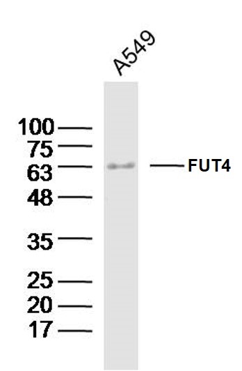
Sample:A549 Cell (Human) Lysate at 40 ug Primary: Anti-FUT4(bs-1702R)at 1/300 dilution Secondary: IRDye800CW Goat Anti-Rabbit IgG at 1/20000 dilution Predicted band size: 58kD Observed band size: 63kD
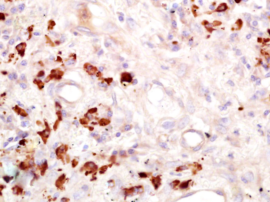
Paraformaldehyde-fixed, paraffin embedded (Human lung cancer); Antigen retrieval by boiling in sodium citrate buffer (pH6.0) for 15min; Block endogenous peroxidase by 3% hydrogen peroxide for 20 minutes; Blocking buffer (normal goat serum) at 37°C for 30min; Antibody incubation with (FUT4) Polyclonal Antibody, Unconjugated (bs-1702R) at 1:400 overnight at 4°C, followed by operating according to SP Kit(Rabbit) (sp-0023) instructionsand DAB staining.
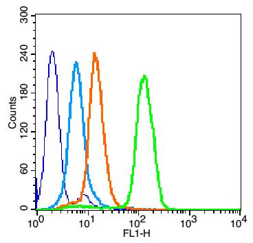
Overlay histogram showing HL 60 cells stained with bs-1702R (Green line). The cells were fixed with 90% methanol (5 min) and then permeabilized with 0.01M PBS-Tween for 20 min. The cells were then incubated in 1x PBS / 10% normal goat serum to block non-specific protein-protein interactions followed by the antibody (bs-1702R,1μg/1x10^6 cells) for 30 min at 22℃. The secondary antibody used was fluorescein isothiocyanate goat anti-rabbit IgG (H+L) (bs- 0295G-FITC , Brillant blue line) at 1/200 dilution for 30 min at 22℃. Isotype control antibody was rabbit IgG (polyclonal,bs-0295P,Orange line) (1μg/1x10^6 cells) used under the same conditions. Unlabelled sample (blue line) was also used as a control. Acquisition of 20,000 events were collected using a 20mW Argon ion laser (488nm) and 525/30 bandpass filter.
Blank control:HL-60.
Primary Antibody (green line): Rabbit Anti-FUT4 (bs-1702R)
Dilution: 1μg /10^6 cells;
Isotype Control Antibody (orange line): Rabbit IgG .
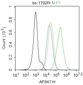
Secondary Antibody : Goat anti-rabbit IgG-AF647
Dilution: 1μg /test.
Protocol
The cells were fixed with 4% PFA (10min at room temperature)and then permeabilized with 0.1% PBST for 20 min at room temperature. The cells were then incubated in 5%BSA to block non-specific protein-protein interactions for 30 min at room temperature.Cells stained with Primary Antibody for 30 min at room temperature. The secondary antibody used for 40 min at room temperature. Acquisition of 20,000 events was performed.
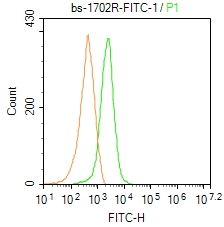
Blank control: Mouse spleen.
Primary Antibody (green line): Rabbit Anti-FUT4/FITC Conjugated antibody (bs-1702R-FITC)
Dilution: 1μg /10^6 cells;
Isotype Control Antibody (orange line): Rabbit IgG-FITC .
Protocol
The cells were fixed with 4% PFA (10min at room temperature)and then permeabilized with 0.1% PBST for 20 min at-20℃. The cells were then incubated in 5% BSA to block non-specific protein-protein interactions for 30 min at room temperature. The cells were stained with Primary Antibody for 30 min at room temperature. Acquisition of 20,000 events was performed.
|
| 1、抗体溶解方法 | |
| 2、抗体修复方式 | |
| 3、常用试剂的配制 | |
| 4、免疫组化操作步骤 | |
| 5、免疫组化问题解答 | |
| 6、Western Blotting 操作步骤 | |
| 7、Western Blotting 问题解答 | |
| 8、关于肽链的设计 | |
| 9、多肽的溶解与保存 | |
| 10、酶标抗体效价测定程序 | |


