

| 产品编号 | bs-0996R |
| 英文名称 | EphB2 Rabbit pAb |
| 中文名称 | 酪氨酸蛋白激酶受体B2抗体 |
| 别 名 | BDPLT22; CAPB; DRT; EK5; EPHT3; ERK; Hek5; PCBC; Tyro5; Cek5; ETECK; Nuk; Prkm5; Qek5; Sek3; EPHB2_HUMAN; EPHB2; Developmentally-regulated Eph-related tyrosine kinase; ELK-related tyrosine kinase; EPH tyrosine kinase 3; EPH-like kinase 5 (EK5 | hEK5); Ren |
 |
Specific References (3) | bs-0996R has been referenced in 3 publications.
[IF=11.47] Choi, Won Hoon, et al. "Open-gate mutants of the mammalian proteasome show enhanced ubiquitin-conjugate degradation." Nature Communications 7 (2016). WB ; Human.
[IF=2.63] Yang Xuesong. et al. Intervention Mechanism of Hunag-Lian Jie-Du Decoction on Canonical Wnt/β-Catenin Signaling Pathway in Psoriasis Mouse Model. EVID-BASED COMPL ALT. 2022;2022:3193572 WB ; Mouse.
[IF=2.303] Junjun Ling. et al. EPHB2 as a recurrence-related gene and a prognostic indicator in nasopharyngeal carcinoma: A bioinformatics screening and immunohistochemistry verification. HISTOL HISTOPATHOL. 2022 Apr 20;18459 IHC ; Human.
|
| 研究领域 | 肿瘤 细胞生物 免疫学 信号转导 转录调节因子 激酶和磷酸酶 细胞膜受体 |
| 抗体来源 | Rabbit |
| 克隆类型 | Polyclonal |
| 克 隆 号 | |
| 交叉反应 | Human,Mouse,Rat (predicted: Cow,Chicken,Dog) |
| 产品应用 | WB=1:500-2000,Flow-Cyt=3ug/Test
not yet tested in other applications. optimal dilutions/concentrations should be determined by the end user. |
| 理论分子量 | 108 kDa |
| 检测分子量 | 120 |
| 细胞定位 | 细胞浆 细胞膜 |
| 性 状 | Liquid |
| 浓 度 | 1mg/ml |
| 免 疫 原 | KLH conjugated synthetic peptide derived from human EPHB2: 551-650/1055 |
| 亚 型 | IgG |
| 纯化方法 | affinity purified by Protein A |
| 缓 冲 液 | 0.01M TBS (pH7.4) with 1% BSA, 0.02% Proclin300 and 50% Glycerol. |
| 保存条件 | Shipped at 4℃. Store at -20℃ for one year. Avoid repeated freeze/thaw cycles. |
| 注意事项 | This product as supplied is intended for research use only, not for use in human, therapeutic or diagnostic applications. |
| PubMed | PubMed |
| 产品介绍 |
This gene encodes a member of the Eph receptor family of receptor tyrosine kinase transmembrane glycoproteins. These receptors are composed of an N-terminal glycosylated ligand-binding domain, a transmembrane region and an intracellular kinase domain. They bind ligands called ephrins and are involved in diverse cellular processes including motility, division, and differentiation. A distinguishing characteristic of Eph-ephrin signaling is that both receptors and ligands are competent to transduce a signaling cascade, resulting in bidirectional signaling. This protein belongs to a subgroup of the Eph receptors called EphB. Proteins of this subgroup are distinguished from other members of the family by sequence homology and preferential binding affinity for membrane-bound ephrin-B ligands. Allelic variants are associated with prostate and brain cancer susceptibility. Alternative splicing results in multiple transcript variants. [provided by RefSeq, May 2015] Function: Receptor for members of the ephrin-B family. Phosphorylates ARHGEF15, leading to its ubiquitination and degradation by the proteasome which promotes EFNB1-dependent synapse formation. Can function in aspects of retinal ganglion cell axon guidance to the optic disk even when lacking its tyrosine kinase domain. Acts as a tumor suppressor. Subunit: Heterotetramer upon binding of the ligand. The heterotetramer is composed of an ephrin dimer and a receptor dimer. Oligomerization is probably required to induce biological responses. Interacts (via PDZ-binding motif) with GRIP1 and PICK1 (via PDZ domain). Interacts with ARHGEF15; mediates ARHGEF15 phosphorylation, ubiquitination and degradation by the proteasome. Interacts with AQP1; involved in endolymph production in the inner ear. Subcellular Location: Cell membrane; Single-pass type I membrane protein. Cell projection, axon. Cell projection, dendrite. Tissue Specificity: Brain, heart, lung, kidney, placenta, pancreas, liver and skeletal muscle. Preferentially expressed in fetal brain. DISEASE: Defects in EPHB2 may be a cause of susceptibility to prostate cancer (PC) [MIM:176807]. It is a malignancy originating in tissues of the prostate. Most prostate cancers are adenocarcinomas that develop in the acini of the prostatic ducts. Other rare histopathologic types of prostate cancer that occur in approximately 5% of patients include small cell carcinoma, mucinous carcinoma, prostatic ductal carcinoma, transitional cell carcinoma, squamous cell carcinoma, basal cell carcinoma, adenoid cystic carcinoma (basaloid), signet-ring cell carcinoma and neuroendocrine carcinoma. Note=EPHB2 mutations have been found in a prostate cancer cell line derived from a brain metastasis. Similarity: Belongs to the protein kinase superfamily. Tyr protein kinase family. Ephrin receptor subfamily. Contains 2 fibronectin type-III domains. Contains 1 protein kinase domain. Contains 1 SAM (sterile alpha motif) domain. SWISS: P29323 Gene ID: 2048 Database links: Entrez Gene: 2048 Human Entrez Gene: 13844 Mouse Omim: 600997 Human SwissProt: P29323 Human SwissProt: P54763 Mouse Unigene: 523329 Human Unigene: 250981 Mouse |
| 产品图片 |
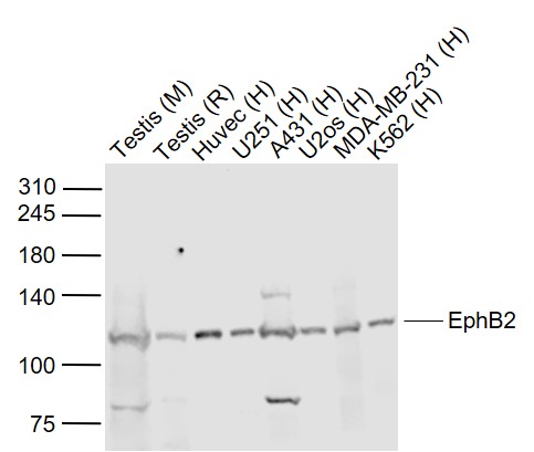
Sample:
Lane 1: Testis (Mouse) Lysate at 40 ug
Lane 2: Testis (Rat) Lysate at 40 ug
Lane 3: Huvec (Human) Cell Lysate at 30 ug
Lane 4: U251 (Human) Cell Lysate at 30 ug
Lane 5: A431 (Human) Cell Lysate at 30 ug
Lane 6: U2os (Human) Cell Lysate at 30 ug
Lane 7: MDA-MB-231 (Human) Cell Lysate at 30 ug
Lane 8: K562 (Human) Cell Lysate at 30 ug
Primary: Anti-EphB2 (bs-0996R) at 1/1000 dilution
Secondary: IRDye800CW Goat Anti-Rabbit IgG at 1/20000 dilution
Predicted band size: 125 kD
Observed band size: 120 kD
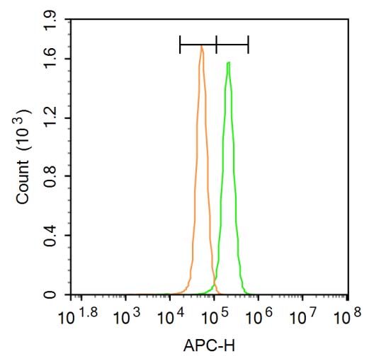
Blank control: A431.
Primary Antibody (green line): Rabbit Anti-EphB2 antibody (bs-0996R)
Dilution: 3μg /10^6 cells;
Isotype Control Antibody (orange line): Rabbit IgG .
Secondary Antibody: Goat anti-rabbit IgG-AF647
Dilution: 3μg /test.
Protocol
The cells were fixed with 4% PFA (10min at room temperature)and then permeabilized with 20% PBST for 20 min at room temperature. The cells were then incubated in 5%BSA to block non-specific protein-protein interactions for 30 min at room temperature .Cells stained with Primary Antibody for 30 min at room temperature. The secondary antibody used for 40 min at room temperature. Acquisition of 20,000 events was performed.
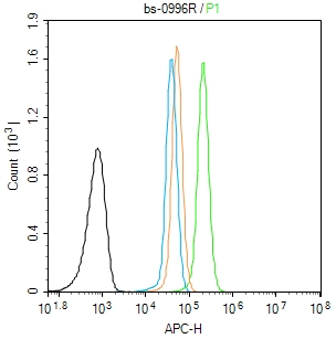
Blank control (Black line): A431 (Black). Primary Antibody (green line): Rabbit Anti-EphB2 antibody (bs-0996R) Dilution: 1μg /10^6 cells; Isotype Control Antibody (orange line): Rabbit IgG . Secondary Antibody (white blue line): Goat anti-rabbit IgG-AF647 Dilution: 1μg /test. Protocol The cells were fixed with 4% PFA (10min at room temperature)and then permeabilized with 0.1% PBST for 20 min at room temperature. The cells were then incubated in 5%BSA to block non-specific protein-protein interactions for 30 min at room temperature.Cells stained with Primary Antibody for 30 min at room temperature. The secondary antibody used for 40 min at room temperature. Acquisition of 20,000 events was performed.
|
| 1、抗体溶解方法 | |
| 2、抗体修复方式 | |
| 3、常用试剂的配制 | |
| 4、免疫组化操作步骤 | |
| 5、免疫组化问题解答 | |
| 6、Western Blotting 操作步骤 | |
| 7、Western Blotting 问题解答 | |
| 8、关于肽链的设计 | |
| 9、多肽的溶解与保存 | |
| 10、酶标抗体效价测定程序 | |


