

| 产品编号 | bs-1529R |
| 英文名称 | CD46 Rabbit pAb |
| 中文名称 | 膜辅蛋白抗体 |
| 别 名 | Mcp; AHUS2; MIC10; TLX; TRA2.10; MCP_HUMAN; CD46; Trophoblast leukocyte common antigen; MCP_MOUSE; MCP_RAT; |
| 研究领域 | 免疫学 糖蛋白 |
| 抗体来源 | Rabbit |
| 克隆类型 | Polyclonal |
| 克 隆 号 | |
| 交叉反应 | Human,Mouse |
| 产品应用 | WB=1:500-2000,IHC-P=1:100-500,IHC-F=1:100-500,IF=1:100-500,Flow-Cyt=1μg /test
not yet tested in other applications. optimal dilutions/concentrations should be determined by the end user. |
| 理论分子量 | 43 kDa |
| 检测分子量 | 52 |
| 细胞定位 | 细胞膜 |
| 性 状 | Liquid |
| 浓 度 | 1mg/ml |
| 免 疫 原 | KLH conjugated synthetic peptide derived from human CD46: 251-355/355 <Extracellular> |
| 亚 型 | IgG |
| 纯化方法 | affinity purified by Protein A |
| 缓 冲 液 | 0.01M TBS (pH7.4) with 1% BSA, 0.02% Proclin300 and 50% Glycerol. |
| 保存条件 | Shipped at 4℃. Store at -20℃ for one year. Avoid repeated freeze/thaw cycles. |
| 注意事项 | This product as supplied is intended for research use only, not for use in human, therapeutic or diagnostic applications. |
| PubMed | PubMed |
| 产品介绍 |
The protein encoded by this gene is a type I membrane protein and is a regulatory part of the complement system. The encoded protein has cofactor activity for inactivation of complement components C3b and C4b by serum factor I, which protects the host cell from damage by complement. In addition, the encoded protein can act as a receptor for the Edmonston strain of measles virus, human herpesvirus-6, and type IV pili of pathogenic Neisseria. Finally, the protein encoded by this gene may be involved in the fusion of the spermatozoa with the oocyte during fertilization. Mutations at this locus have been associated with susceptibility to hemolytic uremic syndrome. Alternatively spliced transcript variants encoding different isoforms have been described. [provided by RefSeq, Jun 2010]. Function: Acts as a cofactor for complement factor I, a serine protease which protects autologous cells against complement-mediated injury by cleaving C3b and C4b deposited on host tissue. May be involved in the fusion of the spermatozoa with the oocyte during fertilization. Also acts as a costimulatory factor for T-cells which induces the differentiation of CD4+ into T-regulatory 1 cells. T-regulatory 1 cells suppress immune responses by secreting interleukin-10, and therefore are thought to prevent autoimmunity. A number of viral and bacterial pathogens seem to exploit this property and directly induce an immunosuppressive phenotype in T-cells by binding to CD46. Subunit: Interacts with C3b and C4b. Binds to Measles virus H protein, to Human herpesvirus 6 GH protein and to human adenovirus B/D PIV/fiber protein, and acts as a receptor for these viruses. Binds to Streptococcus pyogenes M protein and to type IV pili from Neisseria, and may act as a receptor for these pathogenic bacteria. Subcellular Location: Cytoplasmic vesicle, secretory vesicle, acrosome inner membrane; Single-pass type I membrane protein. Note=Inner acrosomal membrane of spermatozoa. Internalized upon binding of Measles virus, Herpesvirus 6 or Neisseria gonorrhoeae, which results in an increased susceptibility of infected cells to complement-mediated injury. In cancer cells or cells infected by Neisseria, shedding leads to a soluble peptide. Tissue Specificity: Expressed by all cells except erythrocytes. Post-translational modifications: N-glycosylated on Asn-83; Asn-114 and Asn-273 in most tissues, but probably less N-glycosylated in testis. N-glycosylation on Asn-114 and Asn-273 is required for cytoprotective function. N-glycosylation on Asn-114 is required for Measles virus binding. N-glycosylation on Asn-273 is required for Neisseria binding. N-glycosylation is not required for human adenovirus binding. Extensively O-glycosylated in the Ser/Thr-rich domain. O-glycosylation is required for Neisseria binding but not for Measles virus or human adenovirus binding. In epithelial cells, isoforms B/D/F/H/J/L/3 are phosphorylated by YES1 in response to infection by Neisseria gonorrhoeae; which promotes infectivity. In T-cells, these isoforms may be phosphorylated by Lck. DISEASE: Defects in CD46 are a cause of susceptibility to hemolytic uremic syndrome atypical type 2 (AHUS2) [MIM:612922]. An atypical form of hemolytic uremic syndrome. It is a complex genetic disease characterized by microangiopathic hemolytic anemia, thrombocytopenia, renal failure and absence of episodes of enterocolitis and diarrhea. In contrast to typical hemolytic uremic syndrome, atypical forms have a poorer prognosis, with higher death rates and frequent progression to end-stage renal disease. Note=Susceptibility to the development of atypical hemolytic uremic syndrome can be conferred by mutations in various components of or regulatory factors in the complement cascade system. Other genes may play a role in modifying the phenotype. Patients with CD46 mutations seem to have an overall better prognosis compared to patients carrying CFH mutations. Similarity: Contains 4 Sushi (CCP/SCR) domains. SWISS: P15529 Gene ID: 4179 Database links: Entrez Gene: 4179 Human Entrez Gene: 17221 Mouse Omim: 120920 Human SwissProt: P15529 Human SwissProt: O88174 Mouse Unigene: 510402 Human Unigene: 12884 Mouse Unigene: 163242 Rat MCP为一单链穿膜糖蛋白,是补体系统的一种新的调节蛋白,是内源性辅助因子活性蛋白,属于RCA基因簇的成员。CD46通过GPI锚固定于细胞上。MCP的细胞分布广范,在粒细胞、血小板、T细胞(Th、Ts、Tc)、B细胞、NK细胞、造血细胞系、成纤维细胞、表皮细胞、内皮细胞及星状胶质细胞等都有分布。 |
| 产品图片 |
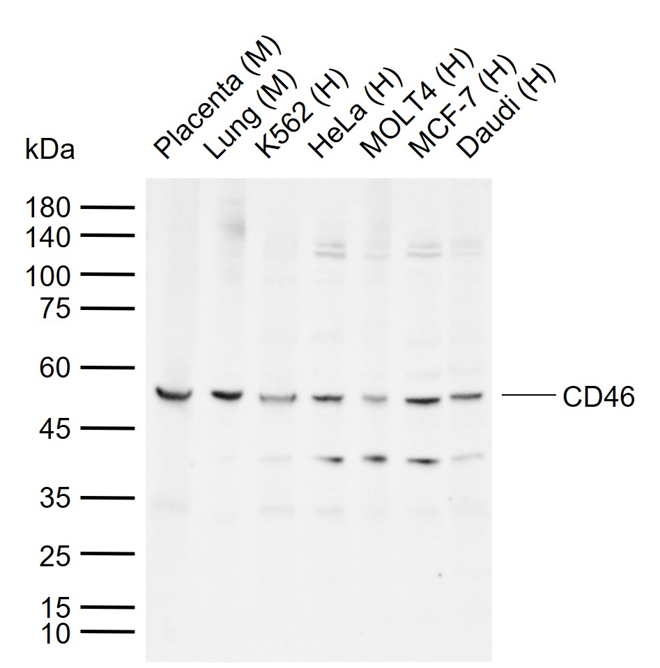
Sample:
Lane 1: Mouse Placenta tissue lysates
Lane 2: Mouse Lung tissue lysates
Lane 3: Human K562 cell lysates
Lane 4: Human HeLa cell lysates
Lane 5: Human MOLT4 cell lysates
Lane 6: Human MCF-7 cell lysates
Lane 7: Human Daudi cell lysates
Primary: Anti-CD46 (bs-1529R) at 1/1000 dilution
Secondary: IRDye800CW Goat Anti-Rabbit IgG at 1/20000 dilution
Predicted band size: 43 kDa
Observed band size: 52 kDa
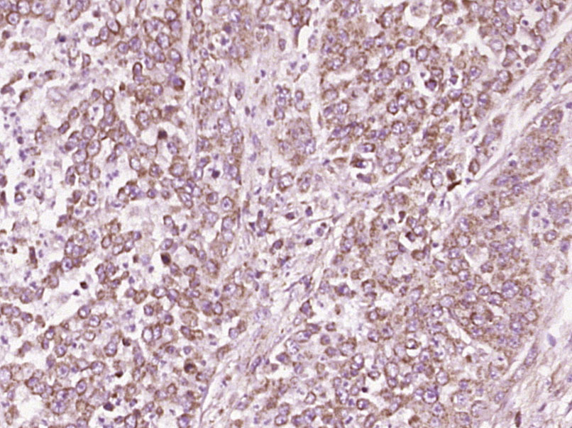
Paraformaldehyde-fixed, paraffin embedded (Human colon carcinoma); Antigen retrieval by boiling in sodium citrate buffer (pH6.0) for 15min; Block endogenous peroxidase by 3% hydrogen peroxide for 20 minutes; Blocking buffer (normal goat serum) at 37°C for 30min; Antibody incubation with (CD46) Polyclonal Antibody, Unconjugated (bs-1529R) at 1:400 overnight at 4°C, followed by operating according to SP Kit(Rabbit) (sp-0023) instructionsand DAB staining.
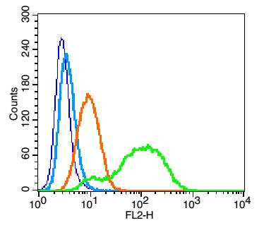
Blank control: U937 (blue). Primary Antibody: Rabbit Anti- CD46 antibody(bs-1529R), Dilution: 1μg in 100 μL 1X PBS containing 0.5% BSA; Isotype Control Antibody: Rabbit IgG(orange) ,used under the same conditions ); Secondary Antibody: Goat anti-rabbit IgG-PE(white blue), Dilution: 1:200 in 1 X PBS containing 0.5% BSA.
Protocol
The cells were fixed with 2% paraformaldehyde (10 min). Primary antibody (bs-1529R, 1μg /1x10^6 cells) were incubated for 30 min on the ice, followed by 1 X PBS containing 0.5% BSA + 1 0% goat serum (15 min) to block non-specific protein-protein interactions. Then the Goat Anti-rabbit IgG/PE antibody was added into the blocking buffer mentioned above to react with the primary antibody at 1/200 dilution for 30 min on ice. Acquisition of 20,000 events was performed.
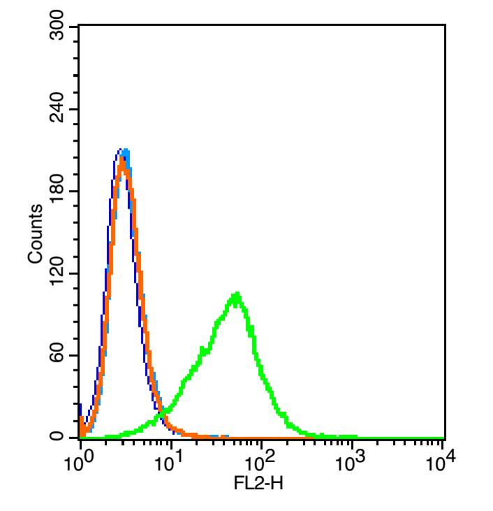
Blank control: 293T(blue).
Primary Antibody:Rabbit Anti-CD46 antibody(bs-1529R), Dilution: 1μg in 100 1μL 1X PBS containing 0.5% BSA(green);
Isotype Control Antibody: Rabbit IgG(orange), used under the same conditions );
Secondary Antibody: Goat anti-rabbit IgG-PE(white blue), Dilution: 1:200 in 1 X PBS containing 0.5% BSA.
protocol
The cells were washed twice with phosphate-buffered saline (PBS).The cells were then incubated in 1 X PBS containing 0.5% BSA + 10% goat serum (15 min) to block non-specific protein-protein interactions followed by the antibody (bs-1529R, 1μg/1x10^6 cells) for 30 min on ice. The secondary antibody used was Goat Anti-rabbit IgG/PE antibody at 1/200 dilution for 30 min on ice.Acquisition of 20,000 events was performed.
|
| 1、抗体溶解方法 | |
| 2、抗体修复方式 | |
| 3、常用试剂的配制 | |
| 4、免疫组化操作步骤 | |
| 5、免疫组化问题解答 | |
| 6、Western Blotting 操作步骤 | |
| 7、Western Blotting 问题解答 | |
| 8、关于肽链的设计 | |
| 9、多肽的溶解与保存 | |
| 10、酶标抗体效价测定程序 | |


